Anti-proliferating Effects and Gene Expression Profiles through Antioxidant Activity of Porphyra yezoensis Fractions on Human HepG2 Cell Lines
Youn Jeong Oh
1, Jung Min Kim
2and In Seok Bang
1*
1
Departmentof Biological Science and the Research Institute for Basic Sciences, Hoseo University, Asan 31499, Korea
2
Genoplan, Inc. & NAR Center, Inc., Teheran-ro 216, Seoul 06221, Korea
Received November 13, 2017 /Revised December 5, 2017 /Accepted December 19, 2017
In this study, the total polyphenol contents, antioxidant activities and anti-proliferation effects of HepG2 cell lines in organic slovent fractions obtained from the main methanolic extract of P. yezoensis were analyzed. The polyphenol content of the CHCl
3fraction was 10.3 μg/mg, slightly less than 13.08 μg/mg of the water fraction, but ED50 estimated by measuring DPPH free radical scavenging activity exhibited the highest 16.96 μg/ml in the CHCl
3fraction. The proliferation effects of CHCl
3and EtOAc fraction toward HepG2 cells inhibited in a dose-dependent manner, showed 90% inhibition when treated for 24 hr at 900 μg/ml of CHCl
3fraction. Meanwhile gene expression patterns in HepG2 cells treated 50 μg/ml of CHCl
3fraction were identified with microarray analysis. Concerning the efficacy of P. yezoensis, gene ontology analysis explored the genes associated with response to molecule of bac- terial origin, vitamin D metabolic process, and response to nutrient. Thus IL6R, CYP1A1 were selected as significant genes based on expression patterns of HepG2 cells, and pathway analysis indicates that ARNT might be considered as a upstream regulator. Also, expression analysis of IL6R and CYP1A1, activity of upstream regulator ARNT in HepG2 cells was confirmed based on Western blotting analy- sis at the protein level after being treated with 50 and 100 μg/ml of CHCl
3fraction.
Key words : Antioxidant activity, gene ontology, HepG2, microarray, P. yezoensis
*Corresponding author
*Tel : +82-41-540-9595, Fax : +82-41-548-6231
*E-mail : [email protected]
This is an Open-Access article distributed under the terms of the Creative Commons Attribution Non-Commercial License (http://creativecommons.org/licenses/by-nc/3.0) which permits unrestricted non-commercial use, distribution, and reproduction in any medium, provided the original work is properly cited.
Journal of Life Science 2018 Vol. 28. No. 2. 176~186 DOI : https://doi.org/10.5352/JLS.2018.28.2.176
서 론
김(purple laver)은 식물분류학적으로 식물계(Plantae), 홍 조식물문(Rhodophyta), 홍조강(Rhodophyceae), 원시홍조아 강(Bangiophycidae), 보라털(김)목(Bangiales), 보라털(김)과 (Bangiaceae), 김속(Porphyra)에 속하는 식용 해조로 해의(海 衣), 자채(紫菜), 청태(靑苔), 감태(甘苔)라고도 한다[3, 10]. 김 은 전 세계적으로 약 80여종이 있으며 우리나라에는 둥근돌김 (P. suborbiculata), 긴잎돌김(P. pseudolinearis), 미역김(P. denta-
ta) 등 20여 종 이상이 분포하고, 양식하는 김은 참김(P. tenera),방사무늬돌김(P. yezoensis), 모무늬돌김(P. seriata) 그리고 잇바 디돌김(P. dentata)의 4종이 있다[7]. 우리나라 외에도 일본, 중 국, 대만 등 일부 아시아 국가에서 양식되고 있으며[3], 식용의 역사 및 주 소비지역은 우리나라와 일본 등의 아시아 지역에 서 많이 애용되어 왔다[25]. 최근에 김은 다양한 생리활성 기능 이 보고되어 새로운 약의 개발, 건강 식단 등의 개발연구로 그 관심과 가치가 높아지고 있다[2, 4, 22]. 또한 식품으로서
우수한 기호성과 영양 생리학적인 면에서 주목을 받게 됨으로 주 소비지역이 아시아 지역으로 치중되었던 과거와 달리 세계 화 추세를 통하여 점차 국제적인 인지도가 증가하고 있다[28].
김은 식이섬유, 타우린(taurine), 다중불포화지방산, 필수아 미노산, 마이코스포린 유사 아미노산(mycosporine-like ami- no acid, MAAs), 폴리페놀 뿐만 아니라 각종 무기질과 비타민, 그리고 상대적으로 높은 양의 단백질이 함유되어 있다[8]. 식 이섬유의 주요 당은 isofloridoside, floridoside 등의 유리당과 세포벽 구성성분으로 불용성 다당인 hemicellulose, 그리고 세 포간 충전물질로서 수용성 산성 다당인 porphyran으로 이루 어져 있다[26]. 특히 porphyran은 항종양[17, 18, 27], 항염[26, 30], 항산화[24, 36], 항고지혈증과 고콜레스테롤성[11]에 대한 활성, 그리고 혈압[16, 32] 및 면역[9] 조절 등의 다양한 생물학 적 활성이 보고되고 있다. 김의 타우린(taurine)은 혈중 콜레스 테롤을 낮추며, ω-3지방산인 에이코사펜타엔산(eicosapentae- noic acid, EPA)은 혈액을 맑게 하는 것으로 알려졌다 [13].
필수 아미노산은 김이 가지는 독특한 맛을 나타내며[20], MAAs는 UV에 노출된 피부세포 보호[15]와 지방세포의 억제 [14] 효과가 있다. 또한 폴리페놀, 플라보노이드 등에 의한 항 산화 활성[6]이 보고되었다.
김에 함유된 다양한 생리활성물질에 의한 인체 내의 여러
가지 조절 기능에 관한 활발한 연구가 진행되고 있으나, 세포
내에서의 작용 기전에 대해서는 연구된 바가 없다. 본 연구는
생리활성 기능 성분을 활용한 고부가가치의 기능성 식품 소재
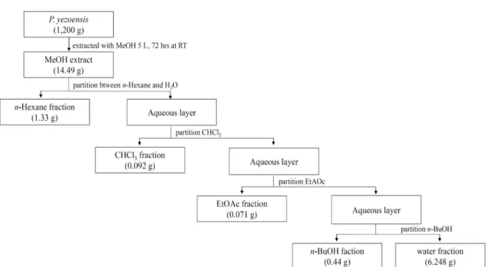
Fig. 1. Schematic diagram of extraction and fractionation of P. yezoensis.
화 기술개발의 일환으로 P. yezoensis의 methanol (MeOH) 추 출에 의한 유기 용매별 분획물로부터 폴리페놀 함량, 항산화 활성 그리고 인간 간암세포주인 HepG2 세포의 증식 억제를 평가하였다. 또한 이를 토대로 P. yezoensis의 chloroform (CHCl
3) 분획물이 HepG2 세포에 미치는 작용 양상을 DNA microarray로 확인하여 김이 HepG2 세포의 유전자 발현에 미치는 영향을 규명하고자 하였다.
재료 및 방법
재료 및 시약
P. yezoensis를 충남 서천군 소재의 양식장에서 생물 상태의
잎과 줄기를 제공 받아 세척 후 쇄절하여 실험에 사용하였다.
추출 및 분획물의 유기용매로는 MeOH, n-hexane, CHCl
3, ethyl acetate (EtOAc), n-butanol (n-BuOH)을 Duksan Co.
(Korea)로부터 구입하여 사용하였다. HepG2 (human hep- atocellular carcinoma cells) 세포는 호서대학교 혈관 생물학 연구실에서 제공받았으며, MEM (Minimum Essential Med- ium)과 DPBS (Dulbecco's Phosphate-Buffered Salines)는 WELGEN (Korea)에서 구입하였다. FBS (Fetal Bovine Serum) 은 Wisent (USA)에서 구입하였고 MTT (3-(4,5-dimethylth- iazol-2-yl)-2,5-diphenyl-2H-tetrazolium bromide), DPPH (1,1- diphenyl-2-picryl-hydrazyl), 0.2 M Folin-Ciocalteu’s phenol reagent, galic acid는 Sigma (USA)에서 구입하였다. Penicillin- streptomycin은 Gibco (USA)에서 항체는 Ab frontier (Korea) 와 Sigma (USA)에서 각각 구입하여 사용하였다.
분획물 제조
젖은 상태의 P. yezoensis 1,200 g에 5 liter의 MeOH을 가하 여 상온에서 72시간 동안 교반하면서 3회 반복 추출한 후 여과 시켜 35℃ 항온수조(Oilbath, Switzerland)에서 회전 감압농축 기(Rotavapor, Switzerland)로 감압하여 MeOH 추출 분획물 을 얻었다. MeOH 추출 이후 용매에 따른 추출 분획물은 n- hexane, CHCl
3, EtOAc, n-BuOH, water 순으로 추출하였다.
두 번째 용매 n-hexane 추출은 얻은 MeOH 분획물에 water와
n-hexane을 1:9:10의 비율로 혼합한 후 분액깔대기에 넣어 강한 혼합과 정지를 반복하여 water 층과 분획 용매가 두 층으로 나뉘도록 방치하여 n-hexane 분획물을 얻었다. 이 후 차례로 CHCl
3, EtOAc, 및 n-BuOH 순으로 분액깔대기에서 각 용매를 이용하여 추출한 후 마지막으로 남은 water 잔류액을 회수 water 추출을 분획화 하였다(Fig. 1).
폴리페놀 함량 측정
P. yezoensis의 MeOH 추출에 의한 유기용매 별 분획물의
폴리페놀 함량은 Folin-Denis 법[37]에 따라 측정하였다. 20
mg/ml 농도의 분획 별 시료 20 μl에 0.2 M Folin-Ciocalteu’s
phenol 용액(Sigma-Aldrich Co., USA) 20 μl를 가하여 실온에
서 3분 동안 반응시킨 다음 20 μl의 10% Na
2CO
3용액과 140
μl의 증류수를 가하여 암소에서 1시간 동안 방치 후 700 nm에
서 흡광도를 측정하였다. 폴리페놀의 함량은 0~500 μg/ml 농
도의 galic acid를 표준물질로 한 표준곡선에 의하여 산출하였
다.
DPPH 자유라디칼 소거능 측정
항산화 활성은 DPPH (1,1-diphenyl-2-picryl-hydrazyl)의 자유라디칼 소거효과(free radical scavenging effect)를 측정하 는 Blois[1]의 방법에 따라 측정하였다. MeOH에 녹인 0.2 mM DPPH 용액 180 μl에 MeOH에 용해시킨 각 분획의 농도 별 시료 20 μl를 혼합하여 실온에서 30분 동안 암실 방치 후 517 nm에서 흡광도를 측정하였다. 양성대조물로는 ascorbic acid (vitamin C)를 실험군과 동일한 농도로 사용하였고 시료의 전 자공여능(electron donating ability, EDA)은 다음의 식으로 계 산하였다.
EDA (%) = (1-A/B) ×100
A: 517 nm에서의 시료첨가군의 흡광도 B: 517 nm에서의 무첨가군의 흡광도
각 분획물의 EDA (%) 값을 바탕으로 흡광도를 50% 감소시 키는데 필요한 시료의 양 ED
50(mg/ml)을 구하고, ascorbic acid와 비교하여 P. yezoensis의 MeOH 추출에 의한 유기용매 별 분획물의 항산화 활성을 검정하였다.
HepG2 세포 배양
HepG2 세포를 10% fetal bovine serum (FBS)와 100 units/
ml penicillin-streptomycin이 포함된 minimum essential me- dia (MEM) 배지를 사용하여 37℃, 5% CO
2배양기에서 배양하 였다. 배양 시 주 2~3회 동일 배지로 갈아준 다음 배양 6~7일 경과 후 PBS로 세척 및 0.05% trypsin-0.02% EDTA로 부착된 세포를 분리 및 원심분리하여 새로운 MEM 배지로 재 부유하 여 세포 수를 1×10
5cell/ml로 조절하여 사용하였다.
HepG2 세포 증식 억제효과 측정
조절된 HepG2 세포를 96 well plate에 각 100 μl를 분주하 여 37℃, 5% CO
2배양기에서 24시간 동안 세포를 부착시킨 후, DMSO에 녹인 P. yezoensis의 각 분획물의 농도 별 200 μl를 첨가하여 48시간 동안 배양하고 각 well에 PBS 완충액에 녹인 10 μl의 MTT 용액(10 mg/ml)을 첨가하여 동일 배양조건에서 2시간 동안 반응시켰다. 이 때 생성된 formazan 결정체에 100 μl의 DMSO (dimethyl sulfoxide)로 용해시켜 ELISA reader (TECAN, USA)로 570 nm에서 흡광도를 측정하여 대조군 세 포수를 100%로 하였을 때의 상대적인 세포성장 억제율을 구 하였다.
Total RNA 추출
P. yezoensis의 MeOH 추출에 의한 CHCl3
분획물 50 μg/ml 과 대조군으로 DMSO가 6시간 또는 24시간 동안 각각 처리된 HepG2 세포를 회수하고, TRIzol® REAGENT (Invitrogen, USA)를 이용하여 total RNA를 추출하였다. Total RNA 정량 및 정성분석은 RNA nano kit (2100 Bioanalyzer System, Agilent Techologies, USA)를 사용하였다.
Microarray 분석
각 시료의 total RNA로부터 target cRNA의 합성(amplifi- cation and labeling) 및 혼성화 반응(nucleic acid hybrid- ization)은 Agilent’s Low RNA Input Linear Amplification Kit PLUS (Agilent Technology)를 이용하여 수행하였다. 대조 군 시료는 cyanine 3, 실험군 시료는 cyanine 5로 각각을 증폭 및 라벨시켰다. 증폭 및 라벨된 cRNA는 RNase Mini Spin Columns (Qiagen, Hilden, Germany)를 이용하여 정제, ND- 1000 spectrophotometer (NanoDrop Technologies, Wilming- ton, DE, USA)를 이용하여 정량하였다. 750 ng cRNA를 이용 하여 hybridization buffer를 만들고 Agilent Whole Human Genome Oligo Microarray (44K)에서 혼성화 반응을 실시하 였다. Hybridization image는 Agilent DNA microarray scan- ner를 이용하여 스캔하였으며, 데이터 정량은 Agilent Feature Extraction software를 이용하였다. 모든 데이터의 normal- ization (LOWESS)과 fold-changed genes의 선택은 GeneSpring GX 7.3 (Agilent Technology)으로 수행하였다. 각 실험군에서 flag-out genes들은 제거하였으며, normalized ratio는 signal channel intensity 중간값을 control channel intensity 중간값 으로 나누어 산출하였다. 유전자 선정(gene selection)을 위한 functional annotation은 Gene Ontology
TMConsortium (http://
www.geneontology.org/index.shtml) DB 정보를 따랐으며, IPA (ingenuity pathways analysis, http://www.ingenuity.
com)를 이용하여 상위 조절자(upstream regulator)를 탐색하 였다.
Western blotting
P. yezoensis의 CHCl3
분획물 50 또는 100 μg/ml이 48시간 동안 처리된 HepG2 세포를 PBS로 washing하여 수확하였다.
세포를 lysis buffer로 용해한 다음 13,000 rpm에서 15분 동안 원심분리하여 상층액을 분리하였다. 30 μg의 상층액을 8%
SDS-PAGE[19]로 분리시킨 후, Towbin 등[34]의 방법에 따라 PVDF membrane으로 150 V에서 1시간 동안 전이시켰다.
Membrane을 3% BSA가 포함된 0.05% PBST (0.05% Tween- 20, Phospho-buffered saline)로 1시간 동안 blocking 한 다음 1차 항체를 4℃에서 16시간 동안 반응시켰다. 다시 PBST로 5분 동안 3회 세척 후 2차 항체를 상온에서 1시간 동안 반응시 켰다. PBST로 5분 동안 3회 세척 후 ECL (ATTO, Japan)로 발광하여 X-ray 필름에 노출시켜 단백질 발현 분석을 실시하 였다.
통계처리
모든 실험은 최소한 3회 이상 실험을 실시하였으며, 실험군
당 평균±표준편차로 표현하였다. 데이터의 통계처리는 SPSS
program (ver. 12.0.01)을 이용하여 일원배치 분산분석(one-
way ANOVA)을 시행하였으며, 신뢰수준 p<0.05에서 평균값
Table 1. Yields of each organic solvent fraction obtained from the total MeOH extract
1)of P. yezoensis
Solvent fractions Dried weight (g) Yield (%)
2)n-hexane
CHCl
3EtOAc n-BuOH
H
2O
1.330 0.092 0.071 0.440 6.248
0.110 0.008 0.006 0.037 0.520
1)
MeOH extract (14.490 g) obtained from 1,200 g of P. yezoensis.
2)
yield (%) = Dried weight of solvent fractions (g) Dried weight of P. yezoensis (g) ×100
Table 2. Contents of total phenolics in each fraction of P. ye- zoensis
Solvent fractions Total phenolics (μg/mg)
1)n-hexane
CHCl
3EtOAc n-BuOH
H
2O
9.33±0.439 10.34±0.156 7.04±0.038 6.11±0.025 13.08±0.533
1)
Results are presented as mean ± SD (n=3).
Table 3. Absorbance change by DPPH radical scarvenging activity of each fraction of P. yezoensis
Sample Control Fractions
Conc.(mg/ml) Vitamin C n-Hexane CHCl
3EtOAc n-BuOH Water
20 10 5 1
0.223±0.003
a0.241±0.001
a0.275±0.007
a0.338±0.007
a1.649±0.013
a1.651±0.064
a1.825±0.017
a1.829±0.005
a0.853±0.021
a1.325±0.052
a1.562±0.018
a1.731±0.042
a1.325±0.101
a1.591±0.035
a1.856±0.038
a1.815±0.087
a1.238±0.064
a1.594±0.043
a1.770±0.021
a1.840±0.015
a1.535±0.027
a1.765±0.031
a1.807±0.037
a1.915±0.030
aBlank: 1.996. Values are mean ± SD, n=3.
a
significantly different (p<0.01) by ANOVA test.
들에 대한 유의성을 검증하였다.
결과 및 고찰
분획물의 수율
생물 상태의 P. yezoensis 1,200 g에서 얻어진 14.490 g의 MeOH 추출로부터 n-hexane (1.33 g), CHCl
3(0.092 g), EtOAc (0.071 g), n-BuOH (0.440 g), 그리고 water (6.248 g) 순으로 유기용매의 극성이 낮은 것부터 높은 순서로 순차 분획물을 얻었다. 각 분획물의 용매 별 수율은 water 분획물에서 0.52%
로 가장 높았고, 다음으로 n-hexane (0.11%), n-BuOH (0.037%), CHCl
3(0.008%), EtOAc (0.006%) 순으로 CHCl
3과 EtOAc 분 획물에서는 비교적 수율이 낮게 나타났다(Table 1).
폴리페놀 함량 및 항산화 활성
Galic acid를 표준물질로 환산 및 정량화 한 P. yezoensis의 분획물 별 폴리페놀 함량은 water (13.08 μg/mg), CHCl
3(10.34 μg/mg), n-hexane (9.33 μg/mg), EtOAc (7.04 μg/mg), 그리고 n-BuOH (6.11 μg/mg) 분획물 순으로 나타났다(Table 2). 식물에 함유되어 있는 페놀성 화합물은 항산화, 항암, 정장, 콜레스테롤 저하 작용 등 다양한 생리활성 기능을 가지고 있 는데 이는 화합물 내 phenolic hydroxyl group이 단백질과 같은 생체 내 거대분자와 쉽게 결합하는 성질을 가지고 있어 이러한 생리활성을 나타내는 것으로 알려져 있다[33]. 체내 존 재하는 자유라디칼(free radical)은 생체조직과 반응하여 질병 유발 및 노화과정을 유발하는데, phenolic hydroxyl group은 유지의 자유라디칼 수용체로서 유지 산화의 초기 단계에 생성 된 자유라디칼들이 안정된 화합물을 형성하도록 하여 산화억 제 작용을 한다[29]. 따라서 페놀성 화합물의 함량과 항산화 활성과의 관계는 식물의 항산화 연구에 있어서 필수적인 요소 라 할 수 있다[12].
P. yezoensis의 각 분획물과 DPPH의 반응 후 감소하는 흡광
도를 측정하여 얻은 자유라디칼 소거 활성법을 이용하여 항산 화 활성을 측정하였다(Table 3). 항산화제의 대표적 물질로 알 려진 ascorbic acid와 P. yezoensis의 용매 별 분획물을 1~20 mg/ml 농도로 각각 동일하게 처리하였을 때, P. yezoensis의 용매 별 분획물은 농도가 증가할수록 자유라디칼 소거활성이 증가하는 경향을 보이므로 농도 의존적인 항산화 활성을 나타 내었다. 전자공여능(EDA)에 의해 얻은 분획물 별 ED
50은 CHCl
3분획물에서 16.96 mg/ml으로 가장 높았으며, 다음으 로 n-BuOH, EtOAc, water, n-hexane 분획물 순으로 각각 27.51 mg/ml, 31.23 mg/ml, 50.17 mg/ml, 79.26 mg/ml의 ED
50값을 보였다(Fig. 2). 임 등[23]은 해조류 추출물의 폴리페 놀 함량이 높을수록 항산화 활성이 높다는 것을 보고하였다.
본 실험에서도 폴리페놀 함량이 높은 CHCl
3분획물에서 항산 화 활성이 뛰어남을 나타나는 유의한 결과를 얻었으며, 이는 자유라디칼 소거능과 폴리페놀 함량 간의 밀접한 연관을 나타 낸다. 한편 신 등[31]의 유기 용매별 분획물에 대한 활성 실험 에서는 n-hexane 분획물에서 높은 활성을 보였는데, 본 실험 과의 추출 및 분획 방법의 차이로 인한 결과로 여겨진다.
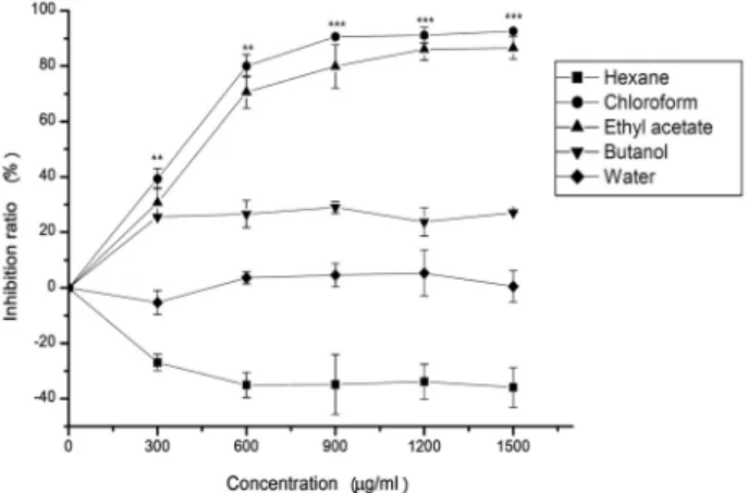
HepG2 세포 증식 억제효과
인간 간암세포주인 HepG2 세포의 증식억제에 대한 P. ye-
zoensis의 효과를 각 분획물의 농도를 300 μg/ml에서부터 300Fig. 2. DPPH free radical scavenging activity in various solvents fraction from P. yezoensis extract. ED
50values were de- fined as the sample concentrations required for 50% the radical to be scavenged. The data are presented as the mean±SD of three independent experiments performed in triplicate.
*p<0.05,
**p<0.01, and
***p<0.001 vs. the control.
Fig. 3. Inhibitory effect on cell growth of the fractions from P.
yezoensis extract on HepG2 cells. Cell viability was ana- lyzed using the MTT assay. The data are presented as the mean ± SD of three independent experiments per- formed in triplicate.
**p<0.01 and
***p<0.001 vs. the control. The significances in statistics are shown only in the results of CHCl
3fraction.
단위로 1,500 μg/ml까지 희석하여 24시간 동안 처리 한 후 MTT assay로 확인하였다(Fig. 3). CHCl
3분획물에서 각 농도 에 따라 39%, 80%, 90%, 91%, 92%의 높은 암세포 증식억제 효과를 나타내었고, 다음으로 EtOAc 분획물에서 30%, 70%, 80%, 86%, 86%의 암세포 증식억제 효과를 보였다. n-BuOH과 water 분획물에서는 HepG2 세포의 증식억제가 나타나지 않 았고, 반면 n-hexane 분획물은 600 μg/ml에서부터 약 30%의 미미한 암세포 증식효과를 보였다. 이는 신 등[31]이 P. ye-
zoensis의 MeOH 추출에 의한 용매별 분획물이 암 세포의 증식을 억제한다는 연구 결과와 일치한다. 이러한 결과로 미루어
볼 때 P. yezoensis의 MeOH 추출에 의한 CHCl
3분획물과 EtOAc 분획물은 암세포의 증식을 억제하는 생리활성물질이 존재하며, 이를 토대로 한 김 추출물이 암 세포의 생육을 억제 시키는 세포독성 연구[5]는 김에 함유되어 있는 화합물의 생리 적 효과 및 기능 연구[21]와 더불어 향후 새로운 암 예방 및 항암 식의약품 소재로의 개발 가능성을 제시한다.
유전체 발현 분석
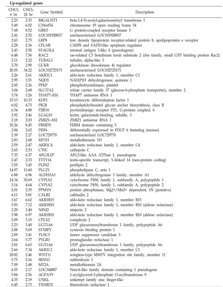
P. yezoensis의 효능 검증을 위한 분자생물학적 연구의 일환
으로 CHCl
3분획물이 처리된 HepG2 세포에 microarray 분석 에 의한 gene expression profile을 분석하였다. Time-depend- ent manner로 2-fold changed gene을 hierarchical cluster 분 석 결과, 초기 6시간에서 변화된 유전자 일부는 24시간에서도 유사한 발현패턴이 있는 것으로 나타났다. 따라서 이후의 분 석에서는 6시간 및 24시간 모두에서 변화된 유전자를 분석하 였다. HepG2 세포주에 CHCl
3분획물의 처리는 75개의 유전 자가 2 배 이상으로 발현의 증가를 보였고, 39개의 유전자에서 발현이 감소하였다(Table 4). 2 배 이상 발현 차이가 있는 유전 자를 대상으로 P. yezoensis의 효능 예측을 위한 Gene Ontology 분석으로 비타민 D 대사 과정(vitamin D metabolic process), 항균작용에 대한 반응(response to molecule of bacterial ori- gin), 그리고 영양물질에 대한 반응(response to nutrient)에 관련된 유의 유전자를 탐색하였다(Fig. 4). 이들 유의 유전자는 CYP1A1 (cytochrome P450, family 1, subfamily A, polypep- tide 1), CYP24A1 (cytochrome P450, family 24, subfamily A, polypeptide 1), IL6R (interleukin 6 receptor) 등의 유전자가 중복하여 포함되어 있었다(Fig. 5). 특히 CYP1A1 유전자의 경 우 6시간에서 14 배, 24시간에서 31 배로 발현이 증가되었다.
LOC102725171 유전자는 현재까지 기능이 잘 알려져 있지 않 은 유전자로 6시간에서 21 배, 24시간에서 34 배로 발현이 증 가된 것으로 나타나 후속 연구를 통하여 그 기능의 규명이 요구된다. Vigna 등[35]은 비타민 D 대사 과정과 항균작용에 대한 반응 관련 기능에서 비타민 D는 골격 형성에 필요한 칼 슘의 흡수에 관여하며, 포만감을 느끼게 하는 호르몬인 렙틴 (leptin)의 분비와 연관이 있고, 또한 체중감소에 영향을 줄 수 있다고 보고하였다. 따라서 김은 비타민 D의 대사를 촉진 하는 기능이 있을 것으로 예측되며, 언급한 영양물질에 대한 반응으로부터 식품영양학적 작용에 기여할 것으로 예상된다.
또한 항균 및 면역작용을 설명할 수 있는 예측된 기능은 항균 작용에 대한 반응으로 설명될 수 있다. 이들 유전자들은 신호 전달 경로(signal transduction pathways)의 상위 조절자인 ARNT (Aryl Hydrocarbon Receptor Nuclear Translocator)에 의해 조절됨이 IPA 분석으로부터 확인 되었다(data not shown).
단백질 발현 분석
상기 Gene Ontology 분석에서 P. yezoensis의 효능 예측을
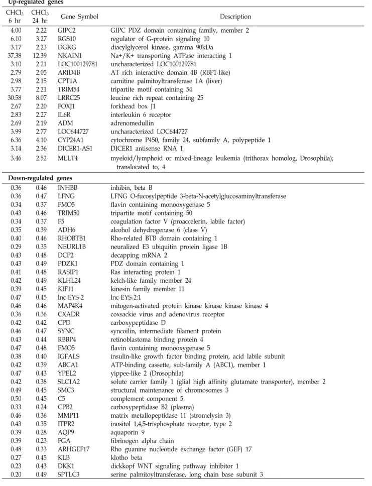
Table 4. The list of altered genes (2-fold changed genes) by CHCl
3fraction from P. yezoensis extract in the HepG2 cells Up-regulated genes
CHCl
36 hr
CHCl
324 hr Gene Symbol Description
2.23 2.33 B4GALNT1 beta-1,4-N-acetyl-galactosaminyl transferase 1 5.49 6.02 C19orf54 chromosome 19 open reading frame 54 7.68 8.52 GRK5 G protein-coupled receptor kinase 5 2.41 2.52 LOC101928837 uncharacterized LOC101928837
2.63 2.75 LRP8 low density lipoprotein receptor-related protein 8, apolipoprotein e receptor 2.28 2.36 CFLAR CASP8 and FADD-like apoptosis regulator
2.43 2.50 STAG3L4 stromal antigen 3-like 4 (pseudogene)
6.48 7.56 RAC2 ras-related C3 botulinum toxin substrate 2 (rho family, small GTP binding protein Rac2) 2.11 2.22 TUBAL3 tubulin, alpha-like 3
2.70 2.90 GCKR glucokinase (hexokinase 4) regulator 20.55 34.22 LOC102725171 uncharacterized LOC102725171
2.26 2.61 AKR1C1 aldo-keto reductase family 1, member C1 2.95 3.51 NQO1 NAD(P)H dehydrogenase, quinone 1 2.05 2.26 PFKP phosphofructokinase, platelet
3.04 2.68 SLC37A2 solute carrier family 37 (glucose-6-phosphate transporter), member 2 3.74 3.26 THAP7-AS1 THAP7 antisense RNA 1
15.13 10.33 KDF1 keratinocyte differentiation factor 1
6.02 4.73 PIGB phosphatidylinositol glycan anchor biosynthesis, class B 6.54 4.88 P2RY6 pyrimidinergic receptor P2Y, G-protein coupled, 6 2.92 2.46 LGALS3 lectin, galactoside-binding, soluble, 3
2.18 2.03 ZMIZ1-AS1 ZMIZ1 antisense RNA 1 10.26 8.45 FRMD5 FERM domain containing 5
2.84 2.62 DEF6 differentially expressed in FDCP 6 homolog (mouse) 2.39 2.27 LOC729770 uncharacterized LOC729770
2.83 2.68 MT1H metallothionein 1H
2.59 2.47 AKR1C4 aldo-keto reductase family 1, member C4
2.65 2.53 CTSC cathepsin C
7.35 6.37 AFG3L1P AFG3-like AAA ATPase 1, pseudogene
2.47 2.33 TTTY14 testis-specific transcript, Y-linked 14 (non-protein coding)
3.55 3.45 PLIN2 perilipin 2
14.97 13.60 PLCZ1 phospholipase C, zeta 1
6.88 6.96 ALDH3A1 aldehyde dehydrogenase 3 family, member A1 14.34 30.69 CYP1A1 cytochrome P450, family 1, subfamily A, polypeptide 1
3.14 4.64 CYP1A2 cytochrome P450, family 1, subfamily A, polypeptide 2 2.01 2.35 PPM1N protein phosphatase, Mg2+/Mn2+ dependent, 1N (putative)
4.11 5.81 CALB2 calbindin 2
3.67 6.62 AKR1B15 aldo-keto reductase family 1, member B15
3.93 7.12 AKR1B10 aldo-keto reductase family 1, member B10 (aldose reductase)
2.29 3.40 NINJ2 ninjurin 2
3.98 6.97 AKR1B10 aldo-keto reductase family 1, member B10 (aldose reductase)
2.09 3.15 CPLX2 complexin 2
2.23 3.40 UGT1A6 UDP glucuronosyltransferase 1 family, polypeptide A6 2.88 5.69 STXBP1 syntaxin binding protein 1
2.09 3.41 TUSC3 tumor suppressor candidate 3 2.64 5.77 PTGR1 prostaglandin reductase 1
2.81 6.63 UGT1A6 UDP glucuronosyltransferase 1 family, polypeptide A6 2.66 7.34 AKR1C1 aldo-keto reductase family 1, member C1
20.82 2.46 WNT11 wingless-type MMTV integration site family, member 11
5.75 2.16 SRXN1 sulfiredoxin 1
7.09 2.48 MT2A metallothionein 2A
4.55 2.17 LOC344887 NmrA-like family domain containing 1 pseudogene 5.84 2.56 AGPAT9 1-acylglycerol-3-phosphate O-acyltransferase 9 4.35 2.19 UNKL unkempt family zinc finger-like
6.45 2.73 TXNRD1 thioredoxin reductase 1
Table 4. Continued Up-regulated genes
CHCl
36 hr
CHCl
324 hr Gene Symbol Description
4.00 2.22 GIPC2 GIPC PDZ domain containing family, member 2 6.10 3.27 RGS10 regulator of G-protein signaling 10
3.17 2.23 DGKG diacylglycerol kinase, gamma 90kDa 37.38 12.39 NKAIN1 Na+/K+ transporting ATPase interacting 1
3.10 2.21 LOC100129781 uncharacterized LOC100129781
2.79 2.05 ARID4B AT rich interactive domain 4B (RBP1-like) 2.98 2.15 CPT1A carnitine palmitoyltransferase 1A (liver) 3.77 2.21 TRIM54 tripartite motif containing 54
30.58 8.07 LRRC25 leucine rich repeat containing 25
2.67 2.20 FOXJ1 forkhead box J1
2.83 2.27 IL6R interleukin 6 receptor
2.69 2.19 ADM adrenomedullin
3.99 2.77 LOC644727 uncharacterized LOC644727
6.36 4.10 CYP24A1 cytochrome P450, family 24, subfamily A, polypeptide 1 3.14 2.36 DICER1-AS1 DICER1 antisense RNA 1
3.46 2.52 MLLT4 myeloid/lymphoid or mixed-lineage leukemia (trithorax homolog, Drosophila);
translocated to, 4 Down-regulated genes
0.36 0.46 INHBB inhibin, beta B
0.36 0.47 LFNG LFNG O-fucosylpeptide 3-beta-N-acetylglucosaminyltransferase 0.34 0.37 FMO5 flavin containing monooxygenase 5
0.43 0.46 TRIM50 tripartite motif containing 50
0.34 0.37 F5 coagulation factor V (proaccelerin, labile factor) 0.35 0.39 ADH6 alcohol dehydrogenase 6 (class V)
0.40 0.46 RHOBTB1 Rho-related BTB domain containing 1 0.29 0.35 NEURL1B neuralized E3 ubiquitin protein ligase 1B
0.43 0.48 DCP2 decapping mRNA 2
0.43 0.49 PDZK1 PDZ domain containing 1 0.41 0.48 RASIP1 Ras interacting protein 1 0.42 0.49 KLHL24 kelch-like family member 24 0.39 0.45 KIF11 kinesin family member 11
0.47 0.45 lnc-EYS-2 lnc-EYS-2:1
0.46 0.46 MAP4K4 mitogen-activated protein kinase kinase kinase kinase 4 0.36 0.36 CXADR coxsackie virus and adenovirus receptor
0.42 0.42 CPD carboxypeptidase D
0.46 0.47 SYNC syncoilin, intermediate filament protein 0.43 0.44 RBBP4 retinoblastoma binding protein 4 0.47 0.48 FMO5 flavin containing monooxygenase 5
0.38 0.40 IGFALS insulin-like growth factor binding protein, acid labile subunit 0.42 0.39 ABCA1 ATP-binding cassette, sub-family A (ABC1), member 1 0.47 0.43 YPEL2 yippee-like 2 (Drosophila)
0.42 0.38 SLC1A2 solute carrier family 1 (glial high affinity glutamate transporter), member 2 0.49 0.45 SMC3 structural maintenance of chromosomes 3
0.50 0.45 C5 complement component 5
0.33 0.24 CPB2 carboxypeptidase B2 (plasma)
0.46 0.36 MMP11 matrix metallopeptidase 11 (stromelysin 3) 0.43 0.35 ITPR2 inositol 1,4,5-trisphosphate receptor, type 2
0.39 0.28 AQP9 aquaporin 9
0.39 0.23 FGA fibrinogen alpha chain
0.48 0.33 ARHGEF17 Rho guanine nucleotide exchange factor (GEF) 17
0.27 0.45 KLB klotho beta
0.23 0.43 DKK1 dickkopf WNT signaling pathway inhibitor 1
0.20 0.49 SPTLC3 serine palmitoyltransferase, long chain base subunit 3
Fig. 5. Hierarchical clustering of CHCl
3fraction from P. yezoensis extract on gene expression profiles in human HepG2 cell lines. The list of related genes with vitamin D metabolic process, response to molecule of bacterial origin, and re- sponse to nutrient in CHCl
3fraction-treated HepG2 cells.
The list shows that CYP1A1, CYP24A1, IL6R, and NQO1 were up-regulated in CHCl
3fraction-treated HepG2 cells.
Red represents genes with increased expression, whereas green represents genes with decreased expression.
A Up-regulated genes
B Down-regulated genes
Fig. 4. Gene ontology from gene expression profiles. Biological function of altered genes in response to CHCl
3fraction from P.
yezoensis in human HepG2 cell lines. Genes up-(A) and down-regulated (B) by more than two-fold were categorized into their main biological functions.
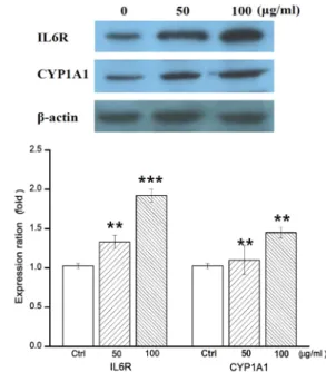
설명할 수 있는 유전자 CYP1A1와 IL6R가 세포 내에서도 동일 한 단백질 발현 양상으로 나타나는지를 확인하기 위하여 Western blotting을 수행하였다. P. yezoensis의 CHCl
3분획물
Fig. 6. Expression of CYP1A1 and IL6R in CHCl
3fraction-treat- ed HepG2 cell lines. Protein expression levels were de- termined by Western blotting. The y-axis indicates the relative expression levels obtained by Western blotting.
The data are presented as the mean ± SD of three in-
dependent experiments performed in triplicate.
**p<0.01
and
***p<0.001 vs. the control.
***
**
Ctrl 50 100 (μg/ml)
ARNT
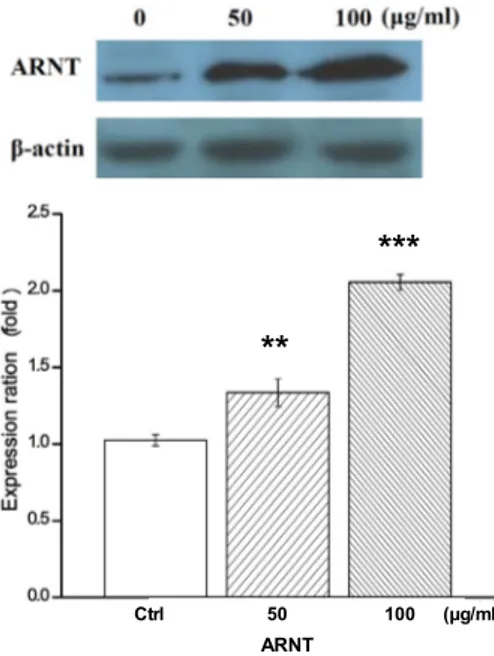
Fig. 7. Expression of up-regulator ARNT of CYP1A1 and IL6R.
The expression level of up-regulator ARNT was in- creased in CHCl
3fraction-treated HepG2 cell lines. The y-axis indicates the relative expression levels obtained by Western blotting. The data are presented as the mean
± SD of three independent experiments performed in triplicate.
**p<0.01 and
***p<0.001 vs. the control.
50, 100 μg/ml이 48시간 동안 처리된 HepG2 세포와 무처리구 (control)를 대조군 단백질 β-actin (loading control)의 발현 양으로 보정, 비교 시 IL6R와 CYP1A1의 단백질량이 증가하는 것을 확인하였다(Fig. 6). 또한 IPA 결과, CYP1A1, CYP24A1, IL6R 및 NQO1 유전자의 상위 조절자인 ARNT의 단백질량이 역시 증가하는 것으로 나타남을 확인하여 ARNT의 활성을 검 증하였다(Fig. 7).
따라서 P. yezoensis의 항산화 활성과 연관하여 인간 간암 세포주에서 효능 예측과 연관된 유전자들은 비타민 D 대사 과정, 항균작용에 대한 반응, 그리고 영양물질에 대한 반응 에 관련된 것들 이었다. 향후 HepG2 세포에서 P. yezoensis 의 효능과 연관되어 있는 IL6R, CYP1A1, 그리고 ARNT에 대하여 추가적인 분자생물학적 분석이 요구된다. 또한 효능 별 적합한 세포주를 재선정하여 추가적인 유전체 분석 및 분자기전 규명 연구를 수행하여 P. yezoensis의 생리활성 효 과에 대한 구체적인 영향을 평가하고 유효단일물질을 탐색 하고자 한다.
감사의 글
본 논문의 재료인 P. yezoensis를 제공하여 주신 충남 서천군 해양수산과에 감사드립니다.
References