C A S E R E P O R T Open Access
Treatment of anterior open bite by
posterior maxillary segmental osteotomy and miniplates: a case report
Sung-Kwon Choi 1 and Kyung-Hwan Kwon 2*
Abstract
Background: Anterior open bite is a challenging malocclusion to correct orthodontic treatment. Anterior open bite associated with over-erupted posterior teeth and long lower facial height should be treated by reduction of posterior dimension for esthetic results. Although the possibility of orthodontic treatment of an anterior open bite has increased with the introduction of skeletal anchorage, there are still cases requiring surgery for various reasons.
Case presentation: This case report covers an anterior open bite of a 25-year-old man successfully treated with the posterior maxillary segmental osteotomy (PMSO) and miniplates. After the pre-surgical orthodontic treatment, the PMSO between canines and first premolars was performed under local anesthesia and miniplates were placed on the zygomatic buttress. As a result of 28 months of treatment, an impaction amount of 3.5 mm was obtained in the maxillary posterior teeth, and the facial esthetics improved at rest and smile.
Conclusion: The impaction of the posterior dentoalveolar segment using the PMSO can be a good treatment option in patients with anterior open bite showing long lower facial height.
Keywords: Anterior open bite, Posterior maxillary segmental osteotomy (PMSO), Skeletal anchorages, Miniplates
Background
Anterior open bite is a challenging malocclusion to treat orthodontically. It could result from various factors such as habits, tongue postures, airway obstruction, vertical skeletal growth problems, and temporomandibular joint disorders [1–3].
Anterior open bite is often associated with over- erupted posterior teeth and long lower face height [4, 5].
In this type of malocclusion, intrusive mechanics are re- quired for optimal esthetic results. Before the introduc- tion of skeletal anchorages, a reduction of the posterior vertical dimension can be achieved by orthognathic sur- gery. The intrusion of posterior teeth became possible by the use of skeletal anchorages. However, the
treatment duration and retention of the treatment re- sults are still issues [6–8].
Posterior maxillary segmental osteotomy (PMSO) is a surgery for repositioning of the posterior dentoalveolar segment. It has been mainly used to make an intermaxil- lary space for the restoration of mandibular teeth [9, 10].
In the area of orthodontics, the segmental surgery can be used to correct the transverse and vertical discrepan- cies of the maxilla [11, 12]. The PMSO has advantages than orthodontic intrusion when there are many teeth to be intruded or when a large amount of intrusion is required.
This case report covers an anterior open bite of a 25- year-old man successfully treated with the PMSO and miniplates.
Case presentation
A 25-year-old male patient visited our Department of Orthodontics with a chief complaint of anterior open
© The Author(s). 2020 Open Access This article is licensed under a Creative Commons Attribution 4.0 International License, which permits use, sharing, adaptation, distribution and reproduction in any medium or format, as long as you give appropriate credit to the original author(s) and the source, provide a link to the Creative Commons licence, and indicate if changes were made. The images or other third party material in this article are included in the article's Creative Commons licence, unless indicated otherwise in a credit line to the material. If material is not included in the article's Creative Commons licence and your intended use is not permitted by statutory regulation or exceeds the permitted use, you will need to obtain permission directly from the copyright holder. To view a copy of this licence, visit http://creativecommons.org/licenses/by/4.0/.
* Correspondence: [email protected]
2
Department of Oral and Maxillofacial Surgery, College of Dentistry, Wonkwang University, Iksan, Republic of Korea
Full list of author information is available at the end of the article
analysis showed a skeletal class II relationship with a prognathic maxilla, normovergent pattern, and labiover- sion of maxillary and mandibular incisors (details are shown in Table 1). All third molars were fully erupted and well occluded.
The patient was diagnosed as skeletal class II and den- tal class I with normovergent pattern, anterior open bite, and proclination of the upper and lower incisors.
The first treatment option was a segmental Le Fort I osteotomy. This procedure can immediately reduce the large amount of posterior vertical dimension and correct two occlusal planes. Since the patient was unable to be hospitalized, he rejected orthognathic surgery under general anesthesia.
The second treatment option was orthodontic intru- sion of maxillary teeth using a skeletal anchorage sys- tem. A large amount of intrusion of all maxillary teeth except central and lateral incisors was needed for optimal esthetic results in this patient. We thought that miniplates were more suitable than mini-implants because miniplates can be placed api- cally enough and provide stable intrusive forces.
Along with miniplate placement, the supplementary
surgery was considered to reduce the treatment time.
A surgical impaction of posterior segments had the advantage of shortening the treatment time and re- tention of treatment results. Since the patient wanted treatment to end quickly, he agreed to this plan with PMSO and informed consent was obtained.
A fixed tongue crib was delivered to control tongue posture. 0.018-inch standard edgewise brackets were bonded, and 0.0175-inch twisted stainless steel wires were inserted in both arches. Brackets on maxillary ca- nines and premolars were bonded to specially designed position to widen the inter-radicular space, and step bends were added between maxillary canine and pre- molar to prevent extrusion of anterior teeth.
Fig. 1 Pre-treatment photographs
IMPA 100.9 96.5 97.4
Interincisal angle 107.9 114.6 112.6
Upper lip E-plane − 0.3 − 0.8 − 1.8
Lower lip E-plane 5.1 4.2 4.1
U1-NF 37.8 38.8 38.8
U6-NF 30.5 27.0 27.4
Four months later, the wires were changed to 0.016- inch stainless steel wires, and open coil springs were inserted between maxillary canines and premolars. After 12 months of alignment, 0.016 × 0.022-inch sectional stainless steel wire was inserted in the maxillary arch, and the patient was referred to the Department of Oral and Maxillofacial Surgery for the PMSO.
The surgery was performed under local anesthesia by dividing the left and right sides in consideration of dining convenience (Fig. 2). After full-thickness muco- periosteal flap was elevated, buccal bone cutting was initiated from inter-radicular space between canine and first premolar, taking care not to damage the root surface. The horizontal cutting was extended to pterygopalatine junction at 5 mm above the apices of molars. A bone fragment of 3 mm in width was re- moved from the horizontal osteotomy line for poster- ior impaction. The palatal bone was cut by curved osteotome without flap elevation to maintain the blood supply. The bone segment was moved apicolat- erally, and T-shaped miniplate was placed on the zygomatic buttress. Posterior teeth were ligated with 0.012-inch dead soft wire to miniplates to hold the bone segment on the place. After surgery, antibiotics
and non-steroidal anti-inflammatory drugs were prescribed.
The patient was followed up 2 days after the sur- gery. Moderate facial swelling and ecchymosis on his maxillary area were identified, but they were relieved after medication and wound irrigation of 2 weeks. A second surgery was performed at the opposite side after a month from the first surgery. The anterior open bite improved immediately after the surgery (Figs. 3 and 4).
A month later from the second surgery, the brackets on maxillary canine were repositioned to normal pos- ition, and 0.016-inch nickel-titanium archwire was placed in the maxillary arch. A mini-implant was placed at the center of the mid-palatal suture for further intru- sion of posterior teeth. Elastic threads were applied from the lingual button on posterior teeth to mid-palatal mini-implant.
The additional intrusion of posterior teeth was contin- ued for 9 months after the surgery. The maxillary right miniplate was loosened after 6 months from the first sur- gery, so we replaced it with a new miniplate. The arch- wires were subsequently changed to 0.014-inch, 0.016- inch, and 0.016 × 0.022-inch stainless steel wire in this
Fig. 2 Intraoperative photographs of posterior maxillary segmental osteotomy (PMSO) and miniplate placement. a Three-mm-wide buccal cortical bone was removed from the horizontal osteotomy line. b The posterior segment was impacted after the palatal bone cut by curved osteotome.
c The miniplate placed on the zygomatic buttress area
Fig. 3 Occlusal changes after the PMSO. a Before the PMSO. b After the PMSO
period. During the finishing stage, the torque of the left maxillary canine and dental midline was corrected.
After 28 months of treatment, a favorable occlusion was obtained, and orthodontic appliances were removed.
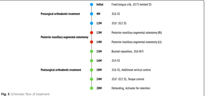
Fixed retainers were bonded on both arches, and the ac- tivator was delivered for retention. A schematic flow of the treatment is shown in Fig. 5.
Treatment results
Tongue habit was gradually improved with the use of tongue crib and was not recognized after the PMSO surgery.
Facial photographs showed the improvement of the patient ’s smile esthetics. Reverse smile arc and reverse lip line were corrected to a consonant smile arc and straight lip line, respectively. The exposure of lower teeth in a smile was decreased, and lip competence was achieved without mentalis strain (Fig. 6).
A post-treatment panoramic radiograph showed good root parallelism, and only mild blunting of the apices of
month retention, treatment results were stable without opening of mandibular plane angle (Fig. 7, Table 1).
Discussion
The introduction of skeletal anchorages has opened a new ground for the treatment of anterior open bite. In- trusion of the posterior teeth using skeletal anchorages provides more stable and esthetic results than extrusion of anterior teeth. However, the intrusion is one of the slowest movements among the various tooth move- ments. Therefore, the treatment duration of anterior open bite is mainly dependent on the rate of molar intrusion.
Various methods for the PMSO have been introduced according to the number of operations and the position of the osteotomy [11 – 13]. In this case, we followed the technique of Tuncer et al. This surgical method has the advantage of maintaining the palatal blood flow by not elevating the palatal flap. In addition, we impacted the posterior segments after removing the buccal cortical bone of 3 mm in width. To secure sufficient inter- radicular space, premolar extraction was also considered, but was dismissed because no posterior movement of the incisor was required.
Fig. 4 Post-surgical panoramic radiograph
Fig. 5 Schematic flow of treatment
Pulp necrosis occurred without apparent root damage on the adjacent tooth to the osteotomy line. Pink discol- oration of right maxillary first premolar was recognized 8 months from the surgery. At that time, the patient did not have any symptoms. After 14 months, however, he complained of pain during occlusion and tenderness to percussion on this tooth. It was diagnosed as pulp ne- crosis, and root canal treatment was performed. Many retrospective studies reported the prevalence of the pulp necrosis after Le Fort I osteotomy ranging 0.5 to 3.4%
[14–17]. Lownie et al. reported that ischemic stress in- duced by surgical procedures could result in inflamma- tory changes in pulp tissue, and there were degenerative changes with vacuolization and atrophy of the odonto- blastic layer [18]. The study using laser Doppler flowme- try showed that the segmental Le Fort I surgery induced a decrease of pulpal blood flow in adjacent to vertical osteotomy line [19]. In this case, osteotomy performed on a narrow inter-radicular space appears to have af- fected the pulpal blood supply.
Fig. 6 Post-treatment photographs
Fig. 7 Superimposition of the cephalometric tracing. Black line, before treatment; red line, after treatment (28 months after initiation of
orthodontic treatment); green line, retention after 49 months
A sufficient impaction of posterior teeth was achieved by PMSO surgery, but the premature contacts on the ca- nines prevented further closure of the mandible (Fig. 3).
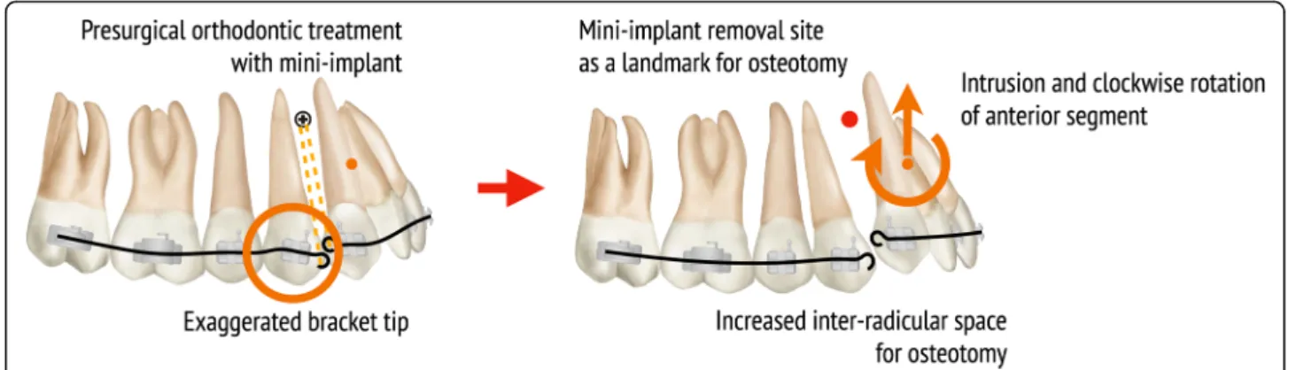
Considerable time was spent for intruding canines after PMSO surgery, and small amount of extrusion of man- dibular posterior teeth occurred during this time. If we had actively intruded the maxillary canines using mini- implants before the PMSO surgery, post-surgical ortho- dontic treatment would have been shortened. To reduce the risk of root damage and shorten the treatment period, it is helpful to actively intrude the anterior seg- ment by using a mini-implant and to improve the axis of the canine and premolar in pre-surgical orthodontic treatment (Fig. 8).
In this case, since the patient mainly had vertical prob- lems including anterior open bite and long lower facial height, the impaction of the posterior teeth was the main focus in the treatment plan. The impaction of the ` through PMSO was judged to be advantageous com- pared to orthodontic intrusion because all the teeth ex- cept the incisors were occluded. Functional occlusion and improvement of facial esthetics were obtained through orthodontic treatment including PMSO, and this treatment results have been maintained over 4 years.
Conclusion
The impaction of the posterior dentoalveolar segment using PMSO can be a good treatment option in patients with anterior open bite showing two occlusal planes. In order to reduce side effects and increase treatment effi- ciency, planned orthodontic movement must be per- formed in the pre-surgical orthodontic treatment, as with orthognathic surgery.
Acknowledgements No acknowledgement.
Authors ’ contributions
SKC drafted the manuscript and critically revised the manuscript. KHK participated in the study design and coordination and helped to draft the manuscript. All authors read and approved the final manuscript.
Funding No funding source.
Availability of data and materials
Not applicable (data sharing not applicable to this article as no datasets were generated or analyzed during the current study).
Ethics approval and consent to participate
This study was conducted after approval from the Institutional Review Board of Wonkwang University Dental Hospital (IRB no. WKDIRB202004-01), and the informed consent was waived.
Consent for publication
Written informed consent was obtained from the patient for the publication of this report and any accompanying images.
Competing interests
The authors declare that they have no competing interests.
Author details
1