Case Report
RECEIVED May 2, 2013, REVISED May 26, 2013, ACCEPTED September 25, 2013 Correspondence to Jong-Moon Chae
Department of Orthodontics, Wonkwang University Daejeon Dental Hospital 77 Dunsan-ro, Seo-gu, Daejeon 302-120, Korea
Tel: 82-42-366-1103, Fax: 82-42-366-1115, E-mail: [email protected]
CC
This is an open access article distributed under the terms of the Creative Commons Attribution Non-Commercial License (http://creativecommons.org/licenses/
by-nc/3.0) which permits unrestricted non-commercial use, distribution, and reproduction in any medium, provided the original work is properly cited.
Minimum Presurgical Orthodontic Treatment with Two Jaw Surgery Combined with Anterior Segmental Osteotmy in Skeletal Class II Malocclusion: A Case Report
Jong-Moon Chae, Jun-Young Paeng 1
Department of Orthodontics, School of Dentistry, University of Wonkwang, Wonkwang Dental Research Institute, Wonkwang Bone Regeneration Research Institute, The Korean Orthodontic Research Institute Inc.,
1Department of Oral and Maxillofacial Surgery, Kyungpook
National University Hospital
Abstract
This case report describes the treatment of a 23-year-old woman who had lip protrusion with gummy smile and mentalis muscle strain. Orthognathic surgery was performed in conjunction with orthodontics. Minimum dental decompensation was performed with presurgical orthodontics followed by an anterior segmental osteotomy for the majority of dental decompensation.
Counterclockwise rotation of the maxillomandibular complex was applied by LeFort I osteotomy, and bilateral sagittal split ramus osteotomies with anterior segmental osteotomy to achieve overall facial balance. The active treatment period was 15 months. Stable occlusion and skeletal relationship were observed after a 10-month follow-up period.
Key words: Minimum presurgical treatment, Skeletal Class II, Anterior segmental osteotomy, Counterclockwise rotation
Introduction
Orthognathic surgery is typically recommended to pa- tients if an orthodontic treatment alone is not sufficient to correct any malocclusion and, at the same time, camou- flage is not an option[1]. The goal of orthognathic surgery is to maximize or optimize skeletal changes upon reducing the pretreatment dental compensations to improve facial esthetics, function, and posttreatment stability[2]. Skeletal Class II malocclusion problems are usually due to a man- dibular deficiency or downward-backward rotation of the mandible caused by excessive vertical growth of the maxilla. The surgical management, therefore, consists of
mandibular advancement, superior repositioning of the maxilla, or a combination of the two[3]. In the conventional orthognathic surgery planning, the anteroposterior discrep- ancies are corrected by advancement or setback of the jaws along the existing occlusal plane. Otherwise, if the maxilla is manipulated in a vertical direction, the mandible will autorotate and alter the angle of occlusal plane[4].
Thus, selective alteration of the occlusal plane allows a maxillofacial surgeon to re-establish the correct and proper jaw function with respect to the cranial base and, con- sequently, offer an improved esthetic result for patients with dentoskeletal deformities[5].
Recently, surgical-orthodontic management of either sur-
Jong-Moon Chae: Minimum Pregsurgical Orthodontic Treatment 317
Fig. 1. Pretreatment facial and intraoral photographs.
gery-first or minimum presurgical orthodontic treatment has been proposed to address the issue of facial esthetics from the beginning of treatment. Then again, orthognathic treatment with an anterior segmental osteotomy in compar- ison to conventional orthodontic treatment can markedly reduce the presurgical orthodontic treatment period for the correction of a maxillary protrusion and decompensation of lower dentition[6-8].
The objective of this article was to present the treatment of a skeletal Class II malocclusion in a patient with gummy smile and a high mandibular plane angle. The counter- clockwise rotation of maxillomandibular complex with an- terior segmental osteotomy and genioplasty provided good maxillary incisor exposure and excellent smile arc, and improved the patient’s facial balance.
Case Report
The patient was a 23-year-old woman with chief con- cerns of lip protrusion, gummy smile, and retrognathic mandible. On exam and in facial photographs, the patient’s face revealed good symmetry but had a convex profile with marked protrusion of the lips, mentalis strain, and excessive lower anterior facial height. Intraoral exam re- vealed Class II canine and Class I molar relationships with minor crowding and poor oral hygiene. The patient had a deep overbite of 7.5 mm and a large overjet of 5.5 mm.
The dental midline was coincident with the facial midline (Fig. 1, 2).
The lateral cephalogram (Fig. 3) and its tracing con-
firmed a severe underlying skeletal problem. The skeletal
pattern was deemed hyperdivergent as evidenced by a
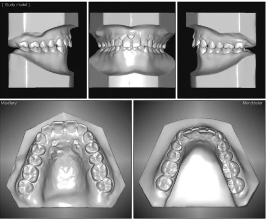
Fig. 2. Pretreatment dental casts.
Fig. 3. Pretreatment radiographs.
Frankfort-mandibularplane angle (FMA) of 37
o. The ANB angle of 6.5
o(SNA, 83
o; SNB, 76.5
o) reflected a class-II skeletal problem. The Z-angle of 50.5
oquantified the over- all facial imbalance. The angle between Frankfort horizon- tal and the maxillary incisor axis of 106
oindicated linguo-
version of the maxillary incisors (Table 1). The lips were incompetent, and the lower lip was strained to compensate for the vertical discrepancy. Consequently, the labiomental sulcus was eliminated by the upward tension (Fig. 1). A panoramic radiograph showed a full complement of denti- tion as well as restoration of several teeth (Fig. 3). On exams as well as imaging studies, there were no significant signs or symptoms of temporomandibular disorders.
In pre-surgical counseling interviews, the patient was
offered two options. The first was that of an orthodontic
intervention with extraction of the four premolars, and di-
rectional force technology with micro-implant anchor-
age[9]. The idea behind this treatment option was that mi-
cro-implants would provide absolute anchorage not only
in the retraction of the anterior teeth of both maxilla and
mandible, but also, in the intrusion of the maxillary anterior
and posterior teeth as well as intrusion of the mandibular
posterior teeth. This would induce a horizontal mandibular
response followed by a more balanced facial profile. A
genioplasty would be necessary to reduce the long lower
facial height and advance the chin. The downside of this
option was that it would take a long period of treatment
and compromise the overall facial esthetics.
Jong-Moon Chae: Minimum Pregsurgical Orthodontic Treatment 319
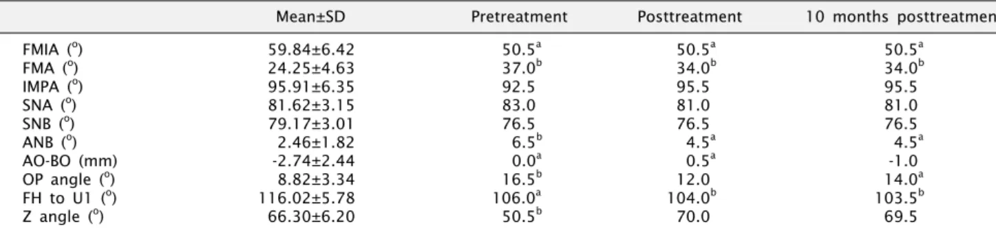
Table 1. Cephalometric measurements
Mean±SD Pretreatment Posttreatment 10 months posttreatment
FMIA (
o) 59.84±6.42 50.5
a50.5
a50.5
aFMA (
o) 24.25±4.63 37.0
b34.0
b34.0
bIMPA (
o) 95.91±6.35 92.5 95.5 95.5
SNA (
o) 81.62±3.15 83.0 81.0 81.0
SNB (
o) 79.17±3.01 76.5 76.5 76.5
ANB (
o) 2.46±1.82 6.5
b4.5
a4.5
aAO-BO (mm) -2.74±2.44 0.0
a0.5
a-1.0
OP angle (
o) 8.82±3.34 16.5
b12.0 14.0
aFH to U1 (
o) 116.02±5.78 106.0
a104.0
b103.5
bZ angle (
o) 66.30±6.20 50.5
b70.0 69.5
Mean±standard deviation (SD) is based on the data from lateral cephalometrics in Korean with normal occlusion by Korean Association of Orthodontists.
FMIA, angle between Frankfort plane and mandibular incisor axis; FMA, angle between Frankfort plane and mandibular plane; IMPA, angle between lower incisor axis and mandibular plane; SNA, angle between SN and NA; SNB, angle between SN and NB; ANB, difference between the SNA and SNB angles; AO-BO, distance between perpendiculars drawn from point A and point B onto the occlusal plane;
OP, occlusal plane; OP angle, angle between Frankfort plane and OP; FH, Frankfort horizontal plane; UI, maxillary incisor axis; FH to UI, angle between Frankfort plane and maxillary incisor axis; Z angle, angle between FH and profile line tangent to the chin and the vermilion border of both lips.
a