A Study on Skeletal Morphology of Anterior Open
Bite Patients by Cone-beam CT
by
Dong Joo Lee
Major in Speedy Orthodontics
Department of Clinical Dentistry
Graduate School of Clinical Dentistry
A Study on Skeletal Morphology of Anterior Open
Bite Patients by Cone-beam CT
by
Dong Joo Lee
A Dissertation Submitted to Graduate School of Clinical Dentistry,
Ajou University in Partial Fulfillment of the Requirements for the
Degree of Master of Science in Dentistry
Supervised by
Kyu Rhim Chung, M.D., Ph.D.
Major in Speedy Orthodontics
Department of Clinical Dentistry
Graduate School of Clinical Dentistry
This certifies that the dissertation
of Dong Joo Lee is approved.
SUPERVISORY COMMITTEE
____________________
Kyu Rhim Chung
_______________________
Kwang Woo Baek
_______________________
Kyung Yen Nahm
Graduate School of Clinical Dentistry
Ajou University
-ABSTRACT-
A Study on Skeletal Morphology of Anterior Open Bite Patients
by Cone-beam CT
The purpose of this study was to assess whether there is a statistically significant difference between the anterior open bite of a control group and an experimental group using measurements on a Cone-beam CT. The subjects for this study were chosen from the at large pool of patients who visited the Orthodontic Department of Dentistry at Ajou University Hospital in Suwon city, South Korea between Jan, 2011 and Jun, 2013 and underwent an initial Cone-beam CT (CBCT) examination. The patients with anterior open bite were included in the experimental group, while the patients lacking an anterior open bite were included in the control group. The tomographic images from Cone-beam CT were converted to DICOM files, and the skeletal and dentoalveolar measurements were processed by Ondemand 3D (Cybermed, Seoul, Korea). There was statistical significance in the SN-MP, SN-PP, LPDH, PFH/AFH ratios, but no significance was found in UPDH between the groups. Higher significance was found in SN-MP and PFH/AFH ratio suggesting that the skeletal pattern, rather than dentoalveolar growth, can be regarded as a major factor causing anterior open bite in many cases. One of the interesting results was that LPDH, not UPDH, showed significant difference between the two groups.
TABLE OF CONTENTS
ABSTRACT···i TABLE OF CONTENTS···ii LIST OF FIGURES···iii LIST OF TABLES···iv I. INTRODUCTION···1A. CONE-BEAM CT (CBCT) FOR THE DIAGNOSIS OF ANTERIOR OPEN BITE···1
B. ETIOLOGY AND CLASSIFICATION OF ANTERIOR OPEN BITE···3
C. TREATMENT OF ANTERIOR OPEN BITE···4
II. MATERIALS AND METHODS···5
A. MATERIALS···5 B. METHODS···10 III. RESULTS···18 IV. DISCUSSION···26 V. CONCLUSION···28 REFERENCES···29 국문요약···32
LIST OF FIGURES
Fig. 1. Reference points used in this study···8
Fig. 2. Linear dentoalveolar parameters used in the study···9
Fig. 3. Angular and linear measurements on CBCT image using Ondemand 3D Software (Cybermed, Seoul, South Korea)···12
Fig. 4. Angular measurement on magnified CBCT image···13
Fig. 5. Linear measurement on magnified CBCT image···14
LIST OF TABLES
Table 1. Inclusion and exclusion criteria of samples··· ·7
Table 2. Linear and angular measurements of skeletal··· · 16
Table 3. Linear and angular measurements of dentoalveolar··· · 17
Table 4. Mean and Standard deviation variables found in this study which were known
as significant indicators of anterior open bite (SN-MP, SN-PP, UPDH,
LPDH, PFH/AFH Ratio)··· 20
Table 5. 13 Mean and Standard deviation variables found in this study which show
skeletal and dentoalveolar tendency of the subjects (SN-Mn occlusal plane
angle, SN-Mx occlusal plane angle, PFH, LPFH, UPFH, AFH, LAFH,
UAFH, LADH, UADH, IMPA, U1-SN angle, Interincisal angle)··· 21
Table 6. T-test result of SN-MP··· 22
Table 9. T-test result of LPDH···24
Ⅰ
. INTRODUCTION
A. Cone-beam CT (CBCT) for the diagnosis of anterior open bite
Cone-beam computed tomography (CBCT) has gradually become favorable to many dental practitioners (Scarfe et al: 2008; Hechler et al: 2008). With 3D reconstruction views and detailed tomographic images, it can give quantification and visualization of bone structure and morphology with accuracy. Many recent studies showed that submilimeters to less than only 5mm of difference was found between linear measurements on CBCT images and direct physical measurements (Lascala et al: 2004; Berco et al: 2009). Since CBCT images present a better and more accurate view of the alveolar bone and periodontium (Nakajima et al: 2009), it is used by several researchers for linear measurements of the alveolar bone (Nahm et al: 2012).
Conventional cephalometric images are two dimensional and have different magnification ratios between the right and the left, which cause error in landmarks marking. This gives not only instrumental error but also personal error. Another variable which inhibits accurate measurements is the variation amongst head positions of patients during the scan. Even though the disadvantages and limitations of conventional cephalometric images are well-known, it had been used broadly for skeletal and dentoalveolar analysis because there is large accumulation of data and statistical results from previous researches for comparison.
Several researchers have analyzed cephalometric images comparing the anterior open bite to the normal bite. It is crucial to figure out whether there is skeletal tendency of anterior open bite to decide on the way of treatment (orthognathic surgery, orthodontic treatment or the combination of both).
the same time. Furthermore, follow up CBCT images, compared to the original CBCT images, can provide a clearer view of changes in the skeletal and dentoalveolar aspects during the retention period. Given the fact that the usage of CBCT has increased recently, more researches based on it should be undertaken.
B. Etiology and classification of anterior open bite
The diagnosis of anterior open bite is often determined based on dental condition. However, various researches in previous years presented evidence that the etiology of the anterior open bite is often skeletal, not only dental, and sometimes it is the combination of both. As many cases of anterior open bite arise from combination of both factors, the classification is often difficult to make.
The dental open bite is often found in the anterior region and it is associated with normal facial growth pattern, anterior-inclined and intruded incisors. The skeletal open bite is often related to as excessive vertical growth of the dentoalveolar region or specific skeletal patterns. Previous research, based on cephalometric anaylsis, identified steep mandibular plane as the key skeletal tendency in the anterior open bite (Ellis et al: 1984). Also, it has been reported that, in patients with anterior open bite, there is significant correlation to the severity of anterior open bite between the mandibular plane angle, ramus height of the mandible, antero-posterior dimension of the maxilla and movement of the front part of the dorsal tongue during deglutition (Fujiki et al: 2004).
Anatomic and physiologic etiology should not be underestimated. Tongue size and position are well known to affect skeletal and dental components (Kawakami et al: 2004). Mouth-breathing, as a result of upper airway obstruction, may cause anterior open bite, but its direct relationship has not been proven (Vaden and Pearson: 2002). Some researchers had found that habits such as finger sucking and tongue thrusting could cause anterior open bite or attribute to its degree (Popovich and Thompson: 1973; Straub: 1960).
C. Treatment of anterior open bite
Depending on the etiology and patient’s growth, anterior open bite can be treated only by orthodontic treatment if the problem is only limited to the dental area. If cases are carefully selected and properly treated, prognosis is quite promising. The treatment procedure is mainly done by fixed appliance for dental movement, often with the camouflage method. With patients with growth potential, a functional appliance inhibiting posterior alveolar bone growth can be helpful and promise better retention (Cusimano et al: 1993). Various extraction modalities have been suggested to treat anterior open bite, which aim to extrude anterior segment and reduce the inclination of both upper and lower incisors to increase overbite. However, correction of malocclusion by extrusion of upper anterior teeth could be detrimental to the patients with excessive anterior vertical height causing gummy smile. Also extrusive teeth movement is questionable considering the long-term retention and biological oral condition.
Therefore, rather than extrusion of anterior teeth, intrusion of posterior teeth is considered a better way to treat an anterior open bite, as it is often accompanied by excessive anterior facial height. As skeletal anchorages, including mini implant and mini plate, developed in recent years, molar intrusion has become a viable treatment option allowing counterclockwise rotation of the mandible, not only closing the anterior open bite (Sherwood et al: 2002; Park and Kwon: 2004).
On the other hand, in the cases of skeletal open bite, orthognathic surgery might be unavoidable considering long-term retention. The surgical procedure might include either maxilla, mandible or both. Some researchers have found that surgery only on the mandible is often highly instable because of muscle stretches; this is why a 2-jaw surgery, with the repositioning of maxilla, might be preferable (Proffit and Fields: 2006). This is why the exact diagnosis of anterior open bite is so important
Ⅱ
. MATERIALS AND METHODS
A. MATERIALS
The subjects who had gone through initial CBCT examination were chosen for the control and experimental groups among the patients who came to Orthodontic Department of Dentistry at the Ajou University Hospital, Suwon city, South Korea. For the experimental group, 15 patients (5 males, 10 females), who were diagnosed with an anterior open bite over 1mm overbite and overjet, were selected. There were 12 subjects of ClassⅡ malocclusion and 3 subjects of ClassⅠ malocclusion in a half cusp range, in the experimental group. For the control group, 15 patients (7 males, 8 females) with positive overjet to edge-to-edge bite without anterior open bite were selected. There were 6 subjects of ClassⅡ malocclusion and 9 subjects of ClassⅠ malocclusion in the control group. The samples with anterior cross bite and ClassⅢ malocclusion were excluded both from the control and the experimental groups. The samples with the severe facial deformities such as apparent condylar resorption or hemi-facial microsomia shown on CBCT images were also excluded. The SN-GoGn angles of all the samples were between 27-42 degrees on CBCT images. The criteria of the subjects are explained in Table 1. Those criteria followed the ones used in previous studies on cephalometric analysis of anterior open bite (Tsang et al: 1997; Richard et al: 2003). The mean age was 22.4 for the control group, and 19.3 for the experimental group. In all subjects, all of the second molars were completely erupted and occluded with molars on the opposite arch. The youngest subject was 15 and the oldest was 34 years old. Prior to running the t-test, Skew and Kurtosis calculations demonstrated that the population data was normally distributed; additionally, the samples were randomly selected.
The CBCT Scans (Dinova3, Willmed Ind. Co., Ltd, South Korea) were taken using the manufacturer’s recommended parameters of a 200×190mm field of view, 50-120 kVP; 4-10 mA; 24s scan time and 0.3mm slice thickness.
CBCT image data of the subjects was saved in DICOM (Digital Imaging and Communications in Medicine) files and were loaded into Ondemand 3D software (Cybermed, Seoul, South Korea). The DICOM image files were automatically built-up to 3D reconstruction views. The cephalometric landmarks used in this study are listed in Fig. 1 and Fig. 2. The cross-sectional views, which were highly resolute and showed apparent landmarks, were selected and magnified for the measurements.
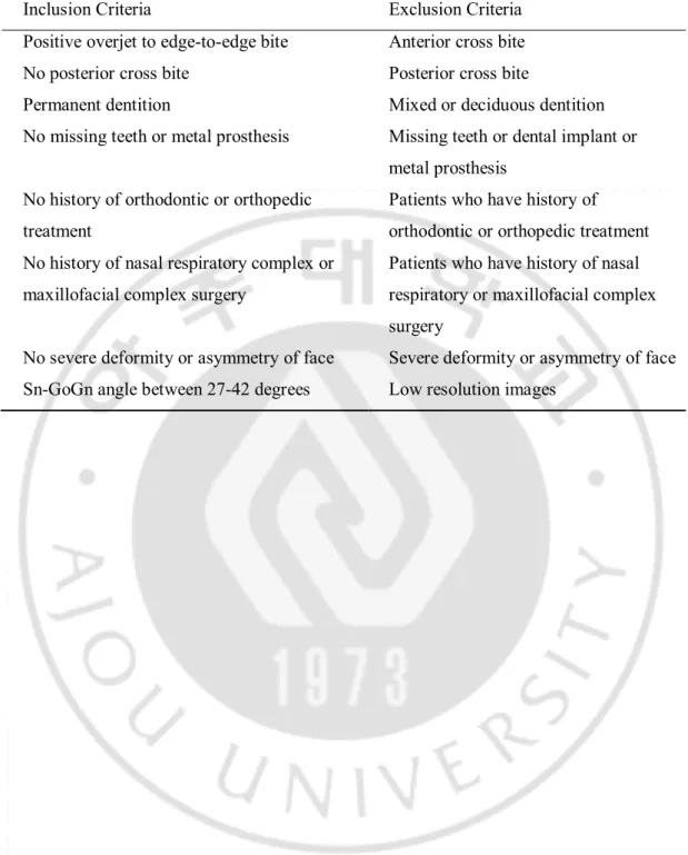
Table 1. Inclusion and exclusion criteria of samples.
Inclusion Criteria Exclusion Criteria Positive overjet to edge-to-edge bite Anterior cross bite No posterior cross bite Posterior cross bite
Permanent dentition Mixed or deciduous dentition No missing teeth or metal prosthesis Missing teeth or dental implant or
metal prosthesis No history of orthodontic or orthopedic
treatment
Patients who have history of orthodontic or orthopedic treatment No history of nasal respiratory complex or
maxillofacial complex surgery
Patients who have history of nasal respiratory or maxillofacial complex surgery
No severe deformity or asymmetry of face Severe deformity or asymmetry of face Sn-GoGn angle between 27-42 degrees Low resolution images
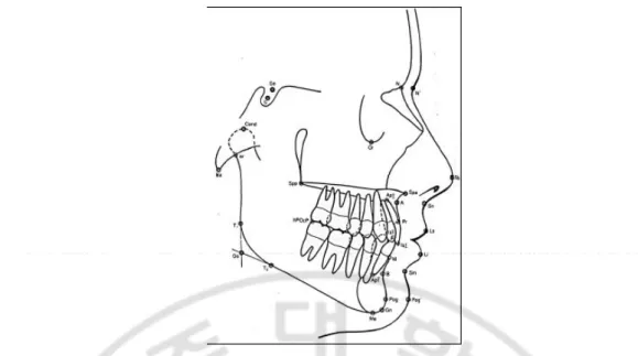
Fig. 1. Reference points used in this study. Sella - the point representing the midpoint of
the pituitary fossa or Sella turcica. It is a constructed point, Nasion - most anterior point in the midway between the frontal and nasal bones on the frontonasal suture, Point A - the deepest point in the midline between anterior nasal spine and the crest of the maxillary alveolar process, Point B - the deepest point in the midline between the alveolar crest of the mandible and the mental process, Gonion - constructed point at the junction of ramal plane and the mandibular plane, Menton - most inferior midline point on the mandibular symphysis, Gnathion - most antero-inferior point on the symphysis of the chin.
Fig. 2. Linear dentoalveolar parameters used in the study. LPDH (mm), UPDH (mm),
B. METHODS
For each patient, the linear and angular measurements were taken. Once the appropriate cross-sectional image was selected, the measurements were conducted by ruler and angular tools on Ondemand 3D software (Fig. 3), (Fig.4), (Fig. 5). A total of 18 cephalometric measurements were selected based on references from previous researches (Yi et al: 1997; Richard et al: 2003) on cephalometric analysis of anterior open bite. The skeletal linear and angular measurements are listed on Table. 2., and the dentoalveolar measurements are listed on Table. 3. Linear measurements are listed in millimeters, and angular measurements are listed in degrees. Among the 18 cephalometric measurements, the 5 variables (SN-MP, SN-PP, UPDH, LPDH and the PFH/AFH ratio) which were regarded as characteristic figures of anterior open bite in previous researches (Greenlee et al: 2011; Kaku et al: 2009; Tsang et el: 1997) were selected to run statistical analysis. For more accuracy and 3-dimensional analysis on CBCT images, AFH, PFH, UPDH and LPDH were measured twice on right and left sides of the each subject. For this study, the Sella-Nasion line (SN) was used as a reference line for the basal skull instead of the FH plane (FH), because Sella and Nasion were more obvious on the CBCT cross-sectional images on the Ondemand 3D program. Previous cephalometric researches used either SN, FH or both (Tsang et al 1997; Yoo and Kim: 2002; Richard et al: 2003).
For statistical analysis, the mean and standard deviation were calculated for each of the variables. Two samples t-test was planned using a regular significance level of P < .05 to find any significant difference between the two groups. To apply the t-test, a normal distribution of samples is necessary, which was verified by Skew and Kurtosis calculations, respectively. The Two samples t-test was performed to compare the 5 variables in each of the control and experimental groups. The 5 variables are SN-MP, SN-PP, UPDH, LPDH and the PFH/AFH ratio which were regarded as characteristic figures of anterior open bite
analysis for PFH/AFH, UPDH and LPDH were performed twice from the data set each from right side and left side. All statistical analyses were performed by Microsoft Exel 2007 (Microsoft, USA) and SPSS Ver. 18 (IBM, USA).
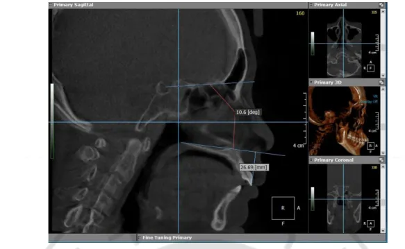
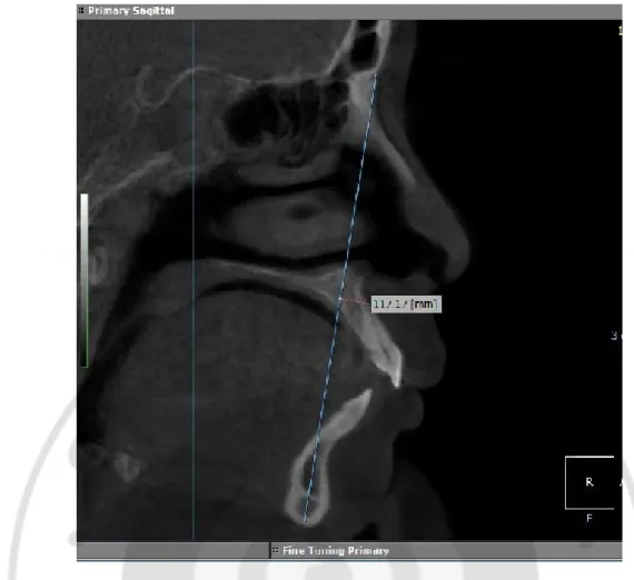
Fig. 3. Angular and linear measurements on CBCT image using Ondemand 3D software (Cybermed, Seoul, South Korea). Measurement image of SN-PP (degree) and
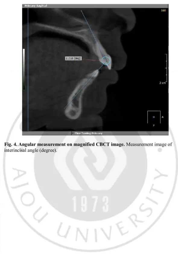
Fig. 4. Angular measurement on magnified CBCT image. Measurement image of
Fig. 5. Linear measurement on magnified CBCT image. Measurement image of AFH
Fig. 6. Dentoalveolar measurements on magnified CBCT image. Measurement image of
UPDH (the distance in millimeters between PP and mesio-buccal cusp of upper right first molar) as an example of dentoalveolar measurement. UPDH was measured twice from the upper right first molar and the upper left first molar. For UPDH, mark PP first as a reference
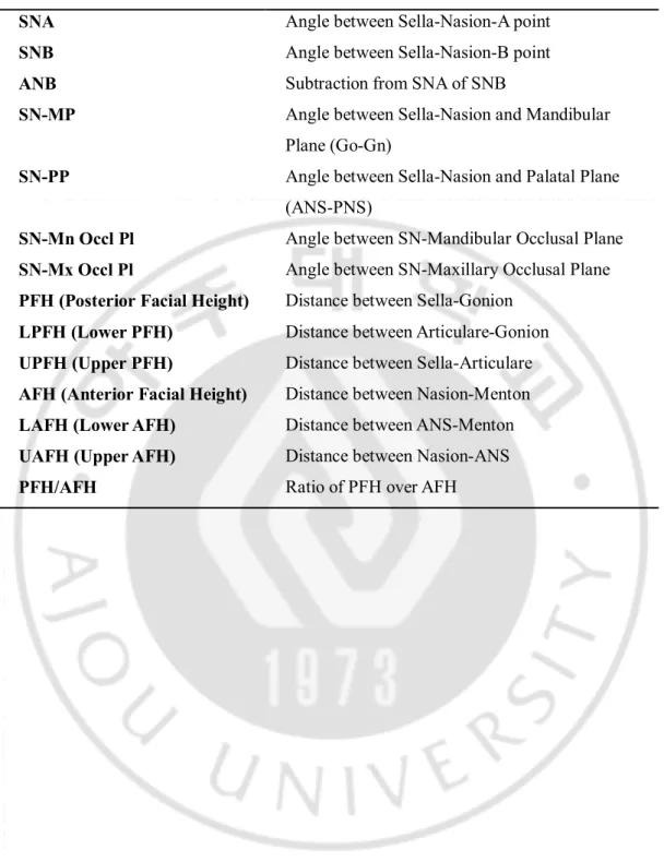
Table 2. Linear and angular measurements of skeletal
SNA Angle between Sella-Nasion-A point
SNB Angle between Sella-Nasion-B point
ANB Subtraction from SNA of SNB
SN-MP Angle between Sella-Nasion and Mandibular
Plane (Go-Gn)
SN-PP Angle between Sella-Nasion and Palatal Plane
(ANS-PNS)
SN-Mn Occl Pl Angle between SN-Mandibular Occlusal Plane
SN-Mx Occl Pl Angle between SN-Maxillary Occlusal Plane
PFH (Posterior Facial Height) Distance between Sella-Gonion
LPFH (Lower PFH) Distance between Articulare-Gonion
UPFH (Upper PFH) Distance between Sella-Articulare
AFH (Anterior Facial Height) Distance between Nasion-Menton
LAFH (Lower AFH) Distance between ANS-Menton
UAFH (Upper AFH) Distance between Nasion-ANS
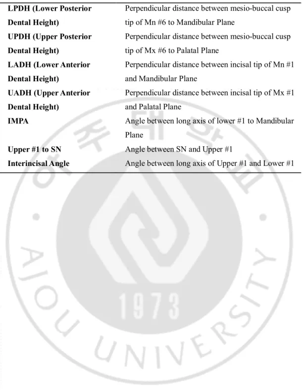
Table 3. Linear and angular measurements of dentoalveolar LPDH (Lower Posterior
Dental Height)
Perpendicular distance between mesio-buccal cusp tip of Mn #6 to Mandibular Plane
UPDH (Upper Posterior Dental Height)
Perpendicular distance between mesio-buccal cusp tip of Mx #6 to Palatal Plane
LADH (Lower Anterior Dental Height)
Perpendicular distance between incisal tip of Mn #1 and Mandibular Plane
UADH (Upper Anterior Dental Height)
Perpendicular distance between incisal tip of Mx #1 and Palatal Plane
IMPA Angle between long axis of lower #1 to Mandibular
Plane
Upper #1 to SN Angle between SN and Upper #1
Ⅲ
. RESULTS
The mean and standard deviation values of the 5 variables which were known as significant indicators of anterior open bite (SN-MP, SN-PP, UPDH, LPDH, PFH/AFH Ratio) are listed on Table 4. The rest of the mean and standard deviation variables found in this study (SN-Mn occlusal plane angle, SN-Mx occlusal plane angle, PFH, LPH, UPFH, AFLH, LAFH, UAFH, LADH, UADH, IMPA, U1-SN angle, Interincisal angle) show skeletal and dentoalveolar tendency of the subjects (Table 5). There are 2 sets of statistical result from the measurements of the right and the left for UPDH, LPDH and PFH/AFH ratio.
The result of the t-test of 5 variables and its meaning is as follows.
1. The SN-MP (SN to mandibular plane angle) was statistically larger in the experimental group than the control groups (Table 6).
2. The SN-PP (SN to palatal plane angle) was statistically different between the experiment group and the control groups (Table 7).
3. No statistically significant difference was found in UPDH (Upper Posterior Dental Height) between the control and experimental groups (Table 8), regardless of the data sets. The P value was 0.60 and 0.35 in order of the right and the left.
4. There was a statistically significant difference in the LPDH (Lower Posterior Dental Height) between the control and experimental groups (Table 9). Even though the level of significance was slightly higher on the left side, both data sets showed statistical significance.
5. The ratio between the posterior facial height and the anterior facial height (PFH/AFH ratio) was statistically much less and significant in patients with anterior open bite (Table 10). Both of the data sets from the right and left showed the same result.
The null hypothesis was rejected for 4 variables: SN-MP, SN-PP, LPDH and the PFH/AFH ratio. Higher significant difference was found in the SN-MP and PFH/AFH ratio, suggesting that skeletal tendency is the important factor of anterior open bite. Though the p-value was not as statistically significant as with the SN-MP and the PFH/AFH ratio, the SN-PP and LPDH were also found to have significant differences. Slightly bigger significance was found in LPDH from the left data set but the difference was a negligible quantity. The UPDH was not found to have a statistically significant difference between the control and experimental groups in both data sets from the right and left, conflicting previous researchers using conventional cephalometric images (Tsang et al: 1997; Yoo and Kim: 2002), which found very much or moderate significance.
Table 4. Mean and Standard deviation variables found in this study which were known as significant indicators of anterior open bite (SN-MP, SN-PP, UPDH, LPDH, PFH/AFH Ratio). UPDH, LPDH, PFH/AFH Ratio were measured twice from the both
right and left side.
SN-MP SN-PP UPDH (Rt) UPDH (Lt) LPDH (Rt) LPDH (Lt) PFH/AFH Ratio (Rt) PFH/AFH Ratio (Lt) Control Mean 33.63 9.29 22.92 22.78 32.33 32.3 0.65 0.65 Median 33.9 8.5 22.4 22.22 32.55 32.46 0.64 0.63 St. Dev 2.6 2.12 1.5 2.2 1.55 1.44 0.03 0.04 Skew 0.15 0.64 0.72 0.73 -1.21 0.22 0.14 0.54 Kurtosis -0.91 -0.79 -0.5 -0.8 1.31 -0.96 -1.14 -1.21 SN-MP SN-PP UPDH (Rt) UPDH (Lt) LPDH (Rt) LPDH (Lt) PFH/AFH Ratio (Rt) PFH/AFH Ratio (Lt) Test Mean 38.54 7.23 23.31 23.4 30.17 30.14 0.54 0.54 Median 37.8 7.8 22.89 23.46 29.9 30.17 0.54 0.54 St. Dev 3.12 2.11 2.45 1.48 2.7 1.45 0.03 0.03 Skew 0.22 -0.73 0.05 -0.39 0.06 0.01 -0.56 -0.26 Kurtosis -0.81 -0.12 -1.5 -0.62 -1.24 -0.96 0.24 0.11
Table 5. 13 Mean and Standard deviation variables found in this study which show skeletal and dentoalveolar tendency of the subjects (Mn occlusal plane angle, SN-Mx occlusal plane angle, PFH, LPFH, UPFH, AFH, LAFH, UAFH, LADH, UADH, IMPA, U1-SN angle, Interincisal angle).
SN-Mn Occpl SN-Mx Occpl
PFH LPFH UPFH AFH LAFH UAFH LADH UADH IMPA U1-SN Inter-incisal Angle Control Mean 15.89 13.87 77.73 46.24 31.49 120.47 67.49 52.98 40.90 29.76 91.52 103.80 123.05 Median 15.90 13.60 77.59 46.28 31.41 121.31 66.01 51.72 40.83 29.02 91.30 104.40 125.30 St Dev 3.95 4.12 4.67 3.70 2.02 7.20 5.10 3.10 2.68 2.81 2.65 3.96 11.18 Skew (0.24) 0.13 (0.26) 0.17 0.75 0.33 0.93 (0.30) 0.20 0.25 0.07 (0.64) 0.29 Kurtosis (0.00) (0.45) (0.66) 0.00 1.09 0.62 1.15 (0.12) (0.79) (1.50) (1.39) 0.79 (0.34) SN-Mn Occpl SN- Mx Occpl
PFH LPFH UPFH AFH LAFH UAFH LADH UADH IMPA U1-SN Inter -incisal Angle Test Mean 19.07 16.98 66.85 43.26 23.60 123.61 68.59 55.03 40.33 27.03 89.46 108.27 110.03 Median 19.50 17.80 68.68 42.80 25.40 122.41 68.23 54.54 39.38 26.83 89.30 109.50 109.90 St Dev 2.28 2.49 5.13 6.60 7.15 5.10 4.31 2.39 2.55 1.83 3.42 6.62 8.41 Skew (0.60) (0.47) (0.38) 1.14 (1.14) 0.71 0.48 0.07 0.26 0.21 0.61 (0.62) 0.34 Kurtosis 0.09 (1.09) (0.47) 2.65 2.69 (0.77) (0.83) (0.01) (0.98) (0.93) 0.67 (0.53) (0.73)
Table 6. T-test result of SN-MP. High significant difference was found in SN-MP. Variable 1 Variable 2 Mean 33.63333 38.54 Variance 6.746667 9.718286 Observations 15 15 Pooled Variance 8.232476 Hypothesized Mean Difference 0
Df 28 t Stat -4.6833 P(T<=t) one-tail 3.3E-05 t Critical one-tail 1.701131 P(T<=t) two-tail 6.59E-05 t Critical two-tail 2.048407
Table 7. T-test result of SN-PP. A significant difference was found in SN-PP.
Variable 1 Variable 2
Mean 9.286667 7.233333
Variance 4.476952 4.456667
Observations 15 15
Pooled Variance 4.46681
Hypothesized Mean Difference 0
Df 28
t Stat 2.660672
Table 8. T-test result of UPDH from the data sets from right and left. No significant
difference was found in UPDH.
Right Side Data Set Variable 1 Variable 2
Mean 22.91533333 23.30533333
Variance 2.245926667 5.992969524
Observations 15 15
Pooled Variance 4.119448095 Hypothesized Mean Difference 0
df 28 t Stat -0.526230143 P(T<=t) one-tail 0.30143604 t Critical one-tail 1.701130934 P(T<=t) two-tail 0.602872081 t Critical two-tail 2.048407142
Left Side Data Set Variable 1 Variable 2
Mean 22.766 23.40133333
Variance 4.721282857 2.198940952
Observations 15 15
Pooled Variance 3.460111905 Hypothesized Mean Difference 0
df 28 t Stat -0.935378107 P(T<=t) one-tail 0.178795578 t Critical one-tail 1.701130934 P(T<=t) two-tail 0.357591157 t Critical two-tail 2.048407142
Table 9. T-test result of LPDH from the data sets from right and left. A Significant
difference was found in LPDH.
Right Side Data Set Variable 1 Variable 2
Mean 32.32933333 30.17
Variance 2.410220952 7.304471429
Observations 15 15
Pooled Variance 4.85734619 Hypothesized Mean Difference 0
df 28 t Stat 2.683186078 P(T<=t) one-tail 0.006050671 t Critical one-tail 1.701130934 P(T<=t) two-tail 0.012101341 t Critical two-tail 2.048407142
Left Side Data Set Variable 1 Variable 2
Mean 32.30533333 30.13666667
Variance 2.078269524 2.119809524
Observations 15 15
Pooled Variance 2.099039524 Hypothesized Mean Difference 0
df 28 t Stat 4.099332331 P(T<=t) one-tail 0.000160887 t Critical one-tail 1.701130934 P(T<=t) two-tail 0.000321774 t Critical two-tail 2.048407142
Table 10. T-test result of PFH/AFH ratio from the data sets from right and left. High
significant difference was found in the PFH/AFH ratio.
Right Side Data Set Variable 1 Variable 2
Mean 0.645818203 0.540653113
Variance 0.000880434 0.001038072
Observations 15 15
Pooled Variance 0.000959253 Hypothesized Mean Difference 0
df 28 t Stat 9.298985752 P(T<=t) one-tail 2.33485E-10 t Critical one-tail 1.701130934 P(T<=t) two-tail 4.66971E-10 t Critical two-tail 2.048407142
Left Side Data Set Variable 1 Variable 2
Mean 0.649474971 0.542836584
Variance 0.001315621 0.001173739
Observations 15 15
Pooled Variance 0.00124468 Hypothesized Mean Difference 0
df 28 t Stat 8.277807385 P(T<=t) one-tail 2.61548E-09 t Critical one-tail 1.701130934 P(T<=t) two-tail 5.23096E-09 t Critical two-tail 2.048407142
Ⅳ
. DISCUSSION
In this study, higher significance was found in the SN-MP and PFH/AFH ratios, which means the skeletal pattern can be considered as the major factor causing anterior open bite. This confirms results from previous researches on cephalometric analysis of anterior open bite (Tsang et al: 1997; Richard et al: 2003; Scarfe and Farman: 2008).
An interesting result of this study was that LPDH, not UPDH, showed significant difference between the two groups; however, UPDH has been known to be significantly significant differences in anterior open bite patients as compared to normal patients based on several researches using conventional cephalometric images (Tsang et al: 1997; Yoo and Kim: 2002). Even though CBCT images are known to have better resolution and accuracy for linear measurements of the alveolar bone, a further study is required using CBCT with more samples to assess the relationship between the anterior open bite and UPDH or LPDH. Since the usage of CBCT is becoming more popular, it is expected that research on more samples will be carried out in the near future.
Nowadays, many practitioners treat anterior open bite cases with the intrusion of maxillary molars. The development of a skeletal anchorage, such as a mini implant or mini plate, made it possible to intrude molars effectively. Whether a patient has significantly bigger LPDH or UPDH, both maxillary and mandibular molars can be intruded allowing the mandible to rotate anticlockwise, which is a favorable movement of mandible in most of anterior open bite cases. It would be very helpful to know in the beginning of treatment which molars in specific arch should be targeted. In that sense, CBCT is especially useful for treatment planning of anterior open bite to decide between surgery and orthodontic treatment with molar intrusion.
rotated mandibular plane and bigger Gonial angle were found, as in other races. Also he suggested anterior open bite was more a function of mandibular plane change rather than tipping of the palatal plane since palatal plane bored the same relationship with the Frankfort plane as the non-open bite group.
In his cephalometric analysis comparing 3 groups of patients subdivided by severity of anterior open bite (according to the distance between maxillary and mandibular anterior teeth), W. M. Tsang et al (1997) found that the palatal plane, mandibular occlusal plane, upper anterior dental height and upper posterior dental height correlate significantly with the severity of anterior open bite.
Further study on larger samples with detailed classification based on severity of anterior open bite is needed to assure dentoalveolar factors of anterior open bite. Also, it is necessary to have long-term research on changes in alveolar bone height after intrusive movement of molars using CBCT images. With accumulation of pre-treatment and post-treatment CBCT data sets, the changes in alveolar bone height throughout years can be examined accurately to find whether the molar intrusion is retentive enough for optimal prognosis after the treatment.
Ⅴ
. CONCLUSION
Significant differences were found in the SN-MP, SN-PP, LPDH, PFH/AFH ratios, but no significance was found in UPDH between the groups. Higher significance was found in the SN-MP and PFH/AFH ratios, suggesting that skeletal pattern, rather than dentoalveolar growth, can be regarded as a major factor causing anterior open bite in many cases.
REFERENCES
1. Berco M, Rigali PH Jr, Miner RM, DeLuca S, Anderson NK, Will LA: Accuracy and reliability of linear cephalometric measurements from cone-beam computed tomography scans of a dry human skull. Am J Orthod Dentofac Orthop 136: 17.e1-9; discussion 17-18, 2009
2. Cusimano C, McLaughlin RP, Zernik JH: Effects of first bicuspid extractions on facial height in high-angle cases. J Clin Orthod 27: 594-608, 1993
3. Yoo EH, Kim DU: Compensation of female adults with openbite tendancy. Korea J Orthod 32(1): 1-7, 2002
4. Ellis E, McNamara JA: Components of adult Class 3 open-bite malocclusion. Am J Ortho 86: 277-290, 1984
5. Fujiki T, Inoue M, Miyawaki S, Nagasaki T, Tanimoto K, Takano-Yamamoto T: Relationship between maxillofacial morphology and deglutitive tongue movement in patients with anterior open bite. Am J Ortho Dentofacial Ortho 125: 160-167, 2004 6. Greenlee GM, Huang GJ, Chen SS, Chen J, Koepsell T, Hujoel P: Stability of
treatment for anterior open-bite malocclusion: a metal-analysis. Am J Orthod Dentofacial Orthop 139: 154-169, 2011
7. Hechler SL: Cone-beam CT: applications in orthodontics. Dent Clin North Am 52: 809-823, 2008
8. Huang J, Bumann A, Mah J: Three-dimensional radiographic analysis in orthodontics. J Clin Orthod. 39: 421-428, 2005
9. Iscan HN, Sorisoy L: Comparison of the effects of passive posterior bite-blocks with different construction bites on the craniofacial and dentoalveolar structures. Am J Orthod Dentofac Orthop 112(2): 171-178, 1997
11. Kawakami M, Yamamoto K, Noshi T, Miyawaki S, Kirita T: Effect of surgical reduction of the tongue on dentofacial structure following mandibular setback. J Oral Maxillofac Sur 62: 1188-1192, 2004
12. Nahm KY, Kang JH, Moon SC, Choi YS, Kook YA, Kim SH and Huang JC: Alveolar bone loss around incisors in Class 1 bidentoalveolar protrusion patients: a retrospective three-dimensional cone beam CT study. Dentomaxillofac Rad 41: 481-488, 2012 13. Lascala CA, Panella J, Marques MM: Analysis of the accuracy of linear measurements
obtained by cone beam computed tomography (CBCT-NewTom). Dentomaxillofac Radiol 33: 291-294, 2004
14. Nakajima K, Yamaguchi T, Maki K: Surgical orthodontic treatment for a patient with advanced periodontal disease: evaluation with electromyography and 3-dimensional cone-beam computed tomography. Am J Orthod Dentofacial Orthop 136: 450-459, 2009
15. Park HS, Kwon TG: Sliding mechanics with microscrew implant anchorage. Angle Orthod 74: 703-710, 2004
16. Popovich F, Thompson GW: Thumb and finger sucking: its relation to malocclusion. Am J Orthod 63: 148-155, 1973
17. Proffit WR, Fields HW: Contemporary Orthodontics-E-book Missouri: Elsevier Health Sciences, 2006
18. Richard A, Glynda R, Ceib P, Camilla T: A Cephalometric Comparison of Black Open-Bite Subjects and Black Normals. Angle Orthod 73: 3, 2003
19. Scarfe WC, Farman AG: What is cone-beam CT and how does it work? Dent Clin North Am 52: 707-730, 2008
20. Sherwood KH, Burch JG, Thompson WJ: Closing anterior open bites by intruding molars with titanium miniplate anchorage. Am J Orthod Dentofacial Orthop 122:
593-21. Straub WJ: Malfunction of the tongue: Part 1. The abnormal swallowing habit: its cause, effects, and results in relation to orthodontic treatment and speech therapy. Am J Orthod 46: 404-424, 1960
22. Tsang WM, Cheung LK, Samman N: Cephalometric characteristics of anterior open bite in a southern Chinese population. Am J Orthod Dentofac Orthop 113(2): 165-172, 1998
23. Vaden JL, Pearson LE: Diagnosis of the vertical dimension. Semin Ortho 8: 120-129, 2002
24. Tsang WM, Cheung LK, Samman N: Cephalometric parameters affecting severity of anterior open bite. Int J. Oral Maxiilofac Surg 26: 321-326, 1997
-국문 요약-
Cone-beam CT 를 이용한 전치부 개방교합 환자의
골격적 특징에 대한 연구
아주대학교 임상치의학대학원 임상치의학과 이 동 주 (지도교수: 정 규 림)본 연구에서는 Cone-beam Computed tomography 영상을 이용하여 전치부 개방교합 소견을 보이는 실험군과 전치부 개방교합 소견이 없는 대조군의 계측을 통하여 두 군간의 두개골과 치조골 영역에서 유의한 차이가 있는지 알아보고자 하였다. 2011 년 1 월~2013 년 6 월 사이 아주대 의료원 치과 교정과에 내원하여 초진시에 Cone-beam CT 촬영을 시행한 환자 중 1mm 이상의 전치부 개방교합을 보이는 15 명의 환자를 실험군으로, 전치부 개방교합을 보이지 않으며 1 급 부정교합을 보이는 15 명의 환자를 대조군으로 선정하였다. Cone-beam CT 영상을 먼저 DICOM(Digital Imaging and Communications in Medicine) 파일로 변환하여 Ondemand 3D 프로그램으로 재구성한 상에서 선과 각도를 측정하였다.
항목에서는 유의한 차이가 없었다. 특히 SN-MP, PFH/AFH ratio 에서 상대적으로 큰 유의성이 관찰되는 바, 치조골의 과다 성장보다는 두개 안면의 골격 패턴이 전치부 개방교합의 발생에 더 관련성이 있는 것으로 확인된다.