pISSN 2466-1384 eISSN 2466-1392 大韓獸醫學會誌 (2017) 第 57 卷 第 4 號 Korean J Vet Res(2017) 57(4) : 253~255 https://doi.org/10.14405/kjvr.2017.57.4.253
253
<Case Report>
Diagnostic imaging of congenital pulmonary aplasia in a dog
Soochan Kim, Hojung Choi, Youngwon Lee*
College of Veterinary Medicine, Research Institute of Veterinary Medicine, Chungnam National University, Daejeon 34134, Korea (Received: September 4, 2017; Revised: September 22, 2017; Accepted: September 26, 2017)
Abstract: A 2-year-old, female Pomeranian dog was referred for dyspnea. Thoracic radiographs revealed left-sided mediastinal shift, increased soft tissue opacity in the caudal aspect of left thorax with loss of the left diaphragmatic silhouette, and dorsal elevation of mediastinal structures and heart from the sternum by lung tissue. The left main bronchus was visualized as an air-bronchogram and observed to abruptly discontinue at the level of the 10th rib. Thoracic computed tomography (CT) revealed absence of the left lung parenchyma and left pulmonary vessels with a rudimentary left main bronchus. The case was congenital pulmonary aplasia diagnosed via radiography and CT.
Keywords: computed tomography, dogs, pulmonary aplasia, radiography
Pulmonary aplasia is a rare congenital anomaly, which is unilateral or bilateral absence of the lung tissue [1, 13]. Uni- lateral aplasia usually has been reported in humans, because bilateral aplasia is not viable [13]. The etiology of pulmo- nary aplasia is unknown, and there is no predominance of affected side lobe or gender [2]. In humans, pulmonary apla- sia occurs during the embryogenic period, when the primi- tive lung is formed [1]. According to the previous report in humans, the pulmonary anomaly has been classified into three groups by Schneider and Schwalbe [14]. In type 1 (agenesis), the main bronchus, lung tissue, and pulmonary vasculature are completely absent; in type 2 (aplasia), the rudimentary bronchus is present, but alveolar tissue and pul- monary vessels do not exist; and in type 3 (hypoplasia), there are deformed bronchi and a reduced amount of lung tissue [14].
Pulmonary aplasia usually presents with variable neonatal respiratory distress [4]. The most common manifestations include neonatal respiratory distress, recurrent pneumonia and cyanosis [5, 16], and occasionally patients who live up to the adulthood live without any symptoms [6, 16]. In humans, diagnostic imaging has a great effect on the diagnosis of pul- monary aplasia. Contrast-enhanced computed tomography (CT) and magnetic resonance (MR) imaging are recently the most reliable choice of imaging modalities, showing the absence of lung parenchyma, bronchial tree and pulmonary vessels [7, 8]. To the authors’ knowledge, this case is the first report of congenital pulmonary aplasia in dogs.
A 2-year-old intact female Pomeranian dog weighing 3.2 kg was presented with forceful breathing and worsen lethargy one day before presentation. According to the owner, the
symptoms of lethargy and wheezing sound had started from three months after the birth, and there was no history of trauma. On physical examination, the patient showed respira- tory distress. The results of laboratory examination were within normal range.
Thoracic radiographs showed left sided mediastinal shift and left main bronchus as an air-bronchogram, which run along about 3 cm length and abruptly discontinued, consis- tent with lung collapse secondary to suspected pneumonia (Fig. 1). In addition, thoracic radiographs revealed increased soft tissue opacity in the caudal aspect of left thorax with loss of the left diaphragmatic silhouette, which suggested perito- neal pericardial diaphragmatic hernia, diaphragmatic hernia, or hiatal hernia. To diagnosis of diaphragmatic hernia, ultra- sonography was performed to identify diaphragmatic hernia and observed the liver located adjacent to heart without echogenic lung surface made by pulmonary gas. Because of suspicion of diaphragmatic hernia on an ultrasound scan and patient’s fatal respiratory distress, exploratory laparotomy was performed immediately. However, the diaphragm was intact and the liver normally located in the abdominal cavity.
To further evaluate underlying causes of respiratory dis- tress, CT was performed under the general anesthesia right after the operation. Scanning parameters were 120 kVp, 100 mAs, 2 mm slice thickness and 0.938 collimation pitch. Two series of pre- and post-contrast CT images were acquired at 35 sec after injection of 600 mg iodine/kg iohexol (Omnipaque;
GE Healthcare, Ireland) intravenously by power injector (2 mL/sec). Cranioventral mediastinal reflection was not found and right cranial lung lobe expanded into contralateral
*Corresponding author
Tel: +82-42-821-6786, Fax: +82-42-821-6703 E-mail: [email protected]
254 Soochan Kim, Hojung Choi, Youngwon Lee
thorax (Fig. 2). Additional CT findings were stenosis of right main bronchus located juxtaposed to the pulmonary vessel and abnormal arrangement of left sided ribs. The carina and short-blind ending left main bronchus were present, but lung parenchyma, and pulmonary arteries and veins running to left sided lobe absolutely did not exist on the affected side (Fig.
3). Amorphous lesions closest to thoracic wall with increased density (Hounsfield unit −350) were detected incidentally.
The rudimentary main bronchus and absence of the lung parenchyma and the pulmonary vessels were demonstrated on the CT images, confirming the diagnosis of pulmonary aplasia. No additional malformations were detected by CT and echocardiography.
In humans, CT and MRI angiography, bronchography, and pulmonary angiography have been used for patients, who have possibility of atelectasis or agenesis of the lung, to get assessment of the bronchial tree, parenchyma, and vascula- ture [7, 8, 15, 16]. Radiographic findings of thorax in this case were similar to those of pulmonary aplasia in human medicine. Mediastinum pulled toward the opacified side and narrowing between the ribs and elevated hemidiaphragm on
the affected side were observed [16]. Plain radiography pro- vides description of hemithorax white-out ipsilateral lung volume loss with ipsilateral shift of meadiastinal structures.
From these findings, the lung collapse, pulmonary hypopla- sia, and pulmonary agenesis are considered as lists of differ- ential diagnoses. The pleural effusion, diaphragmatic hernia and large pulmonary mass are also thought as other causes of hemithorax white-out, if contralateral mediastinal shift pre- sents [3, 9].
According to the previous studies in humans, CT findings of pulmonary aplasia included rudimentary main bronchus, compensatory enlargement of lung, the absence of the lung parenchyma, and deviation of mediastinal structures to the affected side. Contrast enhanced CT imaging, especially, could support to identify nonexistent pulmonary artery and vein on the affected side [16]. The authors suggest that 3D recon- struction CT images provide detailed diagnostic information about the absence of bronchial tree and lung parenchyma on the affected side lung.
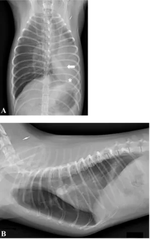
In this case, patient had survived well until adulthood and Fig. 1. Ventrodorsal (A) and right lateral (B) thoracic radio-
graphs. Left sided mediastinal shift and dorsal elevation of the mediastinal structures were present. The left main bronchus abruptly discontinued (arrow), which confirmed rudimentary bronchus on the computed tomography images. Increased soft tissue opac- ity in the caudal aspect of left thorax (asterisk) was also detected.
Fig. 2. Post-contrast lung window computed tomography images of right cranial lung lobe. (A) Transverse plane. (B) Dorsal reconstructed image. The cranioventral mediastinal reflex was not detected and pulmonary infiltration was seen (A). There were marked mediastinal shift to the left and stenosis of right main stem bronchus (arrow in B).
Fig. 3. Ventral (A) and dorsal (B) aspects of volume rendering computed tomography images. Rudimentary main bronchus (white arrow in A) and nonexistent left sided pulmonary vessels were observed.
Pulmonary aplasia 255
presented acute onset dyspnea. It could be associated that contralateral lung may develop as much as twice more alve- oli in response to compensatory hypertrophy [11, 12, 16].
The onset of symptoms in pulmonary aplasia is variable. In human’s reports, presence of this anomaly is usually revealed during infancy because of recurrent chest infections, result- ing from imperfect drainage of lung secretions [10, 15]. In addition, pulmonary aplasia tends to occur lung overinflation and air trapping, causing high susceptibility infection, because of concurrent kinking, compression, or stenosis of major air- ways. Patients with left sided aplasia have a longer life expectancy than those with right sided aplasia, due to pres- ence of tracheobronchial obstruction caused by the aortic arch, aorta, patent ductus arteriosus, aberrant right subcla- vian artery, or dilated left pulmonary artery in the right sided aplasia [13, 14].
In humans, pulmonary aplasia is often observed with other congenital malformations, including diaphragm defects, kid- ney anomalies, cardiovascular, gastrointestinal, or musculosk- eletal systems. The most common malformation is congenital heart disease like atrial or ventricular septal defect, patent ductus arteriosus, and aorta coarctation. Therefore, echocar- diography is essential in cases of pulmonary aplasia to con- firm additional malformations [16]. However, the concurrence of malformation was not present in our case.
This case is a rare congenital pulmonary aplasia diagnosed with radiography and CT in a dog and represents the first report of congenital pulmonary aplasia in dogs. Contrast enhanced CT images can provide important diagnostic infor- mation, including the absence of lung parenchyma, bron- chial tree, and pulmonary vessels on the affected side lung.
References
1. Biyyam DR, Chapman T, Ferguson MR, Deutsch G, Dighe MK. Congenital lung abnormalities: embryologic features, prenatal diagnosis, and postnatal radiologic- pathologic correlation. Radiographics 2010, 30, 1721-1738.
2. Cay A, Sarihan H. Congenital malformation of the lung. J Cardiovasc Surg (Torino) 2000, 41, 507-510.
3. Corne J, Kumaran M. Chest X-ray Made Easy. 4th ed.
Elsevier, St. Louis, 2015.
4. Dembinski J, Kroll M, Lewin M, Winkler P. [Unilateral pulmonary agenesis, aplasia and dysplasia]. Z Geburtshilfe Neonatol 2009, 213, 56-61, German.
5. Frank JL, Poole CA, Rosas G. Horseshoe lung: clinical, pathologic, and radiologic features and a new plain film finding. AJR Am J Roentgenol 1986, 146, 217-226.
6. Fraser RS, Paré JAP. Synopsis of Diseases of the Chest.
2nd ed. pp. 256-286, WB Saunders, Philadelphia, 1994.
7. Glazer HS, Siegel MJ, Sagel SS. Low-attenuation mediastinal masses on CT. AJR Am J Roentgenol 1989, 152, 1173-1177.
8. Keslar P, Newman B, Oh KS. Radiographic manifestations of anomalies of the lung. Radiol Clin North Am 1991, 29, 255-270.
9. Khan AN, Al-Jahdali H, Al-Ghanem S, Gouda A. Reading chest radiographs in the critically ill (Part II): radiography of lung pathologies common in the ICU patient. Ann Thorac Med 2009, 4, 149-157.
10. Kisku K, Panigrahi MK, Sudhakar R, Nagarajan A, Ravikumar R, Daniel J. Agenesis of lung–a report of two cases. Lung India 2008, 25, 28-30.
11. Kurkcuoglu IC, Eroglu A, Karaoglanoglu N, Polat P.
Pulmonary hypoplasia in a 52-year-old woman. Ann Thorac Surg 2005, 79, 689-691.
12. Lee CM, Kim JH, Kang MH, Eom KD, Park HM.
Unusual congenital pulmonary anomaly with presumed left lung hypoplasia in a young dog. J Small Anim Pract 2014, 55, 274-277.
13. Maltz DL, Nadas AS. Agenesis of the lung. Presentation of eight new cases and review of the literature. Pediatrics. 1968, 42, 175-188.
14. Nowotny T, Ahrens BC, Bittigau K, Buttenberg S, Hammer H, Kalache KD, Kursawe R, Maurer T, Schneider M, Wauer RR. Right-sided pulmonary aplasia:
longitudinal lung function studies in two cases and comparison to results from term healthy neonates. Pediatr Pulmonol 1998, 26, 138-144.
15. Roy PP, Datta S, Sarkar A, Das A, Das S. Unilateral pulmonary agenesis presenting in adulthood. Respir Med Case Rep 2012, 5, 81-83.
16. Yetim TD, Bayaro ullari H, Yalçin HP, Ar ca V, Ar ca SG. Congenital agenesis of the left lung: a rare case. J Clin Imaging Sci 2011, 1, 47.
gê
i i