Transgenic Efficiency of FoxN1-targeted Pig Parthenogenetic Embryos
Jae-Hoon Yeo
1,2, In-Sul Hwang
1, Jae Kyung Park
2, Dae-Jin Kwon
1, Seoki Im
1, Eung-Woo Park
1, Jeong-Woong Lee
2, Choon-Keun Park
3and Seongsoo Hwang
1†1
Animal Biotechnology Division, National Institute of Animal Science, RDA, Suwon 441-706, Republic of Korea
2
Department of Animal Biotechnology, Kangwon National University, Chuncheon 200-701, Republic of Korea
3
Research Center of Integrative Cellulomics, Korea Research Institute of Bioscience and Biotechnology, Daejeon 305-806, Republic of Korea
ABSTRACT
The clustered regularly interspaced short palindromic repeats (CRISPR)/CRISPR associated protein (Cas9) system can be applied to produce transgenic pigs. Therefore, we applied CRISPR/Cas9 system to generate FoxN1-targeted pig parthenogenetic embryos. Using single guided RNA targeted to pig FoxN1 genes was injected into cytoplasm of in vitro matured oocyte before electrical activation. In results, regardless of the concentrations of vector, the cleavage rate were significantly (p<0.05) decreased (4 ng/μl, 51.24%; 8 ng/μl, 40.88%; and 16 ng/μl; 45.22%) compared to no injection group (70.44%). The blastocyst formation rates were also decreased in vector injected 3 groups (4 ng/μl, 7.96%; 8 ng/μl, 6.4%; and 16 ng/μl; 9.04%) compared to no injection group (29.07%). In addition, the blastocyst formation rates between sham injected group (13.51%) and no injection group (29.07%) also showed significant difference (p<0.05). The mutation rates were comparable between groups (4 ng/μl, 18.4%; 8 ng/μl, 12.5%; and 16 ng/μl; 20.0%). The sequencing analysis showed that blastocysts derived from each group were successfully mutated in FoxN1 loci regardless of the vector concentrations. However, the deletion patterns were higher than the patterns of point mutation and insertion regardless of the vector concentrations. In conclusion, we described that cytoplasmic micro- injection of FoxN1-targeted CRISPR/Cas9 vector could efficiently generate transgenic pig parthenogenetic embryos in one-step.
(Key words : CRISPR/Cas9 system, FoxN1, pig, parthenogenetic embryos)
*
This work was supported by a grant (No. PJ009457) from the Agenda Program, Rural Development Administration, Republic of Korea.
†
Correspondence : hwangss@korea.kr
INTRODUCTION
Transgenic pigs are very important for the research of agricul- tural and medical science (Whyte and Prather, 2011). However, to produce transgenic pigs, huge endeavor are required from human to material resources because of its complicated and difficult procedures. Regardless of its difficulties, the first trans- genic pig was produced about 3 decades ago by a method of pronuclear microinjection (Hammer et al., 1985). Thereafter, another reliable method for production of transgenic pig was applied using gene-targeted somatic cells such as somatic cell nuclear transfer (Lai et al., 2002). The pronuclear microin- jection and somatic cell nuclear transfer methods are having weakness in the efficiency of productivity and its complicated procedures, respectively. Therefore, some meganucleases such as zinc finger nucleases (ZFNs), transcription activator-like nu-
cleases (TALENs), and clustered regularly interspaced short palindromic repeat (CRISPR)/CRISPR associated protein (Cas9) system (CRISPR/Cas9 system) were developed to improve and simplify the efficiency and procedure. The ZFNs, TALENs, and CRISPR/Cas9 system showed advanced efficiency and simplified procedure (Cong et al., 2013; Gaj et al., 2013;
Hauschild et al., 2011). Additionally, the CRISPR/Cas9 system was reported more recently than others with successful produc- tion of transgenic animals including pig (Hai et al., 2014), monkey (Niu et al., 2014), mouse (Wang et al., 2013), rat (Li et al., 2013), drosophila (Yu et al., 2013) and zebra fish (Hwang et al., 2013).
The present study was designed to investigate whether cy-
toplasmic injection of CRISPR/Cas9 vector can generate the
FoxN1-targeted pig parthenogenetic embryos. Additionally, the
targeting efficiency and developmental competence were inves-
tigated within different concentrations of CRISPR/Cas9 vector.
MATERIALS AND METHODS
1. In Vitro Maturation
All chemicals were purchased from Sigma-Aldrich (St. Louis, MO, USA) unless indicated otherwise. Briefly, prepubertal pig ovaries were obtained from gilts at a local slaughterhouse and transported to the laboratory in saline at around 30 to 35 ℃ within 1 h. Cumulus-oocyte complexes (COCs) were aspirated from follicles (3 ∼6mm diameter) by 18 gauge needle attached to a 10 ml disposable syringe. The follicular fluid with COCs were collected into conical tubes and washed 3 times in tissue culture medium (TCM)-199 containing 0.1% (w/v) polyvinyl alcohol (PVA). After sedimentation, the COCs with several layers of compact cumulus cells were selected for in vitro maturation (IVM). After selection, about 50 ∼70 COCs were transferred into 500 μl of TCM-199 medium (Gibco BRL, USA) supplemented 10% porcine follicular fluid (pFF), 3.05 mM D-glucose, 0.57 mM cysteine, 0.91 mM sodium pyruvate, 0.5 μg/ml FSH, 0.5 μg/ml LH, 10 ng/ml EGF, 75 μg/ml peni- cillin G and 50 μg/ml streptomycin in a four-well dish. The COCs were matured for 22 h with hormone and 22 h without hormone at 39 ℃ under 5% CO
2in air.
2. Construction of CRISPR/Cas9-Mediated Exogenous Vector The genomic DNA was isolated using a DNeasy Blood &
Tissue Kit (Qiagen, Mannheim, Germany) following manufac- turer’s protocol. The region of DNA containing the engineered nuclease target site was amplified by PCR with primer forward 5′-TCCAGCTCTCACCCTTGGAC-3′ and reverse 5′-TCCGA- CCCCAGGATTTGGGG-3′. The amplicons were denatured by heating and annealed to form heteroduplex DNA, which was treated with 5 units of T7 endonuclease 1 (New England Bio- labs, Beverly, MA, USA) for 1 h at 37 ℃ and then analyzed by 2% agarose gel electrophoresis. Then, five guide RNAs (gRNAs) has designed specific to exon 2 in the FoxN1 gene and cloned pX330 vector by addgene (Addgene, Cambridge, MA, USA). To confirm the in vitro DNA cleavage activities of CRISPR/Cas systems, composition of synthetic gRNAs and recombinant Cas9 protein expression in porcine ear fibroblast (pEF) was examined.
3. Confirmation of Mutant Frequency by Sequencing
The genomic DNA isolated from Cas9 targeted oocytes was amplified by PCR with primer forward 5′-TCCAGCTCTCA- CCCTTGGAC-3′ and reverse 5′-TCCGACCCCAGGATTTGG- GG-3′, for the region spanning exon 2. These PCR products were sub-cloned using a TA Cloning Kit (Promega, Madison, WI, USA) following manufacturer’s protocol. Sub-cloned ampli- cons were sequenced with primers used for PCR amplification.
4. Cytoplasmic Injection of Exogenous Vector
After IVM, cumulus cells were removed from oocytes by gentle pipetting after incubating the COCs in PBS supplemen- ted with 0.1% hyaluronidase and 0.1% PVA for 5 min. The exogenous vector were injected into the oocytes with visible first polar body within TCM 199 medium supplemented with 0.59 mM sodium bicarbonate, 3.14 mM HEPES, 30.2 mM so- dium chloride, 50 μg/ml penicillin G, 60 μg/ml streptomycin, 5 μg/ml cytochalasin B. Microinjection was carried out using an inverted microscope (Olympus, Tokyo, Japan) equipped with a micromanipulating arms (Narishige, Tokyo, Japan). The CRISPR/Cas9 vector were diluted in Tris-EDTA buffer (TE buffer) and injected with different concentrations of 0 (sham injection), 4, 8 and 16 ng/μl.
5. Parthenogenetic Activation and In Vitro Culture
Microinjected oocytes were activated in medium containing 0.3 M mannitol, 1.0 mM CaCl
2, 0.1 mM MgCl
2and 0.5 mM HEPES. In between 0.2 mm diameter of electrodes, 2 direct current pulses (1 sec interval) of 1.25 kV/cm were applied for 30 μsec using an Electro Cell Fusion (NEPA gene, Chiba, Japan). Activated embryos were washed and cultured in por- cine zygote medium-3 within four-well dish at 39 ℃ under 5%
CO
2in air. During 7 days of culture period, the embryos were examined the cleavage and blastocyst formation rates.
6. Statistics
Statistical analysis was carried out by statistical analysis sys- tem software (SAS software, SAS Institute, Cary, NC, USA).
The average of cleavage rate and blastocyst formation rate were determined by Duncan’s multiple range tests. All data were showed as mean ± SEM (Standard Error of the sample Mean).
A probability at P<0.05 was considered significant.
RESULTS
1. Design and Construction of CRISPR/Cas9-mediated Vector As shown Fig. 1 (A), five CRISPR/Cas9 sites were designed for targeting FoxN1 exon 2 on chromosome 12 with PAM se- quences for recognizing by Cas9 protein. Then, these five gRNAs have cloned in pX330 vector by Addgene (Cong et al., 2013).
These five gRNAs were transfected into porcine ear fibroblast genomic DNA with CRISPR/Cas9 vector containing different target gRNAs to select optimal heterologous action. In result, foxnt4 (5’-GGACACACCTTTAAGACCCC-3’) was selected for the next experiment because of its optimal cleavage form (2 bands) and size (150 ∼200 bp) by T7E1 assays. These five single gRNAs were driven by the U6 promoter and humanized Streptococcus Pyogenes Cas9 (hSpCas9) driven by hybrid chicken β-actin (CBh) promoter (Fig. 1 (B)).
2. Developmental Competence of CRISPR/Cas9-mediated Vector In- jected Pig Parthenotes
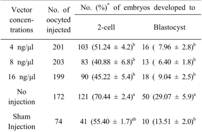
As shown in Table 1, regardless of the concentrations of vector, the cleavage rate were significantly (p<0.05) decreased (4 ng/μl, 51.24%; 8 ng/μl, 40.88% and 16 ng/μl; 45.22%) com- pared to no injection group (70.44%). However, the cleavage
(A)
(B)
Fig. 1. Primers and target site of Sus scrofa FoxN1 sequence (A) and CRISPR/Cas9-mediated vector construction (B).
Five CRISPR/Cas9 target sites and PAM sequences (NGG) are highlighted as green and yellow, respectively (foxnt1 to foxnt5) within red highlights of FoxN1 primers. Target sites has designed specific to exon 2 in the FoxN1 gene and cloned pX330 vector by addgene.
Table 1. Developmental competence of CRISPR/Cas9-mediated vector injected pig parthenotes
Vector concen- trations
No. of oocyted injected
No. (%)
*of embryos developed to
2-cell Blastocyst
4 ng/μl 201 103 (51.24 ± 4.2)
b16 ( 7.96 ± 2.8)
b8 ng/μl 203 83 (40.88 ± 6.8)
b13 ( 6.40 ± 1.8)
b16 ng/μl 199 90 (45.22 ± 5.4)
b18 ( 9.04 ± 2.5)
bNo
injection 172 121 (70.44 ± 2.4)
a50 (29.07 ± 5.9)
aSham
Injection 74 41 (55.40 ± 1.7)
ab10 (13.51 ± 2.0)
bGroups of no injection (Electrical activation only) and sham injection (TE buffer only) were used as control groups for injection and vector, respectively.
*
Percentages are expressed as mean ± SEM of seven repli- cates.
a,b
Different superscripts denote significant differences with in columns (p<0.05).
rate between 3 groups of vector injected and sham injection group (55.4%) showed no difference. The blastocyst formation rates were also decreased in vector injected 3 groups (4 ng/μl, 7.96%; 8 ng/μl, 6.4% and 16 ng/μl; 9.04%) compared to no injection group (29.07%). In addition, the blastocyst formation rates between sham injected group (13.51%) and no injection group (29.07%) also showed significant difference (p<0.05).
3. Knockout Efficiencies and Mutation Patterns
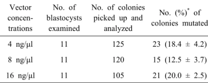
Overall 33 blastocysts from the vector injected groups, 350 colonies were picked up and the sequencing analysis was carried out. As shown in Table 2, the mutation rates were com- parable between groups (4 ng/μl, 18.4%; 8 ng/μl, 12.5% and 16 ng/μl; 20.0%). The sequencing analysis showed that blastocysts derived from each group were successfully mutated in FoxN1 loci regardless of the vector concentrations (Fig. 2 (A)). How- ever, the deletion patterns were higher than the patterns of point mutation and insertion regardless of the vector concentrations (Fig. 2 (B)).
DISCUSSION
In pigs, utilization of gene targeting skill to produce a trans-
Table 2. Knockout efficiency of CRISPR/Cas9-mediated vector injected pig parthenotes.
Vector concen- trations
No. of blastocysts
examined
No. of colonies picked up and
analyzed
No. (%)
*of colonies mutated
4 ng/μl 11 125 23 (18.4 ± 4.2)
8 ng/μl 11 120 15 (12.5 ± 3.7)
16 ng/μl 11 105 21 (20.0 ± 2.5)
*