http://crossmark.crossref.org/dialog/?doi=10.14474/ptrs.2020.9.4.230&domain=pdf&date_stamp=2020-12-25
Received: 9 August, 2020 Revised: 18 September, 2020 Accepted: 18 September, 2020 Corresponding author: Suk-Min Lee (ORCID https://orcid.org/0000-0002-6062-956X)
Department of Physical Therapy, College of Health and Welfare, Sahmyook University, 815 Hwarang-ro, Nowon-gu, Seoul 01795, Republic of Korea Tel: 82-2-3399-1632 Fax: 82-2-3399-1639 E-mail: leesm@syu.ac.kr
This is an Open-Access article distributed under the terms of the Creative Commons Attribution Non-Commercial License (http://creativecommons.org/licenses/
by-nc/4.0) which permits unrestricted non-commercial use, distribution, and reproduction in any medium, provided the original work is properly cited.
Copyright © 2020 Korean Academy of Physical Therapy Rehabilitation Science https://doi.org/10.14474/ptrs.2020.9.4.230
pISSN 2287-7576 eISSN 2287-7584
Phys Ther Rehabil Sci 2020, 9 (4), 230-237 www.jptrs.org
Effects of Schroth exercise therapy on curvature and body appearance of patients with lumbar idiopathic scoliosis
Hyung-Joo Lee
a, Suk-Min Lee
baDepartment of Physical Therapy, The Graduate School, Sahmyook University, Seoul, Republic of Korea
bDepartment of Physical Therapy, College of Health and Welfare, Sahmyook University, Seoul, Republic of Korea
Objective: To investigate the physical appearance and therapeutic changes that occur with the performance of Schroth exercise in patients with scoliosis.
Design: Randomized controlled trial.
Methods: Fifteen subjects with maximum curvature of the lumbar who were diagnosed with idiopathic scoliosis had volunteered to participate in the study. Eight subjects were included in the experimental group where they performed the Schroth Therapeutic Exercise and the other seven were included in the control group. The experimental group underwent 2 hours of weekly treatment for 12 weeks, while the control group did not during the same period based on the decisions of patients or guardians. The Mann- Whitney rank test was carried out to compare the treatment results of the two groups, and the comparison within the group was done by Wilcoxon signed-rank test. The vertebral rotation angle (VRA) was by Scoliometer, and difference of rotated and curved portion volume (DV) between both sides on the major curvature portion measured by 3D human body scanning system.
Results: In the experimental group, 12 weeks of Schroth exercise therapy has significant improved in correction rate (CR) in Cobb’s angle (CA), VRA, and DV between both sides on the major curvature portion (p<0.05), while significant differences were not found between the groups regarding weight bearing difference in both feet (WD) and DV (p<0.05).
Conclusions: Schroth exercise performance showed significant changes in the patient’s therapeutic changes (CA, VRA), but the physical appearance (DV, WD) was not significant, indicating that external changes in the treatment goal setting are more difficult goals to achieve.
Key Words: Exercise therapy, Physical appearance, Physical therapy, Schroth method, Scoliosis
Introduction
As a disease leading to systematic changes, scoliosis car- ries 3-dimensional (3D) deformity of the spine with 10° or more changes of the Cobb’s angle (CA) [1]. In particular, it frequently occurs to adolescent females [2], is a systematic disease [3] that correlates even with deformity of the jaw which is far from the vertebrae of scoliosis patients with more than 40° of curvature, and is hardly discovered as it is superficially observed that initiates internally.
According to statistics from Korea Healthcare big data service in 2019, there are currently 94,158 people with
scoliosis. Since scoliosis begins from childhood and deterio- rates over time, it is most important to diagnose and treat in its early phase [4].
Treatments for scoliosis in the past were mainly operations and use of orthoses, and Schroth therapeutic exercise, which was recently introduced to South Korea, has been ap- plied since 1921 - the year it was first devised by Katharina Schroth – and proved to be effective through a number of studies [5].
Rotation-angular-breathing (RAB), which is the most im-
portant technique in the Schroth therapeutic exercise, is a 3D
breathing method, controlling the breath backward, later-
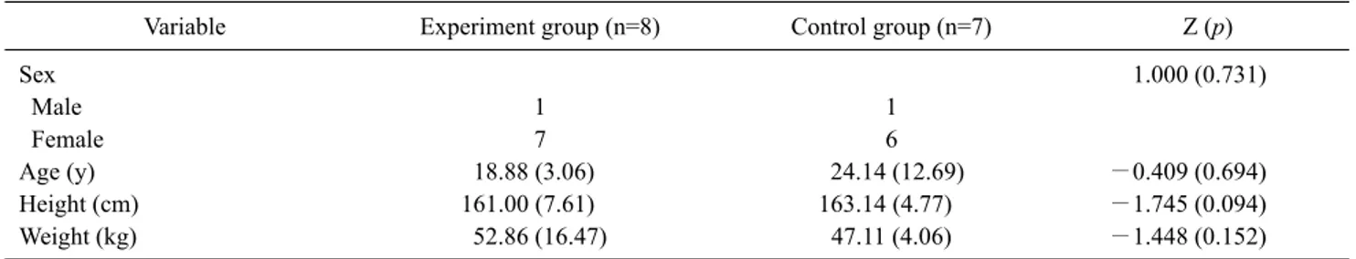
Table 1. General characteristics of participants (N=15)
Variable Experiment group (n=8) Control group (n=7) Z (p)
Sex 1.000 (0.731)
Male 1 1
Female 7 6
Age (y) 18.88 (3.06) 24.14 (12.69) −0.409 (0.694)
Height (cm) 161.00 (7.61) 163.14 (4.77) −1.745 (0.094)
Weight (kg) 52.86 (16.47) 47.11 (4.06) −1.448 (0.152)
Values are presented as number only or mean (SD).
ally, and upward. This breathing method is associated to fill- ing the inverted trunk of the body with air as a balloon is inflated. When this breathing pattern is performed regularly, each breathing serves as a correction exercise, which is a foundation of all Schroth therapeutic exercises, and various movements based on RAB are used in treatments suited for patients with different types of scoliosis.
The main objective measurement variable in research studies on scoliosis is Cobb’s angle (CA). This method has been implemented as a crucial criterion, as well as the most significant variable, for comparison of the curvature rate of the scoliosis. In addition, since the most anticipated sub- jective variable of the patients is appearance [5], research on life quality and appearance among other previous studies have been made through surveys for objective evaluation [6]. Studies on appearance and quality of life have been sup- ported by indices through Scoliosis research society 22 re- vision (SRS 22r) questionnaire provided by Scoliosis re- search society and spinal appearance questionnaire (SAQ).
In addition, scoliosis has a significant difference in the pa- tient’s suspension of both feet. A study by Nachiappan et al.
[7] has shown that weight shift occurs to higher curvature positions compared to ordinary people when standing or walking, and therefore measuring and comparing this differ- ence can also be a variable to evaluate the outcome of treatment.
A study by Schreiber et al. [8] was conducted through sur- veys for 6 months using the Schroth therapeutic exercise, in which the SRS 22r questionnaire evaluated the changes be- tween the 3rd and 6th month and compared with the group that used orthoses and items such as pain and self-awareness of appearance showed significant differences, resulting in high level of quality of life. In the SAQ test, the result showed that no significant differences were found in the overall period, which attributes to the difference between SRS 22r, which evaluates functional aspects such as mental
health and general self-awareness of appearance. The SAQ focuses more on the external aspect, suggesting that Schroth therapeutic exercise reflects the fact that the external changes are more difficult to achieve than the functional as- pects in the treatment of patients with scoliosis.
Among previous studies, those on the functional im- provements proposed significant outcomes from Schroth Therapeutic Exercise in increase of CA and lung capacity [9] and decrease of low back pain [10]. However, studies on quantitative measurement and changes in external aspects like body surface area and volume, which are hardly ob- served physically in the treatment of scoliosis patients, have been so insufficient that our study was set out.
The 3D body type analysis device used for this study can attain 3D topo-graphs and measure the overall outline through calculating the input values to provide computer graphic 3D models, which facilitates objective measure- ment of the body volume and area through quantification of the external volume and area of the trunk, and previous stud- ies that used the 3D body type analysis device focused on the tests for idiopathic scoliosis patients before and after oper- ations [11] and the correlation between kyphotic deformity and low back pain in scoliosis [12], proving that CA is an ex- amination method with a high association [13].
Therefore, this study included the use of x-ray examina- tion and 3-dimensional body type analysis device to inves- tigate for changes in curvature when the Schroth therapeutic exercise was applied to patients with scoliosis.
Methods Participants
Fifteen outpatients with scoliosis of The Goden clinic,
South Korea were selected as subjects for this study after
they have provided their informed consent. The general
characters of subjects were collected, including age, weight,
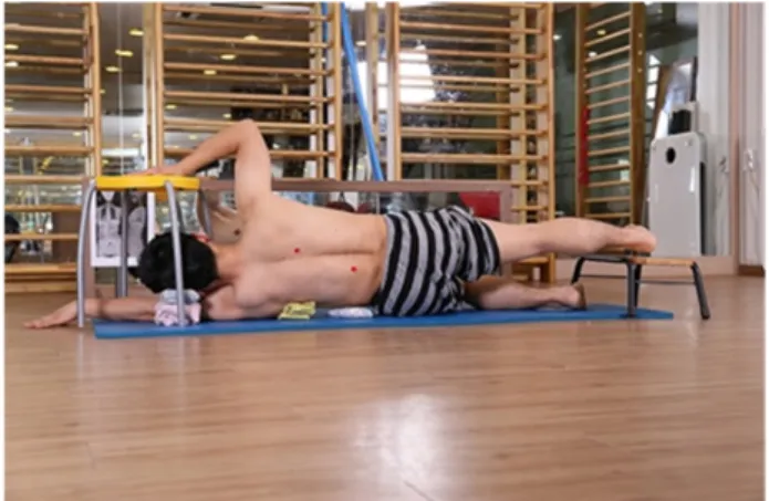
Figure 1. Muscle cylinder.
Figure 2. Cobb’s angle.
Figure 3. 3D body surface examination.
and height, which were followed by a 3D body surface ex- amination for patients with maximum curvature of the lum- bar spine among those with vertebral rotation angle (VRA) and 10° or more of CA together (Table 1).
Intervention
Eight subjects in the experimental group with the in- clusion of the Schroth therapeutic exercise were treated for 2 hours, twice a week, for 12 weeks under the observation of therapists, which was found to be effective in the previous studies [14]. The Schroth exercise was designed to apply a total of 29 movements according to the patient’s angle and type, and the degree of difficulty that the patient can accept.
One of the basic exercises, the muscle cylinder, can be seen in Figure 1.
This study was designed as an equivalent control group pretest-posttest. Among the patients in the experimental group, 2 subjects were excluded from the second measure- ment due to suspension of treatment and 2 additional sub- jects were excluded due to a break period of longer than 1 week. For the control group, the data was collected from those who did not undergo treatment due to cost or other rea- sons while the examinations were conducted with a 12-week gap between January 1, 2014 and September 2016 (Please check the year).
This study was reviewed by the Institutional Review Board of Sahmyook University, Seoul as research to observe research results or phenomena, not to request novel control or intervention that has never existed. The serial number for the review is 2-1040781-AB-N-01-2016043HR.
Measures
After the implementation of this treatment, x-ray and 3D
body surface examinations were conducted again to find the changes in the subjects.
Cobb’s angle
CA was measured by x-ray imaging by means of digital
radiography type imaging equipment (Provision 900; DK
Medical Systems, Seoul, Korea, 2012) (Figure 2).
Table 2. Changes in Cobb’s angle (N=15)
Variable Experiment group (n=8) Control group (n=7) Z (p)
Before (°) 22.11 (7.58) 22.17 (7.27) −0.116 (0.955)
After (°) 15.98 (6.30) 20.08 (7.04)
Change (°) −6.12 (2.99) −2.08 (1.72) −2.540 (0.009)
Z (p) −2.521 (0.012) −2.117 (0.034)
Correction rate (%) 28.14 (11.84) 10.03 (8.98) −2.662 (0.006)
Values are presented as mean (SD).
Difference in ratio volume
The absolute value of the result from one subtracted by the value from the convex portion volume (ℓ) divided by concave portion volume was used for the difference in the ratio of volume because the closer the result of the convex portion volume divided by the concave portion volume goes to 1, the closer to the achievement of symmetry of the trunk volume. This was measured by a 3D body surface examina- tion (Figure 3).
Difference in ratio volume= 1− Convex portion volume Concave portion volume
Vertebral rotation angle
A scoliometer (Scoliometer; Mizuho OSI, Union City, CA, USA, 2013) was used for the VRA, which examined the anterior flexion in standing position. The VRA shows how far the trunk is deviated from the center during rotation with a 3D deformity of the trunk with scoliosis, and it is also an important variable that can be used as a basic test before an x-ray assessment.
Correction rate
Since the closer the CA and the measurement value of the scoliometer converge to zero, the less the alteration, and the CR was expressed as a percentage using the CR used in pre- vious studies [12].
Correction rate= (Before angle−After angle) Before angle *100
The weight-bearing difference in both feet (WD) and dif- ference of rotation volume for tests of WD and difference of the rotated and curved portion volume (DV) produced 3D body surface examination (Medicube; DreamMedics, Seoul, Korea, 2012), the body of the patient was pictured by an in-
frared camera for 3D modeling to measure the volume of each segment on the shoulder, chest, and pelvis, and the vol- ume of the convex portion, which was caused by scoliosis on one side with the navel as the center, was divided by the vol- ume of the concave portion because the closer the result ap- proached to 1, the more the symmetry is achieved. Among the trunk segments, the DV and WD were measured. This assessment helped measure the changes in DV and WD.
Data analysis
PASW Statistics for Windows, Version 18.0 (SPSS Inc., Chicago, IL, USA) was used for all operations and statistics, and descriptive statistics was used for general characteristics.
The Mann-Whitney test was used as a non-parametric test to investigate for the difference between groups, and the Wilcoxon signed rank test was used to investigate for the dif- ferences within groups before and after treatment. The stat- istical significance level α was set to 0.05
Results
Changes in Cobb’s angle
The changes in CA before and after the interventions be- tween the two groups are shown below (Table 2). Although there was no significant difference in angles before and after treatment both in both the experimental and control group (p>0.05), a significance was found in the difference before and after the treatment and the level of correction between the two groups (p<0.05).
Vertebral rotation angle
The changes in VRA by intervention between the two groups are shown below (Table 3). The two groups did not show a statistical significance before treatment, but the di- fference in rotation angle of the experimental group was
−3.38°±2.56° after treatment, suggesting a significant de-
crease between the groups. The changes in the control group
Table 3. Vertebral rotation angle (N=15)
Variable Experiment group (n=8) Control group (n=7) Z (p)
Before (°) 11.75 (3.37) 9.86 (3.93) −0.814 (0.463)
After (°) 8.37 (3.20) 7.42 (2.29)
Change (°) −3.38 (2.56) −2.43 (3.69) −1.049 (0.033)
Z (p) −2.375 (0.018) −1.625 (0.104)
Values are presented as mean (SD).
Table 4. Weight bearing difference in both feet (N=15)
Variable Experiment group (n=8) Control group (n=7) Z (p)
Before (kg) 1.70 (0.67) 2.24 (1.69) −0.291 (0.779)
After (kg) 2.56 (2.08) 2.21 (1.08)
Change (kg) 0.86 (2.00) −0.03 (1.69) −0.926 (0.397)
Z (p) −0.840 (0.401) −1.761 (0.078)
Values are presented as mean (SD).
Table 5. Difference of rotation volume (N=15)
Variable Experiment group (n=8) Control group (n=7) Z (p)
Before (ℓ) 0.07 (0.05) 0.10 (0.14) −0.290 (0.779)
After (ℓ) 0.20 (0.19) 0.13 (0.13)
Change (ℓ) 0.12 (0.22) 0.03 (0.13) −0.810 (0.463)
Values are presented as mean (SD).
was −2.43°±3.36°, but the changes were not significant within the group (p>0.05). After treatment, the difference between the groups and the correction rate showed a sig- nificant difference (p<0.05).
Weight bearing difference in both feet
WD between the two groups before and after the inter- vention was compared by means of the absolute values of the difference in weight on each foot, which can be shown below (Table 4). Before and after the treatment, the ex- perimental group did not show a significant change before and after the experiment, from 1.70±0.67 kg to 2.56±2.08 kg. The control group changed from 2.24±1.69 kg to 2.21±1.08 kg. Without significant difference between the groups before and after the treatment, the average value of the experimental group was 0.86±2.00 kg and there was a decrease by −0.03±1.69 kg in the control group, showing a significant difference between the two groups.
Difference in rotated and curved portion volumes Before and after intervention, the changes in DV between
the two groups are shown below (Table 5). Althouth it was not statistically significant before treatment, DV within the experimental group changed by 0.12±0.22 ℓ after treat- ment showing significant decrease in symmetry (p<0.05). In the control group, the change was 0.03±0.13 ℓ, failing to show a significant change within the group. The discrepancy between the two groups did not show a significant difference.
Discussion
Although the CA after the treatment in the experimental
group and the control group of this study did not show a sig-
nificant difference due to the difference in the curvature de-
gree and peak of each subject with scoliosis, the experi-
mental group and the control group showing significant dif-
ference particularly between the groups in terms of the cor-
rection rate (p<0.001) without any significance within each
group. The same significance and level found in each group
before and after the treatment seemed to attribute to the rea-
son to the effect of the assessment of significance which
does not use the values but the ranking of variables based on the median value of each group that were used for posture recognition, improvement of curvature through the Hawthorne effect, and the Mann-Whitney U method as a nonparametric test.
In terms of correction rate, since it decreased in the ex- perimental group. in the control group showing that im- provements of CA converged to 0 as in the result of the study by Kim et al. [15], the significant result between groups in regards of the correction rate that calculates the degree of improvement by percentage suggests that Schroth ther- apeutic exercise is effective in assisting with correcting the CA for patients with idiopathic scoliosis who have max- imum curvatures on their lumbar spine.
Among previous studies on scoliosis, there has been no experimental research that showed a treatment result con- verging statistically to 0° for the curvature angle, which can attribute reasons to the fact that a previous study, among oth- ers, that implemented a randomized experiment [14], in- stead of showing a significant difference in the result values after treatment for CA, which also revealed significance based on comparing the average values of the groups before and after the treatment , −2.53° in the experimental group and 3.13° in the control group, and that the result of the treat- ment was presented through the correction rate that was used in the previous studies which adopted manual therapy, not the Schroth therapeutic exercises.
Before treatment, the rotation angle was similar in the two groups. After treatment, the results not show a significant difference (p<0.053), but the correction rate signification changed, which represents improvement in scoliosis based on significant reduction of the degree of VRA of the patients because the normal angle converges to 0 as the CA does.
This result can be the same as that of the previous studies, which showed a significant improvement in the ex- perimental group in terms of the VRA of the patients with scoliosis. As the result of improvement in CA and VRA in a previous study [16], which implemented the best selective applications of some of the movements of Schroth, it was confirmed that Schroth therapeutic exercises have a positive effect on surface rotation.
WD was measured by a 3D body surface examination. It provides the difference in weight of both feet obtained from the sensor on which the patient’s feet are placed. After treat- ment, the difference in WD of the control group was close to the significance level. This is similar to a previous study de- signed to have the operation as an independent variable, in
which the pattern showed changes in location and direction of weight-bearing after an operation [6]. As the previous study showed a result that the symmetry was obscured as the patient put more weight on the opposite side than the pre- viously bearing side, the result of the application of Schroth therapeutic exercises can also be interpreted to have affected the patient’s ordinary life through instruction to lead to sit- ting posture and exercise movements which could support the weight on the concave side of the curvature during exercise. Therefore, future studies should further investigate the effects of the differences in weight-bearing on each foot plantar pressure.
The research on the changes in volume of this study launched from a consideration that the symmetry would im- prove as DV decreased proportionally on the convex portion and the concave portion. As mentioned in the definition of the terminology, the closer to 1, the higher the symmetry.
DV in the experimental group showed significant increase after treatment. On the contrary, the control group did not show a significant change.
As Keenan et al. [17] an estimation of the vertebral level and trunk mass of a female idiopathic scoliosis patient through computerized tomography imaging achieved an es- timation that the thickness of the segment with 52.0°±5.9°
for CA changes by the degree of curvature, and Yagi et al.
[18], showed the relation of the iliopsoas muscle and multi- fidus muscle regarding in maintaining spinal stability in standing by comparing the spinal stenosis patient group and the degenerative scoliosis patient group, measuring the area, volume, and cross sectional area by means of radiographs, magnetic resonance imaging, and radiation absorbance, and revealed a significant difference in the cross sectional area of the multifidus muscle attached on the relatively rear side of the spine of the degenerative scoliosis patients compared to the iliopsoas muscle attached to the interior side of the verte- bral body regarding sagittal alignment showing relevance with a significant difference. The muscular strength re- quired to maintain standing under gravity can be revealed through the volume on the body surface, which shows that the Schroth therapeutic exercise affects the convex and con- cave portion on both sidse of the curvature with the navel as the center but rather result in less symmetry.
Among many potential interpretations, the first out of var-
ious influential factors for the application of treatment was
not applied on the day of assessment in order to find the con-
tinuously maintained status. The muscles of the muscu-
loskeletal system are voluntary muscles, which reveal hy-
pertrophy of muscle volume accompanied by the tension right after exercises, but exercises were not performed on the day of assessment, which may have resulted in failure to cre- ate a significant change. Schroth therapeutic exercise trains to direct the vertebral body actively toward the opposite di- rection of the curvature to reduce and, after such move- ments, maintain the curvature by inducing increased tension on the erectors on the concave portion of the curvature, which requires more research on how to increase the sustainment. Thirdly, a radiograph typically divides the two sides by the central sacral line from the center of the spine, but the fact that this study set the center on the navel on the surface for measurement may have affected the result. In consideration that the initial value of the volume on the con- vex portion of the curvature before the experiment was low- er than the volume of the concave portion for a certain pa- tient, the method to measure the body surface volume with the navel as the center may lead to lower significance in judgement of the changes in curvature of patients with sco- liosis, which seems to require more research to find better alternatives. Studies on scoliosis patients through body sur- face assessment are in steady progress, and the major inter- est is given to how to process the attained data and where to put the reference point for measurement [19]. Through this study, more contemplation will be necessary in the selection of variables regarding measuring the volume of the body surface dividing the curvature portion with the navel as the center.
As an equivalent control group pre test-post test design, the most substantial limitation of this study is that the num- ber of the parameters was not achieved as only 7 subjects were recruited for both groups due to the scarcity of patients who were not under treatment whereas a number of patients were looking to have treatment for scoliosis. Scoliosis fre- quently occurs in female adolescents, and they rarely miss treatment while most guardians only seriously consider the location and method of treatment due to the progressive na- ture of the disorder [20]. Most of the patients selected for the control group had financial problems such as the decision of payment by private health insurance or difficulty in agree- ment with family about using the orthosis because the pa- tient was uncomfortable with it or refused to wear it because he or she was too sensitive to outward appearance.
Future studies should research on the correlation between the volume and mass with the measurement result using a body surface device, and more studies are needed to invest- giate the correlation in the asymmetry of the thorax in ac-
cordance with the peak vertebral body curvatures with the maximum curvature at the thoracic vertebrae. In the pre- vious studies, the correlation between the objective differ- ence in trunk volume and the subjective quality of life recog- nized by the patient showed a discrepancy between the ob- jective index and subjective assessment by the patient [6], but since the volume of the body surface can be intuitively recognized outwardly, research is warranted to discover the correlation if more relevant studies are subsequently conducted.
Conflict of interest
The authors declared no potential conflicts of interest with respect to the authorship and/or publication of this article.
References
1. SOSORT guideline committee, Weiss HR, Negrini S, Rigo M, Kotwicki T, Hawes MC, et al. Indications for conservative man- agement of scoliosis (guidelines). Scoliosis 2006;1:5.
2. Miller NH. Cause and natural history of adolescent idiopathic scoliosis. Orthop Clin North Am 1999;30:343-52, vii.
3. Park JO. Correlation between adolescent idiopathic scoliosis and facial asymmetry [Master thesis]. Seoul: Korea University;
2009.
4. Son K. Analysis of the results of thoracolumbosacral orthosis in idiopathic scoliosis and the factors affecting the results [Master thesis]. Jinju: Gyeongsang National University; 2010.
5. Lehnert-Schroth C. Dreidimensionale Skoliosebehandlung:
Atmungs-Orthopädie System Schroth. 8th ed. München: Urban &
Fischer; 2014. German.
6. Yoon JY. The measurement and correlation analysis of pre and postoperative changes in torso deformities using 3-Dimentional body scanner and Scoliosis Research Society outcome Questionnaire (SRS-22) in scoliosis patient [Master thesis].
Ulsan: University Of Ulsan; 2015.
7. Chockalingam N, Dangerfield PH, Rahmatalla A, Ahmed el-N, Cochrane T. Assessment of ground reaction force during scoli- otic gait. Eur Spine J 2004;13:750-4.
8. Schreiber S, Parent EC, Moez EK, Hedden DM, Hill D, Moreau MJ, et al. The effect of Schroth exercises added to the standard of care on the quality of life and muscle endurance in adolescents with idiopathic scoliosis-an assessor and statistician blinded randomized controlled trial: "SOSORT 2015 Award Winner".
Scoliosis 2015;10:24.
9. Park SY, Shim JH. Effect of 8 weeks of Schroth exercise (three-dimensional convergence exercise) on pulmonary func- tion, Cobb’s angle, and erector spinae muscle activity in idio- pathic scoliosis. J Korea Converg Soc 2014;5:61-8.
10. Lebel A, Lebel VA. Severe progressive scoliosis in an adult fe- male possibly secondary thoracic surgery in childhood treated with scoliosis specific Schroth physiotherapy: case presentation.
Scoliosis Spinal Disord 2016;11(Suppl 2):41.
11. Lee CS, Hwang CJ, Kwon J, Choi SH, Lee MY, Yoon SJ, et al.
Surface topography using 3-dimensional whole body scanner in patients with scoliosis. J Adv Spine Surg 2014;4:16-22.
12. Park SY, Kim JH, Lee S, Ahn DK, Lee SB, Ko YJ. Correlation between degree of adolescent [thoracic] kyphosis and low back pain: using 3-Dimension Whole Body Analyzer (BodyCheck 3D BM101R). J Korean Orthop Res Soc 2011;14:9-16.
13. Choi EJ, Kim TY. Correlation of the 3D-surface topography and Cobb’s angle in scoliotic patient’s. J Korean Acad Clin Electrophysiol 2005;3:61-70.
14. Kuru T, Yeldan İ, Dereli EE, Özdinçler AR, Dikici F, Çolak İ. The efficacy of three-dimensional Schroth exercises in adolescent idiopathic scoliosis: a randomised controlled clinical trial. Clin Rehabil 2016;30:181-90.
15. Kim KD, Hwangbo PN. Effects of the Schroth exercise on the Cobb's angle and vital capacity of patients with idiopathic sco- liosis that is an operative indication. J Phys Ther Sci 2016;28:
923-6
16. Weiss HR, Moramarco MM, Borysov M, Ng SY, Lee SG, Nan X, et al. Postural rehabilitation for adolescent idiopathic scoliosis during growth. Asian Spine J 2016;10:570-81.
17. Keenan BE, Izatt MT, Askin GN, Labrom RD, Pettet GJ, Pearcy MJ, et al. Segmental torso masses in adolescent idiopathic scoliosis. Clin Biomech (Bristol, Avon) 2014;29:773-9.
18. Yagi M, Hosogane N, Watanabe K, Asazuma T, Matsumoto M.
The paravertebral muscle and psoas for the maintenance of glob- al spinal alignment in patient with degenerative lumbar scoliosis.
Spine J 2016;16:451-8.
19. Patias P, Grivas TB, Kaspiris A, Aggouris C, Drakoutos E. A re- view of the trunk surface metrics used as Scoliosis and other de- formities evaluation indices. Scoliosis 2010;5:12.
20. Soucacos PN, Zacharis K, Gelalis J, Soultanis K, Kalos N, Beris A, et al. Assessment of curve progression in idiopathic scoliosis.
Eur Spine J 1998;7:270-7.