Cell Death in Lung Epithelial Cells
Eun Kyung Choi, M.D., Yun Seup Kim, M.D., Jae Seuk Park, M.D., Young Koo Jee, M.D., Kye Young Lee, M.D.
Department of Internal Medicine, Dankook Unuversity College of Medicine
폐상피세포에서 흡연추출물-유도성 세포사에 관한 연구
단국대학교 의과대학 내과학교실 최은경, 김윤섭, 박재석, 지영구, 이계영
배 경 : 폐기종에서 발생하는 폐포 파괴의 원인으로서 전통적으로 protease/anti-protease 불균형과 산화성 스트레스가 주요 가설로 여겨져 왔으나 최근 폐포세포의 아포프토시스가 폐포파괴 및 폐기종의 원인이 된다는 이론이 제기되고 있어 서 A549 폐상피세포에서 흡연추출물에 의한 세포사의 특성을 규명하고자 본 연구를 시행하였다.
방 법 : A549 폐상피세포주에서 여러 농도의 흡연추출물 및 억제제를 첨가한 후 MTT assay를 잉용하여 세포생존율을 측정하였다. 세포사 분석은 FACScan을 이용한 DNA 분절확인, 전자현미경 검사, Hoecst/PI 이중염색을 이용한 현광현미 경 검사를 이용하였고 cytochrome c 유리는 면역형광법을 이용하였다. Bcl-2 과발현세포주를 이용하여 bcl-2의 역할을 확인하였고 p53 Western blot 및 HPV-E6 과발현 세포주를 이용하여 p53의 역할을 확인하였다.
결 과 : A549 세포주에서 흡연추출물에 의한 세포사는 FACScan에서 DNA 분절에 의한 subG1 분획의 확인 및 Hoecst/PI 이중염색 및 현광현미경 소견 상 아포프토시스임이 확인되었고 전자현미경 소견상 저농도에서는 아포프토시스가 발생하 지만 고농도에서는 괴사가 발생함을 확인하였다. Cytochrome c가 세포질로 유리됨을 확인하였으나 caspase 억제제에 의 해서 세포사가 차단되지 않았다. 흡연추출물에 의한 세포사는 Bcl-2과발현에 의해 억제되었고 p53활성화를 유도하고 p53 이 기능적으로 knock-out 된 세포주에서 억제되었다.
결 론 : 흡연추출물에 의한 폐상피세포의 세포사는 저농도에서는 아포프토시스를 고농도에서는 괴사를 유도하고 bcl-2 및 p53 경로가 중요한 역할을 담당하며 이는 폐기종 발생기전에 있어서 폐세포 세포사 이론을 뒷바침하는 자료로 활용될 수 있다고 생각된다. (Tuberc Respir Dis 2005; 58:43-53)
Key words : Cigarette smoke extract, Apoptosis, Lung epithelial cells, Emphysema
This work was supported by 2000 Seoul National University College of Medicine Alumni Award.
Address for correspondence : Kye Young Lee, M.D.
Department of Internal Medicine College of Medicine, Dankook University 16-5 Anseo-dong, Cheonan 330-714 South Korea
Phone : +82-41-550-3916 Fax : +82-41-556-3256 E-mail : [email protected] Received : Aug. 30. 2004 Accepted : Dec. 7. 2004
Introduction
Tobacco use is a major cause of death from cancer, cardiovascular disease, and pulmonary disease.
More than 80% of chronic obstructive lung disease in the United States is attributable to cigarette smoking. Cigarette smoking also increases the risk of respiratory infection, including pneumonia, and
results in greater disability from viral respiratory tract infections.
1-3Approximately 92 to 95 percent of the total weight of mainstream smoke is present in the gas phase (nitrogen, oxygen, carbon dioxide).
The remaining gases and particulate matter, including tar, nicotine, acrolein, formaldehyde, and phenol, are the substances of medical importance.
4The influence of these substances on the composition and biological activity of cigarette smoke is not unknown. The mechanism of injury by cigarette smoking is com
plex and appears to include direct injury by oxidant gases
5, increased elastase activity, and decreased antiprotease activity.
6Cigarette smoking produces and increases oxidant burden on the lung and smoke is potentially injurious to airspace epithelial cells, since it has been calcula
ted to contain 10
17oxidant molecules/puff, of which
10
14are oxygen radicals.
7Cigarette smoking has been shown to result in increased airspace epithelial permeability.
8,9It can also suppresses proliferation, attenuates attachment, and augments detachment of the epithelial cells by oxidant-induced injury.
10These oxygen radicals are short-lived. But cigarette smoke condensate continues to produce oxidants which may result in a more persistent oxidant injury
10and may contribute to the development of the lung diseases.
Few studies have examined the signaling path
way of cigarette smoke-induced cellular injuries and cell loss in airspace epithelial cells. Many di
fferent compounds in cigarette smoke can readily react directly to form oxygen radicals and reactive oxygen radicals damage multiple cellular components, including DNA, lipid membranes, and proteins
11, causing mutagenesis and apoptosis.
12Oxygen radicals directly activate the mitochondrial apoptotic path
way and directly induce cytochrome c release through mitochondrial membrane potential loss.
13The cytosolic cytochrome c activates the apoptotic cascade and this is inhibited by the presence of Bcl-2 on mitochondria.
14Cigarette smoke also cause DNA single-strand breaks in the cells.
15Moreover multiple chemical modifications occurred in all four DNA bases in a pattern suggestive of reaction with hydroxyl group or deaminating species in cigarette smoke.
16DNA damage induces the accumulation of p53 and DNA damage-mediated p53 accumulation induces cell cycle arrest by p21 upregulation or apoptosis by increase in the ratio of BAX/Bcl-xL, cytochrome c release, and caspase activation.
17,18Therefore cigarette smoke may affect to mitocho
ndrial pathway, especially Bcl-2 progtein and p53 pathway.
We set out to investigate the cytotoxic effct of cigarette smoke through cigarette smoke extract (CSE) which is particulate fraction containing
most of the toxic organic components of smoke.
Here we demonstrate that CSE induces mainly apoptosis in A549 lung epithelial cell lines at lower concentrations and that CSE-induced apoptosis may be caspase-independent apoptosis. We also determine that Bcl-2 and p53 pathway play a key role in CSE-induced apoptosis.
Materials and Methods Cells and Reagents
A549 cells (alveolar type II cell-derived cell line) purchased from ATCC. Cells were cultured in RPMI 1640 media with 10% fetal bovine serum supple
mented 100 U/ml penicillin and 100 mg/ml streptomycin.
Caspase-3 substrate (Ac-DEVD-pNA; Ac-Asp-Glu- Val-Asp-pNA), Ac-DEVD-cho (N-acetyl-Asp-Glu- Val-Asp-aldehide), Z-VAD-fmk (Z-Vad-Ala-Asp- fluoromethyl-keton) were provided by Alexis (San Diego, CA). Ac-YVAD-cho (N-acetyl-Tyr-Val-Ala- Asp-aldehyde) was obtained from Transduction laboratories (Lexington, KY). Anti-cytochrome c antibody was obtained from PharMingen (San Diego, CA). Anti-bcl-2 antibody and anti-p53 antibody were provided by Santa Cruz Biotechnology (Santa Cruz CA). All other chemicals and proteins were purchased from Sigma.
Preparation of Cigarette Smoke Extract
The CSE was prepared as described previously reported. 19 Commercial cigarettes (Marlboro, Philip Morris, Inc., Richmond, VA) were smoked continu
ously and mainstream smoke was bubbled through
10 ml of phosphate-buffered saline (PBS) that was
prewarmed to 37 C by application of a vacuum to
the flask containing the PBS. Each cigarette was
smoked for 2 min, and one cigarettes were used per
10 ml of PBS solution of the particulate-phase extract of cigarette smoke. The CSE obtained was then filtered through a 0.22-mm milipore filter. Final concentra
tions of this solution are expressed as percent values to total volume (vol %). Solutions raging from 0.5 to 20 % were used in the present studies and the CSE was prepared immediately before each experiment.
Cell viability assay
Cell viability was measured by a MTT assay.
Briefly, untreated cells or cells treated with CSE in a 96-well plate were harvested at the indicated times followed by the addition of 3-(4,5-dimethyl thiazol-2-yl)-2,5-diphenyl tetrazolium bromide (MTT) and then cell were solubilized with 0.1 N acidified CH
3Cl-HCl. The 96-well plate was read at a wav
elength of 590 nm on an iEMS Labsystems plate reader.
Apoptotic assay
Apoptotic cell death tested was performed by three separate methods: FACScan, double staining with Hoechst 33342 and propium iodide, and trans
mission electron microscopy.
The cells incubated with CSE for 48hr in a 6-well plate were harvested and fixed with ethanol. Ethanol- fixed cells are centrifuged at 400g then washed in PBS. The pelleted cells are resuspended in DNA staning reagent, propium iodide (PI;10 ug/ml) followed by FACScan analysis (Becton Dickinson, Flanklin Lakes, NJ).
Morphological changes in the nuclear chromatin of cells undergoing apoptosis were detected by double staining with 2.5 mg/ml bisbenzimide Hoechst 33342 fluorochrome and 5 mg/ml propium iodide, followed by examination on a fluorescence micro
scope. The number of viable, apoptotic, and necrotic cells was quantified with minimum cell counts of
300 cells.(blue intact nuclei: viable, blue fragmented nuclei: early apoptotic, pink fragmented nuclei : late apoptotic, pink intact nuclei : necrotic cells)
Cytochrom c release with immunofluorescence microscopy
A549 cells with CSE treatment that were plated on four well Lab-Tek chamber slides (nunc, Naper
ville, IL) were fixed for 20 min in 4% formaldehyde made in saline and then postfixed in 70% ethanol overnight. Monoclonal antimouse-cytochrome c anti
body diluted 1/100 was used to detect cytochrome c. Protein-antigen-antibody complex was revealed with FITC-a mouse antibody (Zymed) diluted 1/200.
The cell nuclei were stained with 2.5 mg/ml Hoechst 33342 for distinguish from apoptotic cells.
Epi-fluorescence microscopy was done on a Olym
phus IX-FLA inverted reflected light fluorescence microscope. Photographs were obtained with a DP11 microscope digital camera system (Olymphus).
Assessment of caspase-3-like activity
The caspase-3-like activity was measured by colorimetric assay using the peptide-based substrate Ac-DEVD-p-nitroanilide. In brief, caspase-3 fluor
ogenic substrate was incubated with CSE-treated cell lysates for 1 hr at 37 ℃, then pNA liberated from Ac-DEVD-p-nitroanilide was measured using fluorometric plate reader with 405 nm.
Plasmid DNA and Transfection
PcDNA3- bcl-2 expression vector was provided by YJ Oh, Yeon-sei University and HPV-E6 ex
pression vector was provided by Dr. Rosen, Stanford
University. These were stably transfected into A549
cells using lipofectamine-plus (Gibco-BRL, Gaithers
CSE
20%
10%
5%
0%
SubG1 G1
S
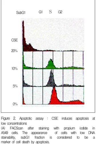
G2Figure 2. Apoptotic assay : CSE induces apoptosis at low concentrations
(A) FACScan after staining with propium iodide in A549 cells. The appearance of cells with low DNA stainability, subG1 fraction is considered to be a marker of cell death by apoptosis.
A B
0 20 40 60 80 100
CSE
cell viability (%)
control 1%
5%
10%
15%
20%
Control CSE 5%
CSE 10% CSE 20%
Figure 1. Dose-dependent CSE-induced cytotoxicity in A549 cells.
(A) MTT assay: A549 cells were incubated with CSE in RPMI 1640 medium for 48 hr. Each dose point represents the average of triplicate measurement of three samples within an experiment. Data represent mean
± SD.
(B) Light microscopy (×100) of A549 cells cultured with CSE in medium for 48 hr, showing cell death.
burg, MD) and resistant clones were pooled after selection in 400-600 mg/ml of G418.
Western blot analysis.
A549 cells were treated with 10% CSE for the indicated times and then lysed in a boiling solution contaning 1% SDS, 1 mM sodium vanadate and 10 mM Tris-HCl pH 7.4. Samples were centrifuged for 5 min to remove insoluble material followed by measurement of protein concentration by the Bradford method 9Bio-Rad Laboratories, Hercules, CA). Samples containing equal protein concentrations were dena
tured by boiling and analyzed by SDS- PAGE, and then transferred to nitrocellulose. The blot was then placed in blocking buffer containing 1% milk, 1%
BSA, 10 mM Tris-HCl pH 7.5, 100 mM Nacl, and
0.1 % Tween 20 for 1 hr at room temperature or
overnight at 4 ℃. The blot was then incubated in
blocking buffer with individual antibodies in a solution
which contained 10mM Tris-Hcl pH 7.5, 100 mM
Figure 2. (B), (C)Induction of A549 cell death was due to apoptosis, because the 10% CSE treatment for 48 hr induced chromatin condensation and nuclear fragmentation but apoptosis and necrosis was mixed in 20% CSE as detected by double staining with Hoechst33342/PI.(blue intact nuclei: viable, blue frag- mented nuclei: early apoptotic, pink fragmented nuclei : late apoptotic, pink intact nuclei : necrotic cells)
B Control CSE 10% CSE 20%
C
0 1 0 2 0 3 0
c o n t r o l C S E 1 0 % C S E 2 0 %
(%) a p o p t o t i c c e l l
n e c r o t i c c e l l
0 10 20 30
control CSE 10% CSE 20%
(%) apoptotic cell
necrotic cell
Nacl, and 0.1 % Tween 20 followed by incubation in blocking buffer contaning a horseradish peroxidase- conjugated anti-rabbit IgG or anti-mouse IgG (Caltag Laboratories, San Francisco, CA) at a dilution of 1:1500 and detected by ECL (Amersham, Arlington Heights, IL) followed by autoradiography.
Results
CSE induces dose-dependent cell death in A549 cells
To determine CSE-induced cell death in A549 cells, A549 cells were grown in 96-well plate and the cells were exposed to serial dilutions of CSE raging from 0.5% to 20% for 48hr. Then the cells were analyzed with MTT assay. MTT assay showed that cell viability was markedly decreased in dose-dependent fashion at 48 hr after CSE incubation. (Figure 1A) Light microscopic features were almost identical with MTT assay. (Figure 1B)
CSE induces apoptosis at low concentrations of CSE
The reduction of cell viability was due to apop
tosis as demonstrated by DNA fragmentation using FACScan for subG1 fraction. (Figure 2A) Double staining with Hoechst 33342/propium idodide and electron microscopy also confirmed that CSE in
duced apoptotic cell death. After 48 hr of treatment low concentrations (10%) of CSE induced nuclear condensation and fragmentation in 15% of the cells, which are hallmarks of apoptosis and 92% of cells of untreated cells were alive. At the same time point, 22% of cells treatment with high concentra
tions (20%) CSE were apoptotic and 24% of cells
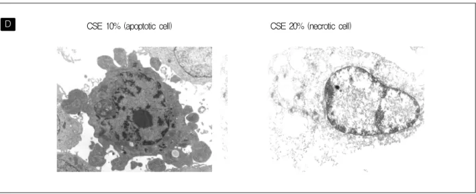
D CSE 10% (apoptotic cell) CSE 20% (necrotic cell)
Figure 2. (D) Apoptotic feature (convolution of cell and nuclear outlines, mass of condensed chromatin) also well demonstrated by electron microscopy 48hr after addition of 10% CSE.
A Control B CSE 10%
Hoechst stain Immunofluorescent stain Hoechst stain Immunofluorescent stain
Figure 3. CSE-induced cytochrome c release. Cytochrome c of normal control cells that had blue intact nuclei with Hoechst stain was showed perinuclear halo with FITC-immunofluorescent stain (A). Dead cells in low concentration (10% ) of CSE showed blue fragmented nuclei and were stained in entire cytoplasm with anti-cytochrome c antibody typical of mitochondrial release in apoptosis (B). Visualization of apoptosis (arrow) in cells 48hr after exposure using antibody that recognizes cytochrome c (green). Nuclei were counterstained with hoechst33342 (blue).
were necrotic cell death. (Figure 2B, 2C) Apoptotic phenomenon that were multiple nuclear fragments, mass of condense chromatin, convolution of cell and nuclear outlines also well demonstrated by electron microscopy 48hr after addition of 10% CSE.
(Figure 2D) These data confirm the finding that CSE induces apoptosis in low concentrations of CSE and indicates that the cell death in high concentrations of CSE most likely is a combination of apoptosis and
necrosis.
CSE induces cytochrome C release in A549 cells
Another cellular event associated with apoptotic cell death is the release of cytochrome c from mi
tochondria. Therefore, we next examined whether
mitochondrial cytochrome c was release into cytosol
using immunofluorescence with monoclonal anti-
A
0 0.5 1 1.5 2 2.5 3
24hr 48hr
ab/mg protein/hr
CSE 0%
CSE 5%
CSE 10%
CSE 15%
CSE 20%
cyclo+ TNF-a
B
0 20 40 60 80 100 120
CSE 5% CSE 10% CSE 15%
cell viability (%)
control ZVAD YVAD DEVD
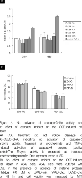
Figure 4. No activation of caspase-3-like activity and no effect of caspase inhibitor on the CSE-induced cell death
(A) CSE treatment did not induce cleavage of Ac-DEVD-pNA, indicating no activation of caspase-3 enzyme activity. Treatment of cycloheximide and TNF-a induced activation of caspase-3 enzyme (positive control).The Enzyme activity is expressed as increased absorbance/mgprotein/hr. Data represent mean ± SD.
(B) No effect of caspase inhibitor on the CSE-induced cell death in A549 cells. A549 cells were cultured with CSE in the presence or absence of cysteine protease inhibitors (40 μM of ZVAD-fmk, YVAD-cho, DEVD-cho) for 48 hr and cell viability was measured by MTT assay. Data represent mean ± SD.
CSE 5% CSE 10% CSE 15%
0 50 100
Control Dexamethasone NAC Captopril NAME
Cell viability(%)
Figure 5. Inhibition of CSE-induced cell death by inhibitor in A549 cells. A549 cells were incubated with CSE (5%, 10%, 15%) in presence or absence of NAC (1 mM), dexamethasone (1 μM), captopril (1 mM) or L-NAME (1 mM) for 48 hr and cell viability was measured by MTT assay. NAC significantly increased cell viability, while dexamethasone, captopril and L-NAME didnt affect cell viability at all. Data represent mean ± SD.
cytochrome c antibody. As release of cytochrome c from the mitochondria an early event in apoptotic pathways, the cells were incubated in the presence of 10% CSE for 24 hr. Localization analysis of cytochrome c by fluorescence microscopy revealed the release of cytochrome c from mitochondria to cytoplasm in apoptotic cells which were nuclear frag
mentation in Hoechst staining. (Figure 3) This redistribution
confirms again apoptotic cell death in 10% CSE.
CSE dose not activate caspase-3 and caspase inhibitors do not prevent CSE-induced cell death in A549 cells
Caspase-3-like activity is known to increase in apoptotic cell death but no increase in caspase- 3-like activity was observed in low concentrations of CSE-treated A549 cells. (Figure 4A) Furthermore the inhibition of caspase activity by inhibitor pro
tects cells from caspase-dependent apoptotic cell death. But the treatment of A549 cells with the peptide-based caspase-3 inhibitor Ac-DEVD-cho, pan-caspase inhibitor ZVAD-fmk, caspase-1 inhibitor Ac-YVAD-cho did not prevent cell death in the presence of low concentrations of CSE that induced apoptotic cell death. (Figure 4B) This results suggests that CSE-induced apoptosis might be caspase- independent apoptosis.
NAC prevents CSE-induced cell death
In order to get the clues for the signaling events,
A A549-neo A549-bcl-2
Bcl-2
B
CSE 1% CSE 5% CSE 10% CSE 15%
0 50 100
A549-neo A549-bcl2
Cell viability(%)
Figure 6. Bcl-2 reduced CSE-induced cell death.
(A) Characterization of A549-bcl-2 cells by bcl-2 western blot analysis. There was bcl-2 induction in A549-bcl-2 cells but no induction in A549-neo cells.
(B) A549-bcl-2 cells and A549-neo cells were cultured with various concentrations of CSE in the RPMI 1640 medium for 48 hr. A549-bcl-2 cells were significantly more resistance to CSE induced cell death than A549-neo cells. Data represent mean ± SD.
A western
CSE(hr) 0 0.5 1 2 4
CSE 1% CSE 5% CSE 10% CSE 15%
0 50 100
A549-neo A549-E6
Cell viability(%)
B
Dox(1uM) - + - +
p53
A549-neo A549-E6
Figure 7. Effect of CSE on p53 expression in A549 cells.
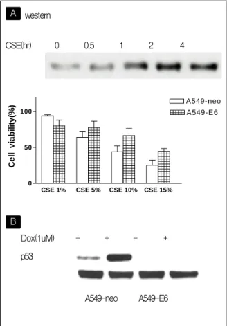
(A) A549 cells were treated with CSE (10%) and harvested 0 hr, 0.5 hr, 1 hr, 2 hr and 4 hr after treatment of CSE for Western blot analysis with a monoclonal p53 antibody. P53 was induced by CSE.
A549-E6 cells and A549-neo cells were cultured with various concentrations of CSE in the RPMI 1640 medium for 48 hr. A549-E6 cells were resistance to CSE induced cell death compared with A549-neo cells. Data represent mean ± SD.
(B) Characterization of A549-E6 cells by p53 Western blot analysis. A549-E6 and A549-neo cells were treated with doxorubicin (1 μM) and harvested 8 hr after treatment of doxorubicin for western blot ana- lysis with a monoclonal p53 antibody. Doxorubicin could induce the expression of p53 in A549-neo cells. In con-
we tried several inhibitor studies including N-
acetylcysteine (a nonspecific antioxidants), captopril which is reported to inhibit apoptosis in lung epi
thelial cells, L-NAME (iNOS inhibitor), and dexame
thasone (anti-inflammatory and anti-fibrotic drug).
CSE-induced cell death was near completely blocked by pretretment with NAC, but the other inhibitors did not show any effect on CSE-induced cell death by MTT assay. (Figure 5) This result suggests that CSE might induce apoptosis through intracellular oxidative stress.
Bcl-2 reduces CSE-induced cell death
Because oxidants seemed to be the major contri
butors of the cytotoxic effects in CSE, we examined the effect of bcl-2 on CSE-induced cell death using stable cell line overexpressing bcl-2 (A549-bcl-2).
A549-bcl 2 cell line was characterized by bcl-2 western blot analysis. (Figure 6A) A549-bcl-2 stable cell line resisted significantly against CSE-induced cell death than control cell line (A549-neo) by MTT assay. (Figure 6B) These findings are highly su
ggestive of the role of mitochondrial pathway in
CSE-triggered death signal in A549 cells.
p53 plays a significant role in CSE-induced cell death
In response to a variety of stimuli, including DNA damage, hypoxia, or ribonucleotide depletion, p53 can be activated, which generally results in cell cycle arrest or apoptosis of the affected cells. Oxidant induces DNA damage and then appears to play a critical role in regulating p53 function. So we ex
amined the role of p53 on CSE-induced cell death using western blot analysis and functional p53 knock-out study employing human papilloma virus E6 (HPV-E6) expression vector. Western blot analysis showed significant p53 activation in time-dependent fashion in the presence of 10% CSE (figure 7A) and CSE-induced cell death was inhibited by stable over
expression of HPV-E6 cell line (A549-E6). (Figure 7B). A549-E6 cell line was characterized by p53 western blot analysis after 1mM doxorubicin treat
ment.(Figure 7A). These results suggest that p53 was important signaling pathway in CSE-induced cell death in A549cells
Discussion
Epithelial cells are important in maintaining the integrity and fluid balance of tissue and in the control of inflammation. Injury to the epithelium may be an important early event following exposure to cigarette smoke and other oxidant gases, mani
festing as an increase in epithelial permeability
8, cellular detatchment
10and inhibition of cellular re
pair processes.
20Thus damage to alveolar cells by cigarette smoke may implicated in the development of several pulmonary disease for example chronic bronchitis, emphysema, asthma. But the mechanism of the epithelial cell injury or epithelial cell loss by cigarette smoke in the some pulmonary disease is unknown. For the explanation of epithelial cell loss,
we built the hypothesis that cigarette smoke itself induce apoptotic cell death in lung epithelial cell death and investigated the mechanisms by which cigarette smoke induced cell death in lung epithelial cells. In this study, we showed that CSE induced apoptosis of A549 type II pnuemocyte cells at lower concentration and necrosis of A549 cells at higher concentrations. Apoptotic cell death was demonstra
ted by three methods, FACScan, Hoechst 33342/PI double staining, Electron microscopy. Also apoptotic feature was confirmed by cytochrome c release from mitochondria at lower concentrations (10%
CSE) which had dominat feature of apoptosis in double staining. Although morphological and molecular changes of the cells clearly showed that CSE concentrations of 10% or less induced apoptosis, a broad-spectrum caspase inhibitor was unable to inhibit CSE-induced apoptosis and CSE did not activate caspase 3 like enzyme. This results suggests that CSE might induce apoptosis through a caspase-independent pathway. In recent report, some proapoptotic proteins such as Bax, a mammalian cell death protein that targets mitochondrial mem
branes, can induce mitochondrial damage and cell death even when caspases are inactivated.
21Such experimental observations argue that a caspase- independent mechanism for commitment to death exists. This mechanism is likely to involve mito
chondria. Mitochondria undergoing PT liberate an
apoptogenic protein, apoptosis including factor (AIF),
which is capable of inducing nuclear apoptosis. AIF
has recently been cloned and characterized. It is a
protein that is translated with a mitochondrial
targeting sequence that is cleaved upon import into
mitochondria. The cleavage exposes a nuclear
targeting sequence that allows the protein to target
to the nucleus and induces nuclear changes upon
release from the mitochondria.
22This mechanism
has been suggested to be independent of a caspase-
mediated pathway for apoptosis. The findings of the present study provide some new information into the important area of the influence of cigarette-smoke extract on caspase-independent apoptotic cell death but further investigations in the molecular basis for the caspase-independent apoptotic process, such as Bax protein or AIF release will be needed.
CSE is known to contain a considerable amount of oxygen-derived free radicals such as superoxide anion, hydroxyl radicals or hydrogen peroxide.
19, 23The present study showed that NAC, scavenger of oxygen-free radicals, attenuated the CSE-induced cell death. Also Bcl-2, antiapoptotic protein, over
expression was same result. This suggests that cell death may result from a direct oxidant effect on epithelial cells and that mitochondrial pathway plays a important role in CSE-induced cell death. Oxidant scavenger and Bcl-2 overexpression are an important protective mechanism against cigarette smoke- induced airspace epithelial pertubation.
In summary this study shows that lower dose cigarette smoke extract has a detrimental effect on cultured epithelial cells causing caspase-in
dependent apoptosis. This effects of CSE are probably oxidant mediated, although the exact mechan ism has not elucidated. And mitochondrial pathway and p53 play a important role in CSE- induced apoptosis in A549 epithelial cells. It remains to be proven whether cigarette smoke could result in a apoptotic process in the primary pulmonary epithelial cells and in the lung airspaces of animal models resulting in direct oxidative injury to airway epithelium.
Abstract
Emphysema is characterized by air space enlarge
ment and alveolar destruction. The mechanism
responsible for the development of emphysema was thought to be protease/antiprotease imbalance and oxidative stress. A very recent study shows that alveolar cell apoptosis causes lung destruction and emphysematous changes. Thus, this study was per
formed to support the evidence for the role of apoptosis in the development of emphysema by characterizing cigarette smoke extract (CSE)-induced apoptosis in A549 (type II pneumocyte) lung epithelial cells. CSE induced apoptosis at low concentration (10% or less) and both apoptosis and necrosis at high concentration (20%). Apoptosis was demonstrated by DNA fragmentation using FACScan for subG1 fraction. Discrimination between apoptosis and necrosis was done by morphologic analysis using fluorescent microscopy with Hoecst 33342/propium iodide double staing and electron microscopy. Cy
tochrome c release was confirmed by using immuno
fluorescence with monoclonal anti-cytochrome c antibody. However, CSE-induced cell death did not show the activation of caspase 3 and was not blocked by caspase inhibitors. This suggests that CSE-induced apoptosis might be caspase-independent apoptosis. CSE-induced cell death was near com
pletely blocked by N-acetylcystein and bcl-2 over
expression protected CSE-induced cell death. This results suggests that CSE might induce apoptosis through intracellular oxidative stress. CSE also activated p53 and functional knock-out of p53 using stable overexpression of HPV-E6 protein inhibited CSE-induced cell death. The characterization of CSE-induced cell death in lung epithelial cells could support the role of lung cell apoptosis in the patho
genesis of emphysema.
References
1. Holbrrok JH, Groundy SM, Hennekens CH, Kannel WB, Strong JP. Cigarette smoking and cardiova
scular diseases: a statement for health professionals by a task force appointed by the steering committee of the American Heart Association. Circulation 1984;70:1114A-7A.
2. Altshuler B. Quantitative models for lung cancer induced by cigarette smoke. Environ Health Perspect 1989;81:107-8.
3. Higgins M Epidemiology of COPD: state of art. Chest 1984;85:3S-8S.
4. Sethi JM, Rochester CL. Smoking and chronic ob
structive pulmonary disease. Clin Chest Med 2000;
21:67-86.
5. Rahman I, MacNee W. Role of oxidants/antioxidants in smoking-induced lung disease. Free Radic Biol Med 1996;21:669-81.
6. Stockely RA. Proteases/antiproteases: pathogenesis and role in therapy. Clin Pulm Med 1998;5:203-10.
7. Church T, Pryor WA. Free-radical chemistry of cigarette smoke and its toxicological implications.
Environ Health Perspect 1985;64:111-26.
8. Jones JG, Lawler P, Crawley CW, Minty BD, Hulands G, Vesll N. Increased alveolar epithelial permeability in cigarette smokers. Lancet 1980;1:
66-8.
9. Burns AR, Hosford SP, Dunn LA, Walker DC, Hogg JC. Respiratory epithelial permeability after cigarette exposure in guinea pigs. J Appl Physiol 1989;66:
2109-16.
10. Lannan S, Donaldson K, Brown D, MacNee W.
Effect of cigarette smoke and its condensates on alveolar epithelial cell injury in vitro. Am J Physiol 1994;266:L92-L100.
11. Nakano M. Free radicals and their biological sign
ificance: present and future. Hum Cell 1992;5:334- 40.
12. Adams JD, Mukherjee SK, Klaidman LK, Chang ML, Yasharel R. Apoptosis and oxidative stress in
the aging brain. Ann N Y Acad Sci 1996;786:135-51.
13. Green DR, Reed JC. Mitochondria and apoptosis.
Science 1998;281:1309-12.
14. Adams JM, Cory S. The Bcl-2 protein family:
arbiters of cell survival. Science 1998;281:1322-6.
15. Leanderson P, Tagesson C. Cigarette amoke-induced DNA damage in cultured human lung cells: role of hydroxyl radicals and endonuclease activation.
Chem Biol Interact 1992;81:197-208.
16. Repine JE, Bast A, Lankhorst I. Oxidative stress in chronic obstructive pulmonary disease. Am J Respi Crit Care Med 1997;156:341-57.
17. Hall PA, Lane DP. Tumor suppressors: a developing role for p53? Curr Biol 1997;7:R144-7.
18. Ko LJ, Prives C. p53: puzzle and paradigm. Genes Dev 1996;10:1054-72.
19. MuroharaT, Kugiyama K, Ohgushi M, Sugiyama S, Yasue H. Cigarette smoke extracts contracts isolated porcine coronary arteries by superoxide anion- mediated degradation of EDRF. Am J Phyiol 1994;
266:H874-80.
20. Wang H, Liu X, Umino T, Skild CM, Zhu Y, Kohyama T, et al. Cigarette smoke inhibits human bronchial epithelial cell repair process. Am J Respir Cell Mol Biol 2001;25:772-9.
21. Xiang J, Chao DT, Korsmeyer SJ. Bax-induced cell death may not require interleukin 1 beta-converting enzyme-like proteases. Proc Natl Acad Sci U S A 1996;93:14559-63.
22. Susin SA, Lorenzo HK, Zamzami N, Marzo I, Snow BE, Brothers GM, et al. Molecular characterization of mitochondrial apoptosis inducing factor. Nature 1999;397:441-6.
23. Zang LY, Stone K, Pryot WA. Detection of free radicals in aqueous extracts of cigarette tar by electron spin resonance. Free Radic Biol Med 1995;19:161-7.