Ⅰ. 서 론
악골의 발육기형 양상은 하악골전돌증, 하악골왜소증, 개교증, 안면비대칭 등의 형태로 나타난다
1). 이 중 하악골전돌증은 한국 인 등 동양인에서 빈도가 높다고 보고되었다
2). 하악골전돌증은 많은 수술법이 개발되어 왔는데 가장 많이 사용되는 술식으로 Obwegeser에 의해 소개되고
3), Dal Point에 의해 발전된
4)하악지 시상분할골절단술과 Winstanly에 의해 소개된 하악지 수직골절 단술이 있으며
5), 모두 수술시 혈관, 신경의 손상을 방지하기 위하 여 하악지 내면의 해부학적 구조에 대해 많은 이해가 요구된다.
그 중 하악지 내면의 소설과 하악공의 위치는 하악지 시상분할 골절단술시 수평 내측골절단선 위치 결정과 하악지 수직골절단
술시 수직골절단선 위치를 선정할 때 중요한 역할을 한다. 하지 만 때로는 정확한 위치를 보지 못하고 일반적인 해부학적 지식 을 기초로 하여 골절단선 위치를 결정해야 하는 경우가 많다. 만 일 이 위치를 잘못 파악하여 신경혈관속의 손상을 주었을 경우 하치조신경 손상에 의한 지각마비의 합병증이나 하치조혈관의 손상으로 인한 심한 출혈을 야기할 수 있다.
이러한 하악지에 대한 해부학적 연구는 국외, 국내의 여러 논 문에서 보고되었다
6-13).
그러나, 외국의 연구와는 인종적인 차이가 있고 대부분의 연구 에서 교합평면에 평행한 선을 소설과 하악공에 적용하여 기준점 을 설정하였으나 실제 건조 하악골내 전체 치아의 보존상태가 완전하지 않고, 수술시 하악의 개구에 의해 교합평면을 적절히 관찰할 수 없으므로 이를 이용한 계측방법은 실제적으로 수술시 참고로 하기에 어려운 점이 많다.
따라서 본 연구에서는 하악 제2대구치가 완전히 맹출된 영구 치열기 한국인 성인 건조 하악골 65개를 대상으로 하여 전방기 준점을 교합평면이 아닌 하악지 전연의 최전방점과 외사선과 치 조골 상방선이 만나는 점을 기준으로 양측 130면의 소설과 하악 공의 위치를 계측하여 악교정 수술시 가능한 합병증을 예방하며
악교정 수술을 위한 한국인 하악지의 해부학적 위치에 관한 연구
우순섭∙조정연∙박원희∙유임학∙이영수∙심광섭 한양대학교 의과대학 치과학교실
Abstract (J. Kor. Oral Maxillofac. Surg. 2002;28:126-131)
우 순 섭
471-701, 경기도 구리시 교문동 249-1 한양대학교 구리병원 치과
Soon-Seop Woo
Dept. of dentistry, Hanyang university Kuri hospital, 249-1, Kuri city, Kyunggi-do, 471-701, Korea Tel : 82-31-560-2660
Email: [email protected]
A STUDY OF MANDIBULAR ANATOMY FOR ORTHOGNATHIC SURGERY IN KOREANS
Soon-Seop Woo, Jung-Yeon Cho, Won-Hee Park, Im-Hag Yoo, Young-Soo Lee, Kwang-Sup Shim
Dept. of Dental Surgery, Medical College of Hanyang University
Anatomical shape of the mandibular ramus, which includes the area from the rear of the mandibular second molar to the mandibular posterior border and from the mandibular sigmoid notch to the inferior mandibular border, must be carefully considered to perform orthognathic surgery. The locations of the lingula and mandibular foramen in medial side of mandibular ramus are one of the most important factors to decide the location of the horizontal medial osteotomy in sagittal split ramus osteotomy and to select the line of vertical osteotomy in intraoral vertical ramus osteotomy.
Sixty-five different Korean human dry mandibles were surveyed.
All mandible have permanent dentition including complete eruption of the mandibular second molar. The locations of the lingula and mandibular foramen in medial side of the ramus were identified and following results were obtained.
Anterior ramal horizontal distance from lingula was 16.13±3.53mm(range:8.6�24.3mm), anterior ramal horizontal distance from mandibular foramen was 23.91±4.79mm(range: 14.1�39.7mm), horizontal width of mandibular foramen was 2.79±0.95mm(range: 1.5
�6.1mm), height of lingula was 10.51±3.84mm(range: 3.1�22.4mm), vertical distance from sigmoid notch to lingula was 19.82±
5.11mm(range: 9.1�35.3mm).
From this study, the result could be used to select the location of osteotomy lines and to decide amount of periosteal elevation to avoid injury of neurovascular bundle, and to accomplish the appropriate split in Korean patients in mandibular orthognathic surgery.
Key words: Manibular ramus, Lingula, Mandibular foramen
적절한 시술이 용이한 골절단선 설정에 도움을 주고자 본 연구 를 시행하였다.
Ⅱ 연구대상 및 방법
1. 연구대상
65개의 하악 제2대구치가 완전히 맹출된 영구치열기 한국인의 건조 하악골 양측 130면의 하악지 내면을 연구대상으로 하였다.
2. 연구방법
하악골 악교정 수술시 관찰가능한 하악골의 내면의 특징적인 점을 기준점으로 설정하였고(Fig. 1), 이 기준점을 이용하여 수직, 수평 기준선을 설정하였으며 (Fig. 2), 이 기준선과 하악골이 만나 는 점 (Fig. 3)을 이용하여 Table. 1의 항목을 계측하였다.
하악골의 내면을 버니어 캘리퍼스(Mitutoyo , Japan)로 계측 을 시행하였으며, 기준선 설정으로 직접 계측이 불가능한 경우 기준자로 Ruler(Kanamata , Japan)와 함께 하악지 내면을 촬영 한(Nicon F-801, Japan) 사진을 이용하여 버니어 캘리퍼스로 항목
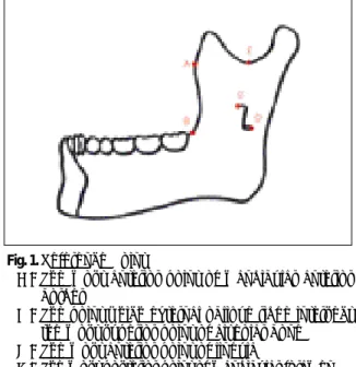
Fig. 1. Reference Point
A : The most anterior point of mandibular anterior border
B : The point which external oblique ridge intersect the most superior point of alveolar bone C : The most anterior point of lingula
D : The most posterior point of mandibular foramen E : The most lower point of sigmoid notch
Fig. 2. Reference Line 1. Vertical Reference Line
1) Vertical reference line of anterior border of ramus:The line from point A to point B 2) Vertical reference line of sigmoid notch:The
parallel line with A-B line from point E 2. Horizontal Reference Line
1) Horizontal reference line of lingula:The right angled line with A-B line which pass by point C 2) Horizontal reference line of mandibular fora-
men:The right angled line with A-B line which pass by point D
Fig. 3. Measuring Point
F : The point of horizontal reference line of lingula intersect anterior border of ramus G : The point of horizontal reference line of
mandibular foramen intersect anterior border of ramus
H : The point of horizontal reference line of lingula intersect vertical reference line of sigmoid notch
I : The point of horizontal reference line of mandibular foramen intersect vertical reference line of sigmoid notch
J : The point of vertical reference line of sigmoid notch intersect inferior border of ramus K : The point of horizontal reference line of lingula
intersect posterior border of ramus
L : The point of horizontal reference line of mand- ibular foramen intersect posterior border of ramus
계측을 시행하였으며(Fig. 4), 각 계측치를 토대로 평균 및 표준편 차 및 최대, 최소 범위를 구하였다.
Ⅲ. 결 과
65개의 하악 제2대구치가 완전히 맹출된 영구치열기의 한국인 건조 하악골을 연구대상으로 하여 양측 130면 하악지 내면의 소 설과 하악공의 해부학적 위치를 계측하여 평균, 표준편차 및 범 위를 계산하였고(Table 2) 하악지내의 상대적인 위치를 구하였 다. (Table 3)
하악지 수평거리 계측에서 하악지 전소설 수평거리(하악지 전 연에서 소설의 최전방점까지의 수평거리)는 16.13±3.53mm [범 위: 8.6�24.3mm], 하악지 전하악공 수평거리(하악지 전연에서 하 악공의 최후방점까지의 수평거리)는 23.91±4.79mm [범위: 14.1
�39.7mm], 하악공 수평폭경은 2.79±0.95mm [범위:1.5�6.1m]였 다.
하악지 수직거리 계측에서 소설 높이(소설의 최상방점에서 하 악공의 최하방점까지의 거리)는 10.51±3.84mm [범위:3.1�
22.4mm], 하악절흔-소설 수직거리(하악절흔의 최하방점에서 소 설의 최상방점까지의 수직거리)는 19.82±5.11mm [범위: 9.1�
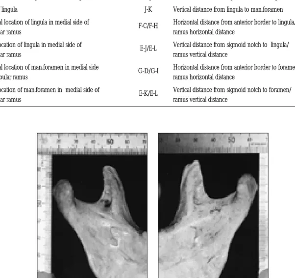
Table 1.Measurement of contents
Anterior ramal horizontal distance from lingula F-C Horizontal distance from anterior border to lingula
Anterior ramal horizontal distance from mandibular foramen G-D Horizontal distance from anterior border to mandibular foramen Horizontal width of mandibular foramen Horizontal width of mandibular foramen
Vertical distance from sigmoid notch to lingula E-J Vertical distance from sigmoid notch to lingula
Height of lingula J-K Vertical distance from lingula to man.foramen
Horizontal location of lingula in medial side of F-C/F-H Horizontal distance from anterior border to lingula/
mandibular ramus ramus horizontal distance
Vertical location of lingula in medial side of E-J/E-L Vertical distance from sigmoid notch to lingula/
mandibular ramus ramus vertical distance
Horizontal location of man.foramen in medial side G-D/G-I Horizontal distance from anterior border to foramen/
of mandibular ramus ramus horizontal distance
Vertical location of man.foramen in medial side of
E-K/E-L Vertical distance from sigmoid notch to foramen/
mandibular ramus ramus vertical distance
Contents line Description
Fig. 4. The Photography of medial side of mandibular ramus with reference ruler
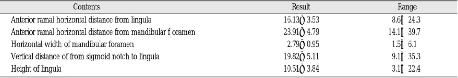
Table 2.Measured Result In Mandibular Ramus
Anterior ramal horizontal distance from lingula 16.13±3.53 8.6�24.3
Anterior ramal horizontal distance from mandibular f oramen 23.91±4.79 14.1�39.7
Horizontal width of mandibular foramen 2.79±0.95 1.5�6.1
Vertical distance of from sigmoid notch to lingula 19.82±5.11 9.1�35.3
Height of lingula 10.51±3.84 3.1�22.4
(Ave±S.D unit: mm)
Table 3.Measured Relative Location In Mandibular Ramus
Horizontal location of lingula in medial side of mandibular ramus 0.39±0.07 0.22�0.54 Vertical location of lingula in medial side of mandibular ramus 0.34±0.05 0.22�0.45 Horizontal location of mandibular foramen in medial side of ramus 0.61±0.08 0.44�0.75 Vertical location of mandibular foramen in medial side of ramus 0.47±0.12 0.35�0.76
Contents Result Range
Contents Result Range
35.3mm]였다.
하악지내 소설의 수평위치(하악지 전체 수평 폭경중 소설의 위치)는 0.39±0.07 [범위: 0.22�0.54], 하악지내 소설의 수직위치 (하악지 전체 수직 고경중 소설의 위치)는 0.34±0.05 [범위: 0.22
�0.45]였고, 하악지내 하악공의 수평위치(하악지 수평 폭경중 하 악공의 수평위치)는 0.61±0.08 [범위:0.44�0.75], 하악지내 하악 공의 수직위치(하악지 수직 고경중 하악공의 수직위치)는 0.47±
0.12 [범위: 0.35�0.76]였다.
Ⅳ. 총괄 및 고찰
소설과 하악공의 위치를 정확히 파악하여야 악교정 수술시 하 치조신경 손상, 출혈의 예방이 용이하고, 골 두께를 유의하여야 불리한 골절 없이 분할이 가능하다. 하지만 하악지 내면을 수술 시 충분한 시야확보가 불가능하고, 수술방법에 따라서는 내면의 박리 없이 골절단을 설정해야 한다.
하악지 수직골절단술은 술식이 내측의 하치조신경을 육안으 로 확인할 수 없고, 외측 상행지를 경험적 판단에 의존하여 골절 단을 시행하기 때문에 골내의 하치조신경을 손상시킬 수 있고, 안쪽의 연조직 및 상악동맥의 손상가능성이 있다.
하악지 시상분할 골절단술후 신경손상에 관해 Turvey가 8.2%
14), Van Merkesteyn는 25.8%
15), Wester mark는 39%의 합병증을 보고한 바 있다
16). 하악지 수직골 절단술시 Bell은 1% 이하
17), Westermark 는 9%
16), Van Merkesteyn는 11.8%의 신경손상을 보고한 바 있다
15).
하악지 시상분할절단술시 가능하면 하악절흔 직하방, 즉 가능 한 상부위치에서 수평골절단을 시행하면 신경혈관속의 손상을 피할 수 있다. 하지만 하악지에서 협측피질골판과 설측피질골판 의 유합되는 점이 하악지의 상방에 존재하여 상방에서의 내측수 평골 절단시 망상골의 개입이 없으므로 근심, 원심 골편의 분할 이 어렵고 불리한 골절의 가능성이 높다
8). Nichoson 등은 근심 골
편의 골절빈도를 1�3%로 보고하였고
10)Van Merkesteyn는 17%
15), O’Ryan F는 1.9�20%의 빈도를 보고 하였다
18). 하악지의 하연으 로 갈수록 망상골의 양이 많아지므로 가능한 하방에서 골절단이 필요하며 이때 기준이 되는 소설과 하악공의 위치를 계측하여 이를 참고로 골절단을 시행하면 불리한 골절의 예방과 충분한 피질골 두께를 얻을 수 있다. 그러나, 하방으로 올수록 신경혈관 속의 손상이 두려워 술자로 하여금 하방에서의 골절단을 회피하 게 만들 수 있다.
Smith는 50개의 동양인 하악지의 피질골 두께를 측정하여 소설 로부터 7.5�13.3mm 상방에서 협측과 설측 피질골판이 합쳐짐 과 시상분할시 이 부위 하방에서 내측 골절단할 것을 주장하였 고
8), 48개의 서양인 하악골을 계측한 다른 연구에서는 소설 7.08
�7.69mm 상방에서 협측과 설측 피질골판의 합쳐짐을 보고하였 다
9).
하악지에서 소설의 위치에 관한 연구는 국외 및 국내의 여러 논문에서 보고되었다. 하악지 전소설 수평거리에 관한 국내의 연구에서 김 등은 242개의 한국인의 하악골을 계측하여 하악지 전소설 수평거리는 유치악의 경우 17.5±2.6mm, 무치악의 경우 17.3±2.4mm를 보고하여
12)본 연구 (16.13±3.53mm)와는 평균 1mm 이상의 차이를 보였으나 이는 기준선 설정의 차이로 생각 된다.
소설 높이에 관한 연구에서 Nicholson은 8.4±2.0mm
10), Bremer 는 8.3mm로 보고하여
19)본 연구 (10.51±3.84mm)와는 평균 2mm 이하의 차이를 보였다.
소설의 수평적 위치에 관한 연구에서 김 등은 하악지 전연에서
후연 사이의 거리를 1로 볼 때 하악지 전연으로부터 0.54 위치에
소설이 존재한다고 보고하여
12)본 연구(0.39±0.07)와는 평균 0.15
이상의 차이를 보였다. 소설의 수직적 위치에 관한 연구에서 김
등은 하악절흔과 하악하연 사이의 거리를 1로 볼 때 하악 절흔으
로부터 유치악은 0.32, 무치악은 0.30으로 보고하여
12)본 연구(0.34
±0.05)와는 비슷하였다. 이 결과는 하악골 시상분할 골절단술시 하악지 내면 박리시 하방까지의 박리량 결정시 도움을 줄 수 있 다.
또한 하악지에서 하악공의 위치에 관한 연구도 여러 논문에서 보고되었다. 하악지 전하악공 수평거리에 관한 연구에서 Hayward는 45개의 아시아 그룹의 하악골을 계측하여 하악지 전 연에서 하악공 중심까지의 수평거리는 19.74±2.16mm로 보고하 여
6)본 연구(23.91±4.79mm)와는 4mm 이상의 차이를 보였으나 본 연구에서는 하악공 최후방위치로 기준점을 설정해서 별 차이 는 없는 것으로 생각된다. 이 계측치들은 하악지 시상분할골절 단술시 하악지 내면 박리시 박리량을 결정하는데 도움을 줄 수 있고 하악지 수직골절단술시 이것보다 후방에서 골절단을 시행 해야 함을 의미한다.
다른 서양인 하악골 계측 연구에서는 평균 20mm의 결과를 보 고하였다
20).
하악공 폭경에 대한 연구에서 Hayward는 아시아 그룹에서 8.31±1.85 mm로 보고하여
6)본 연구(2.79±0.95mm)와는 평균 5mm 이상의 차이를 보였다. 57개의 서양인 하악골을 계측한 다 른 연구에서는 평균 6.1mm (범위: 3-11mm)였고
7), 2-11mm의 범위 를 보고한 연구도 있었다
21). 이는 본 연구 결과와는 상당한 큰 차 이로 이렇게 큰 차이가 나는 것을 설명할 수 없으나, 본 연구에서 는 하악공 내면에서의 직경을 직접 계측하였다.
하악공의 수평적 위치 연구에서 Daw는 하악공의 수평적 위치 를 0.64로 보고했고
7), 이는 본 연구(0.61±0.08)와 비슷한 결과였 다. 다른 서양인 하악골 계측 연구에서는 0.52
10), 0.56
11)으로 보고 했다.
또 다른 연구에서는 하악지의 전후연 사이의 중심에 하악공이 위치한다고 보고했다
22-25).
본 연구결과는 하악지 수직골절단술시 하악지 외측면 박리후 하악지 전후방을 모두 파악할 수 있기 때문에 골절단선이 이보 다 후방에 위치하도록 설정하는데 도움을 줄 수 있을 것이다.
하악공의 수직적 위치에 관한 연구에서 Nichoson 등은 80개의 서양인의 성인 하악골을 계측하여 하악공이 하악절흔과 하악지 의 하연 중심에 위치한다고 보고했으며 다른 연구에서도 중심에 하악공이 위치한다고 보고하여
26,27)본 연구(0.47±0.12)와 차이가 없었고, 또 다른 연구에서는 0.37
7), 0.39
11)으로 보고하여 본 연구와 는 큰 차이를 보였다.
한편 하악공의 위치의 다양함을 보고한 연구가 있었으며
7,10), 하 악공의 위치를 국소화하기 어렵다는 연구도 있었다
28,29). 따라서 이 부분은 경험적, 통계적 판단에만 의존하기보다는 수술전 방 사선 사진을 통한 충분한 분석을 시행하여야 함을 의미한다.
Ⅴ. 결 론
본 연구는 65개의 하악 제 2대구치가 완전히 맹출된 영구치열 기의 한국인 성인 건조 하악골을 대상으로 소설과 하악공의 위 치를 계측하였다. 소설 높이가 평균 10.5 mm의 결과를 보여 내측 수 평골절단시 소설까지 골절단을 시행하여도 감각마비의 위험
이 적음을 알 수 있었다.
하악지내에서 소설은 수평적으로는 전방에서 약 2/5 부위에, 수직적으로는 상방으로부터 약 1/3 부위에 위치함을 알게되어 수술시 골절단선 설정에 도움을 줄 수 있을 것이다.
따라서 본 연구의 이러한 결과는 한국인 악안면 기형환자의 악 교정 수술시 신경손상 및 출혈의 예방과 적절한 골편의 분할이 가능한 골절단 선정의 기준점으로 활용될 수 있으리라 판단되며 향 후 이를 임상적으로 응용할 수 있는 진보적인 연구가 필요하 고 이의 적극적 활용이 악교정 수술의 발전에 참고자료가 될 수 있을 것으로 사료된다.
참고문헌
1. Babcock WW: Surgical treatment of certain deformities of jaw asso- ciated with malocclusion of teeth. JAMA. 53:833 1909.
2. Archer WH: Oral and Maxillofacial Surgery. Philadelphia, WB Saunders company. 2:1448-1449, 1975.
3. Obwegesser H: The surgical correction of mandibular prognathism and retrognathism with consideration of genioplasty. Oral Surg Oral Med Oral Pathol. 10:677-689, 1957.
4. Dal Point JB: L Osteotomie retromolare per la correzione della prog- enia. Minerva chir. 14:1138, 1959.
5. Winstanly RP: Subcondylar osteotomy of the mandible and the intraoral approach. Oral Surg Oral Med Oral Pathol. 6:134-136, 1968.
6. Hayward J, Richardson ER, Malhotra SK: The mandibular foramen its anteroposterior position. Oral Surg Oral Med Oral Pathol. 44:837- 43, 1977.
7. Daw JL, de la Paz MG: The mandibular foramen anstomic study and its relevance to the sagittal ramus osteotomy. J Cranofac Surg.
10:475-479, 1999.
8. Smith BR, Rajchel JL, Waite DE, Read L: Mandibular ramus anatomy as it relates to the medial osteotomy of the sagittal split ramus osteotomy. J Oral Maxillofac Surg. 49:112-6, 1991.
9. Tom WK, Martone CH: A study of mandibular ramus anatomy and its signific anceto sagittal split osteotomy. Int J Oral Maxillofac Surg.
26:176-178, 1997.
10. Nicholson ML: A study of the position of the mandibular foramen in the adult human mandible. Anatomical Record. 212:110-112, 1985.
11. Langston JR, Tebo HG: The incidence and relationship of the lateral ramus prominence to the mandibular foramen. Oral Surg. 44:190- 196, 1977.
12. Kim HJ, Lee HY, Chung IH, Cha IH, Yi CK: Mandibular anatomy related to sagittal split ramus osteotomy in Koreans. Yonsei Med J.
38:19-25, 1997.
13. 최재덕, 이의웅: 한국인 성인 하악골의 상행지 외융기에 대한 방사
선 계측학적 연구. 대한치과의사협회지 17:29-34, 1979.
14. Turvey T: Intraoperative complication of the sagittal osteotomy of the mandibular ramus. J Oral Maxillofac Surg. 43:504-509, 1985.
15. Van Merkesteyn JPR, Groot RH: Intraoperative complications in the sagittal and vertical ramus osteotomies. Int J Oral Maxillofac Surg.
16:665-670, 1987.
16. Westermark A, Bystedt L: Inferior alveolar nerve function after mandibular osteotomies. Br J Oral Maxillofac Surg. 36:425-428, 1998.
17. Bell WH: Mandibular prognathism in Modern Practice in orthog- nathic and reconstuctive surgery. Philadelphia, PA Saunders.
17:1448-1449, 1992.
18. O’Ryan F: Complications of orthognathic surgery. Oral Maxillofac Surg Clin North Am. 2:593-613, 1990.
19. Bremer G: Measurements of special significance in connection with anesthesia of the inferior alveolar nerve. Oral Surg Oral Med Oral Pathol. 5:966-988, 1952.
20. Augier M: Sequelette Cephalique In Traite Anatomie Humaine.
Published under the direction of PA Nicolas Masson Paris. 16:665-
670, 1928.
21. Kaffe I, Ardekian L, Gelerenter I, Taicher S: Location of the mandibular foramen in panoramic radiograph. Oral Surg Oral Med Oral Pathol. 72:662-669, 1994.
22. Basmajian JV: Grant’s Method of anatomy by Regions Descriptive and Deductive. Williams & Willkins Co Baltimore. 10:473, 1980.
23. Hamilton WJ: Textbook of Human Anatomy. The Macmillan Press.
Ltd London. 10:80, 1985.
24. Last RJ: Anatomy Regional and Applied. Churchill Livingstone Edinburgh. 6:572, 1978.
25. Schafer EA, J Symington, and TH Bryce: Quains Elements of
Anatomy. Longmans Green & Co London. 4:104, 1915.
26. Fawcett E: The structure of the inferior maxilla with special refer- ence to the position of the inferior dental canal. J Anat. 29:355, 1895.
27. Miller JA: Studies on the location of the lingula, mandibular foramen and mental foramen. Anat Rec. 115:349, 1953.
28. Moore K: The head in Clinically oriented anatomy. Williams &
Wilkins. 2:794, 1985.
29. Hollinshead WH: Anatomy for surgeons. Harper and Row, New York. 2:393, 1968.