약학회 지 제 43 권 제 6 호 802-808(1999)
Yakhak Hoeji
Vol. 43, No. 6Vibrio vulniGcus cytolysin 의 흰쥐 혈소판 응접 기전
김현철 • 채 수 완 * • 이 병 창 ** • 은 재 순 *
우석 대 학교 약학대 학, *전복대 학교 의 과대 학 약리 학교설, **원광대 학교 의 과대 학 (Received August 31, 1999)
Mechanism of Vibrio vulnificus Cytolysin on Rat Platelet Aggregation
Hyun-Chul Kim, Soo-Wan Chae*, Byung-Chang Lee** and Jae-Soon £ u i/
College of Pharmacy, Woosuk University, Samrye 565-701, Korea
^Departments of Pharmacology, Chonbuk National University Medical School, Chonju 560-182, Korea
**Wonkwang University Medical School, Iksan 570-749, Korea
A bstract —
Vibrio vulnificus
cytolysin has been incriminated as one of the important virulence determinants in
V. vulnificus
infection. In the present study, the effects ofVibrio vulnificus
cytolysin on platelets were examined.Vibrio vulnificus
cytolysin induced platelet aggregation and increased intracellular calcium concentration ([Ca고우]j) of rat platelets. These effects were abolished in Ca요수-free buffer (2 mM EGTA).Cytolysin also potentiated ADP- and collagen-induced platelet aggregation. Lanthanum (2 mM) inhibited cytolysin-induced platelet aggregation. However, another Ca오"*' channel blockers, verapamil (20 p.M) or mefenamic acid (20 |iM) did not block cytolysin-induced platelet aggregation. Osmotic protectants, sucrose (50 mM) and raffinose (50 mM) suppressed platelet aggregation by 35.9% and 63.4%, respectively.
V vulnificus
cytolysin increased membrane conductances of platelet membranes. These results suggest that cytolysin-induced platelet aggregation is mediatedvia
lanthanum sensitive-calcium influx which resulted from the pore formation byV. vulnificus
cytolysinKeywords □
Vibrio vulnificus
cytolysin, platelet aggregation, calcium influx.Vibrio vulnificus는 Hollis도카■ 생화학적 검사룰 통하 여 Vibrio parahaemolyticus Vibrio alginolyticus^
성상이 비숫하나 lactose를 분해하는 균들을 lactose- positive Vibrio라고 명명함으로써 알려지게 되었다. 이 후 Farmer 동2비 Vibrio 속과의 유전적 관계, 표현형 의 유사성에 근거를 두고 Vibrio vulnificus라고 부를 것을 제안하쉬 현재의 공식 균명으로 사용되어지고 있 다. 이 균은 G(-), 호염성간균으로 우리의 식생활과 밀접한 관련이 있는 어패류와 바닷물에 의해 감염되며 감염
시임상증상에 따라 상처 감염군과 원발성 페혈중 군으로 구분된다."^ 이중 특허 원발성 패혈증군의 경우
^본 논문에 관한 문의는 이 저자쎄게로
(전화
) 0652-290-1569 (팩스
) 0652-290-1567간경번이나 hemochromatosis와 같이 면역번움이 서하 되어 있는 환자에서 높은 발병율을 보인다. 그러나, 현 재까지 균 감염시의 발병기전에 대해서는 알려진 바가 없으며 균 감염시의 임상증상으로 머루어 그 exotoxin 이 관여되어 있을 것으로 생각되고 었다.^'^^ V vulnificus가 분비하는 exotoxin으로는 cytolysin,^'구 ^ protease,collagenase,^^ antiphagocytic surface an- tigcn/어 lipase/오 ^ siderophore^^^ 및 phosphoipa- ggi3) 등이 알려져 있으며, cytolysin과 protease가 그 중 중요한 처사인자로 밝혀지고 있다.^^ 그러나 현재까 지 V vulnificus가 분비하는 cytolysin의 병리학적 기 능에 대해서는 논란어 제기되어 왔다
포유동물외 순환 혈액을 이루는 구성 성분 중 가장
작은 세포인 혈소관은 지혈 작용 및 혈전 형성에 중요
Vibrio vulnificus cytolysin익 흰쥐 혈소관 응집 기견
한 역할을 하는 cell fragment로서, 동맥경화증이나 심 맥관게 및 뇌혈관 질환과 같은 성인병의 발병과도 매 우 밀접한 관련이 있는 것으로 알려져 있다.^^^
vulnificus 감염시의 임상증상은 혈관 폐쇄를 일으키는 조직괴사를 유발하며, 또다른 V vuhttfkus 감명시의 임 상소견으로 thrombocytopenia가 보고되기도 하였다.도 ®^
아울러 Khoo 등요지은 Synanceja horrida^ stonu- stoxin에 의한 platelet aggregation과 세포내 calcium 농도([Ca오 '*"]j) 증가를 보고한 바 있고, staphylococcus a-toxin에 의한 혈액응고 촉진에 그리고 zooxanthel- latoxiiwl에 의한 platelet aggregation 유발배 등 여 러가지 득소에 의한 혈소관 기능조절이 알려져 왔다.
그러나, 현재까지 V vulnificus cytolysin의 생체 내 에서의 표적조직 특히 혈소관에 대한 영향에 대해서는 거의 알려진 바가 없다. 이에 본 연구에서는 V vulnificus cytolysin이 혈소관에 미치는 영향을* 규명하고 자 cytolysin에 의한 혈소관 응집, 혈소관 번화 그러고 혈소관 세포막의 전도도 번화률 조사하였다.
실험방법
실험동물 및 시험물질 - 실험 동물로는 대한실험동 물센터에서 구입한 웅성 백서(Sprague-Dawley, 250- 350g)몰 실험실에서 5일 이상 적응시켜 사 S■ 하였다. 혈 소관의 준비는 Rho 등^아의 방법을 변형하지 사용하였 다. 흰쥐의 복대동맥에서 2 .2% sodium citrate률 미 리 채운 주사기에 혈액과 sodium citrate의 비률 9:
l(v/v) 되도록 채혈하고 llOg에서 10분간 원심분리한 상층액을(25^) PRP(platelet-rich plasma)로, 침전물 을 다시 4 X 에서 lOOOg로 20분■간 원심분리한 상층액 을 PPP (platelet-poor plasma)로 이용하였다.
실험에 사용한 Vibrio vulnificus 균주는 미국 M.
H. Kothary(FDA, Washington D,C,, 미국) 박사*로부 터 분주받은 HNCC률, 균은 Kreger 등"넉 방법에 따라 heart infusion diffusate broth에 접중하여 사용 하였다. V vulnificus^} 분비하는 cytolysin은 Kim 등^^^
의 방법에 따라 정제하고, cytolysin의 활성은 머■우스 적혈구의 용혈정도를 이용한 Bemheimer 등^^^ 의 방 법에 준하여 유리된 hemoglobin 능도률 545 nm에서 의 흡광도로 측정하식 50%의 hemoglobin 유리를 일 으킨 cytolysin의 앙을 1 hemolytic unit(HU)로 정의 하였다.
ADP(adenosine diphosphate), collagen등과 같'은' 혈소관 응집인자는 Chrono-Log사(USA), Fura-2 AM 은 Molecular Probe사(USA)에서 그리고 영양액 조제 및 기타 약물은 모두 Sigma사(USA)에서 구입하여 사 용하였다.
혈소관 음점농 측 정 -혈 소 판 응집능 측정에는 aggregometer (Chrono-Log, 미국)를 이용하였으며 응 집정도는 aggregation rate/min으로 표시하였다.
aggregation rate/min
=b/(ax l.2)x 10/6
전기생리학적인 기록 - 혈소관 세포막의 견도도를 측 정하기 위하여 patch-damp technique^^숍 이용하였다.
Sodium salicylate(l mM)룰 처처하여 자발적 응집을 방지한 혈소관을 silicon으로 coating한 bath에 두고, 미세견국과 혈소관 사이에 giga-seal이 형성되면 약 3- 5 분 후 bath에 cytolysin(3 HU)을 가하고 전류의 번화를 관찰하였다.
미세전국(5~10M ^>은* borosilicatefr러관 (1.5*-1.8
X
100 mm, Kimble, 머국)을 이용하여 미세견국제작기 (Narishige PP-83, 일본)에서 준비하였다. Bath(l ml) solution과 미세전국 solution은 모두 pH 7.2, K-5 solution(140 mM KCl, 2 mM MgCl2» 5 mM EGTA, 10 mM HEPES)을 사 §■ 하였다. 기록되는 전류는 신호 증폭기(Axopatch-ID, Axon Instruments, 미국), 디지 탈신호변환장치 (PCM-501ES, Sony, 일본)를 거쳐 버디 오(Gold star, 한국) 테이프에 저장하였다. 저장된 자료 는 A/D converter(Digidata 1200, Axon Instru- ments)롤 거쳐 컴퓨터에 입력하고 pClamp(Ver 5.7.2, Axon Instruments)률 이용하여 분석하였다.
세포내 calcium촉정법- Grynkiewicz,저 Rho 등페 의 방법에 따라 를 측정하였다. 먼저, sodium salicylated mM)률 처치한 혈소관 현탁액(5 X 10® 개/
m/)에 Fura-2 AM(4 빠 )을 가하여 3 7 ^ 에서 40분간
반응시킨 후 Tyrode-HEPES(pH 7.36) solution으로
2-3번 세척하고 혈소관 수률 3 X 10용 개/m/로 재조정
하였다. [Ca오 '*']j는 340 nm와 380과고의 dual excitation
control 1 2 3 10 30 Hemolytic Unit of cytolysin ( HU / ml)
Fig. 2 - Effects of
V vulnificuscytolysin on intracellular Ca
고concentration ([Ca
고■
는]j). Panel A shows typical traces of [Ca
그사'];change recorded in platelets loaded with fura-2. Platelet suspensions were treated as described in Fig. 1. Panel B shows dose-dependent increase of [Ca
고' * ' ] change in response to 30 HU cytolysin was taken as 100
%.Values are given as mean ± s.e.m. of four independent experiments.
J. Pharm. Soc. Korea
미경을 이용한 관찰에서도 1 및 1.5 HU cytolysin에 의한 혈소판 응집은 관찰되지 않았다.
V vulnificus cytolysinO|
혈소판[Ca
^ ] ;에 미치 는 영 향- [Ca2+]j는 혈소판 응집에 중요한 역할을 하 는 인자의 하나로 알려져 왔다.1^''2^2® 혈소관 응접능 의 증가와 함께 y. vulnificus cytolysin(l~30 HU)은 백서 혈소관의 [Ca2'"]i를 농도 의존적으로 증가시켰으 며(Fig. 2k), 이는 혈소관 [Ca2+]j의 증가가 혈소관 응 집의 중요 인자임을 시사하였다. 한편 1 HU vulnificus cytolysin은 혈소판 [Ca키 j은 증가시켰으나 (Fig. 2B) 응집을 일으키지는 못하였다. 이는 혈소관 응집을 유도하는 [Ca^+]i의 증가가 임계점을 가지고 있어서 그 이하의 머세한 [Ca2■그의 변화에서는 응집 을 유도하지 않음을 보여주었다.
V vulniScus cytolysb&\
의 한 증가 기 전- [Ca2그의 증가는 세포 외부로부터의 Ca2+ 유입 증가 wave length에 의한 형광의 버를 emission wave
length 510nm에서 기록하여 나타내었다.
통게처리
-실험결과는 mean+s.e.m으로 나타내었 고 약물반응에 대한 통계학적 처리에는 Student's t- test를 이용하였다.
실 험 결 과 및 고 찰
혈 소 관 웅집에 미 치 는
V vulnificus c y to ly s in
의 영 함- vulnificus cytolysin은 농도 의존적으로(1- 30 HU) 비가역적인 혈소관의 응집을 유도하였다 (n=5, Fig. lA). V vulnificus cytolysin 처리후' 응집 이 시작되기 전까지 lag time이 관찰되었으며, lag time동안 투과도의 감소가 관찰되었다. V vulnificus cytolysin의 농도 중가에 따라 lag time은: 감소하였으 며, aggregation rate는 중가되 었다(Fig. IB). Aggre
gation rate는 실험방법에서 기술한 바와 같다. 한편, V. vulnificus cytolysin의 혈소관에 대한 영향은 2 HU 이상에서 급격허 나타남을 알 수 었었다. 실제 현
A
cyto lysin I H U 2HU 3HU lOHU
•6l _
B I
,o
13 O)
£ O) O)
Log concentration of cytolysin
Fig. 1 - Effects of
Vibrio vulnificuscytolysin on platelet aggregation. Panel A shows typical recordings of aggregation in platelet-rich plasma induced by the cytolysin. Platelet-rich plasma was preincubated for 5 min at 37^C before addition of the cytolysin. Panel B shows dose-dependent increase of aggregation rates induced by the cytolysin. Values are given as mean ± s.e.m. of five independent experiments.
김현철 • 채수완 • 이병창 • 은재순
Time (seconds)
A스08e/0t>£}OPBH
^ ^
M M
M
S
o
B
{% } um
lol M O
co
^i w-
CaCb buffer
I ^
2 mM EGTA buffe200 400
Tim e (seconds)
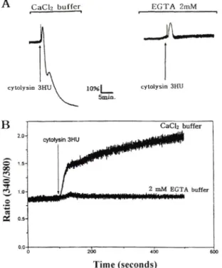
Fig. 3 - Effects of extracellular Ca그'*' on platelet aggre
gation (A) and [Ca고■느]; increase (B) induced by cytolysin (3 HU). V vulnificus cytolysin-induced platelet aggregation and [Ca^"^]| increase were monitored in the presence of extracellular Ca고'*' or in the 2 mM EGTA buffer.
와 세포 내 저장소에서의 유러 증가로 크게 대별해 볼 수 있다
.^^^V vulnificus cytolysin에 의한 혈소관의
응집은
[Ca2+]i의 증가에 따라 농도 의존적으로 일어
남을 확인하였기에
,이후의 실험에서는 혈소관의 응집 에 관여하는
[Ca^^li증가에 동원되는
Ca2객 원천을 조사하였다
.EGTA(2 mM)를 포함한 Ca^'^-free 용액에 서 3 HU V vulnificus cytolysin에 의한 혈소관의 응집은 완전히 차단되었으며
,혈소관의 에도 거 의 번화가 나타나지 않았다 (Fig. 3A). 이는 vulnificus cytolysin에 의한 [Ca미
;증가 및 혈소관 응집이 세포외부로부터의
Ca2+유입에 의한 것임을 보 여주었다
.아울러 혈소판응접 억제와 함께
[Ca2기
i증 가억제가 관찰됨으로써
[C a ^li증가가 혈소판 응집의
중요 인자로 작용하고 있옴을 다시 한번
SK1할 수 었
었다
.한편 EGTA 처리 즉시 [Ca2+]i의 미세한 증가가 관 찰되었다(Fig. 3B). 이는 세포 내 저장소에서 Ca^■"유 리에 의한 것이라고 생각되어지며 추후의 연구가 될요
A
C a C h buffer EG TA 2mM—
버_ 5mm.
cytolysin 3HU
control Ver. Mef. DIDS Sue. Raff. EGTA Lan.
Fig. 4 - Effects of various agents on cytolysin-induced platelet aggregation rate. The following drugs were applied 2-5 min before addition of cytolysin (3 HU);
cyt (cytolysin 3 HU), Ver. (verapamil 20 |iM), Mef.
(mefenamic acid 20 |lM), DIDS (100 |iM), Sue.
(sucrose 50 mM), Raff, (raffinose 50 mM), EGTA 2 mM, Lan. (lanthanum 2 mM). Values are given as mean ± s.e.m. of five independent experiments. *;
Significantly different from control group(p<0.05).
하다고 생각된다. 그러나 앞의 실험결과에서 보는 바 와 같미 세포질 내의 미세한 [Ca이 i의 번화는 혈소관 의 응집에는 영향을 머처지 못하는 것으로 관찰한 바 가 있다. 따라서 세포 내 저장소에서의 미세한 Ca2■"의 유러는 혈소관 응집에 영향을 주지 않을 것으로 사료 된다,
C a ^ channel 및 삼투암 조절물질이 cytolysin의 작용에 미처는 영향 vulnificus cytolysin에 의한 세포 내 Ca2우 유입 경로를 확인하기 위하여 여러가지 통로 차단제롤 사용굵1식 그 효과룰 관찰하였다 (Fig. 4). Voltage-dependent Ca으 channel blocker 인 verapamil(20 iiM )은 cytolysin의 혈소판 응집능에 영향이 없었으며, 이는 cytolysin에 의한 세포내 Ca2""
유입이 voltage-dependent Ca요 '*' channel과 무관함을 보여주었다. 또한 organic antagonist인 mefe
namic acid(20 써 )도 cytolysin에 의한 혈소관의 응집 을 차단하지 못하였다. 그러나 inorganic C a ^ chan
nel blocker인 lanthanum(2 mM)의 전처러에 의해 I스 vulnificus cytolysin에 의한 혈소관응집은 거의 차단됨 을 관찰할 수 었었다. 한편 chloride 통로 차단제인 DIDS 역시 cytolysin에 의한 혈소관 응집에 영향을 주지 않았다. 이는 vulnificus cytolysin에 의한 세
포내 유입이 기존의 세포막 통로와는 다
른 lanthanum 김수성 경로에 의한 것임을 보여주었다.
Vibrio vulnificus cytolysin의 흰쥐 혈소관 응집 기견
^
^
^
^ B
< 0 8 € /0 K >
0
H 1
김현철
•채수완
•이병창
•은재순
기존의 Vibrio toxin둘은 세포막에 oligomer를 형성 하는 것으로 알려져 었으며, 세포막에 선텍성이 없는 pore가 형성되게 되면 삼투압의 불균형이 일어나 세포 의 기능번화가 발생하게 된다. V vulnificus cytolysin 에 의한 혈소관의 응집이 cytolysin의 pore형성과 관 계되어 있는지률 알아보기 위하여 osmotic protec- tants가 cytolysin의 혈소관 응집능에 미처는 영향틀 관찰하였다. Sucrose 및 raffinose를 각각 50 mM썩 5분간 전처처한 후 I스 vulnificus cytolysin(3 HU)에 의한 aggregation rate/min는 대조군의 29.5±4.3에서 각각 18.9+2.0과 10.8±3.2 로 유의하게 감소되었다.
이러한 결과는 cytolysin에 의한 혈소판 응집에 삼투압 의 번화 즉, ^ vulnificus cytolysin에 의한 세포막 pore 형성이 관여하고 있옴을 보여주었다.
Cytolysin의 혈소관움접농에 미치는 온도의 영향 *4스 vulnificus cytolysin에 의한 혈소관 응집에 관여하는 cytolysin의 세포막 결합 및 oligomerization 과정을 알아보기 위하늬, 온도변화에 따른 cytolysin의 혈소관 응집능 번화를 조사하였다. 4 X 에서 vulnificus cytolysin에 의한 혈소관의 응집이나 [Ca미 ; 의 증가는
관찰되지 않았다. 한편 4 X 에서 3 HU cytolysin 처 리에 의해 혈소관 응집이 나타나지 않은 시료를 원심 분러하여 cytolysitti: 제거하고 다시 3TC에서 배양하 면 혈소관 응집과 [Cam'll의 증가가 관찰되었다(Fig.
5). 이러한 걸과는 V. vulnificus cytolysin의 세포막 결합에 온도가 영향을 미치지 않옴을 보여주었고, V vulnificus cytolysin에 의한 혈소관 응집 및 [Ca오기^의 증가가 cytolysin monomer의 세포막 결합 이후 적절 한 온도 이상에서 진행되는 oligomerization에 의해 유도됨을 보여주었다.
A 4X : B
4'€ 3T€cytolysin 3HU
10
%|___
5mio.
Fig. 5 - Effect of temperature on V vulnificus cytolysin- induced platelet aggregation. Platelet suspen
sions were preincubated at 4^C for more than 5 min before addition of V. vulnificus cytolysin (3 HU/m/). In panel B, platelets were resuspended in fresh buffer to remove V vulnificus cytolysin and incubated at 3T*C.
혈소판 세포막 전도도에 머치는 cytolysin의 영향 - V vulnificus cytolysin oligomer에 의한 혈소관 세포 막의 이온투과성 변화가 나타나는지룔 확인하기 위하 여 Vf vulnificus cytolysin에 의한 혈소판 세포막의 전도도 번화률 조사하였다. Patch clamp technique의 whole cell mode에서 I스 vulnificus cytolysin은 혈소 관 세포막을 통한 전류를 생성하였다. 머세전극과 bath 내 용액을 동일한 buffer(K-5 용액)로 채우고 막전압을 -60mV로 고정한 상태에서, bath내에 첨가한 If vulnificus cytoIysin(3 HU/m/) 투여 후 2~5 분에 전류의 증가가 관찰되었으며 전류의 크기는 시간에 비 례하식 증가되었다(Fig. 6). 이때 기록된 단위 전류의 전도도는 34.1 pS이었다. 이러한 결과는 혈소판 세포 막에 형성된 vulnificus cytolysin oilgomer를 통하
\ j>
cytolysin 3HUFig. 6 - Effect of V vulnificus cytolysin on platelet membrane. Trace shows the current obtained from rat platelet membrane after incubation with 3 HU V. vulnificus cytolysin. The bath and pipette solutions were K-5 solution which did not contained CaClg. Membrane potential was held at -60 mV and the filter for tracing reproduction was set at 300 Hz. The number beside the current trace indicates the number of the unit currents. Dot-line indicated in C (control) shows the base line current.
J. Pharm. Soc. Korea
10% I Cytolysin
(IHU)
5 min
Colleen Cytolysin collagetfy
ADP CT+ADP Coll CT+Coll
Fig. 7 - Effects of
V. vulnificuscytolysin (1 HU) on ADP- and collagen-induced platelet aggregation in pla
telet-rich plasma. ADP (1 jxM) and collagen (coll,
1
jig/m/) was added after preincubation with cytolysin (CT, 1 HU) for 3 min.
V vulnificuscyto
lysin (1 HU) potentiated ADP- and collagen-induced platelet aggregation. Values in panel B are given as mean ± s.e.m. of four independent experiments. *;
Significantly different from ADP or collagen treated group(p<0.05).
여 이온 이동이 7|능 하 게 함을 보여 주 었 다 . 그러나 이 러 한
pore
를 통 한 의 이동이[Ca^
■가j 증가의 직접 경 로가 되는지는 확실하지 않으며 이를 위하식V vulnificus cytolysin
이 생성하는pore
의 이온선텍성 연구를 비롯한 추후의 연구가 필요할 것으로 사료된다.CytoIysinOl ADIJ collagen 의 응접농에 미처는 영 향 -V vulnificus cytolysin
외 혈소관 응집능에 미처는 영향 을 알아보기 위하여 혈소 관 웅집 인 자ADP
와collagen
의 혈 소 관 응 집 능 에 머 치 는vulnificus cytolysin
의 영향을 조 사 하 였 다 . 응 집을 일으키지 않는cytolysin
의 농도인1 HU/m/
을 전 처 러 하 고 ,ADP(1
|iM)
또 는collagen(l
을 처리한 결 과 , 대조군에 비 하 ^ 최대응집능(% )의 증 가 와 응 집 시 간 단측이 관결 론
1.
Vibrio
vulnificus cytolysin은 능 도 의 존 적 으 로 혈소관응집을 유도하였으며 아울러 A D P 나 collagen의 응집능에 상 승 효 과 률 나 타 내 었 다 .2.
V. vulnificus
cytolysin은 농 도 의 존 적 으 로 혈 소 관 [C a m 'll" 증 가 시 켰 으 며 , 세포외부의 Ca^■"을 제거 하면vulnificus
cytolysin에 의 한 [Ca타 ]j 증 가 와 함께 혈소관응 집 도 관찰되지 않 았 다 .3. Lanthanum(2 m M )은 1스
vulnificus
cy to lysin 에 의 한 혈소관의 응집 과 [Ca2■가^ 증 7 }률 차 단 하 였 으 나 verapamil(20 |iM) 및 mefenamic add(20 |aM) 는o 공 -
tolysin에 의 한 혈 소 관 응 집 을 차단하지 못 하 였 다 .4 Osm otic protectant언 raffinose(50 m M )와 sucrose (5 0 m M )는
V vulnificus
cytolysin에 의 한 혈 소 판 응 집을 일부 억 제 하 였 다 .5.
V vulnificus
cytolysin은 온도 에 의 존 적 으 로 혈 소 관 응 집 과 증 가 를 유도하며 혈 소 관 세 포 막 의 전도도룰 증 가 시 켰 다 .이 상 의 설험 결 과 는
vulnificus
cyto lysin 이 lanthanum에 감수 성 을 보 이 는 경 로 를 통하여 세 포 밖 의 Ca2누을 세포 내로 유입시키며, 이률 통허여 혈소관 응 집 을 유 도 함 을 보여 주 었 다 . 아울러 I:"vulnificus
cytolysin의 혈 소 관 응 집 은 cytolysin의 세 포 막 pore 형성에 연관된 것으로 사 료 된 다 .문 헌
1) Hollis, D. G., Weaver, R.E,, Baker, C. N. and Thom - berry, C . : Halophilic
Vibrio
species isolated from blood cultures.J. Clin. Microbiol.
3(4), 425 (1976).2) Farmer J. J. 3d.:
Vibrio CBeneckecT) vulnificus,
the bacterium associated with sepsis, septisemia, and the sea.Lancet
2, 903 (1979).3) Blakes, E A., Merson, M. H., Weaver, R. E., Hollis, D. G. and Heublein, E C . : D isease caused by a marine Vibrio. Clinical characteristics and epidemio
logy.
N. Eng. J. Med.
300(1), 1 (1979).4) Oliver, J. D., Warner, R.A. and Cleland D. R . :
Vibrio vulnificus cytolysin
의 흰쥐 혈소관
-s*집 기전
807A
Cytolysin(IH U )
Cytolysin + ADP 1
찰 되 었 다 (Fig. 7). 또 한 , A D P 와 collagen에 의 한 자체 응 집 반 응 은 가역적이었으나 ^
vulnificus
cytolysin 존 재 하 에 서는 비가역적 응집이 관 찰 되 었 다 .0 0 0 0
8642
PQ{%}uoue6ai66v
김현철
•채수완
•이병창
•은재순
Distribution and ecology of
Vibrio vulnificus
and other lactose-fermenting marine vibrios in coastal waters of the southeastern United States.Appl.
Environ. Microbiol
44(6), 1404 (1982).5) Wickboldt, L. G. and Sanders, C. V :
Vibrio vulnificus
infection. Case report and update since 19 70 ./Acad. Dermatol
9(2), 243 (1983).6) Gray, L. D. and Kreger, A. S . : Purification and characterization of an extracellular cytolysin produ
ced by
Vibrio vulnificus. Infect Immun.
48(1), 62 (1985).7) Kreger, A. and Lockwood, D . : Detection of extracellular toxin(s) produced by
Vibrio vulnificus.
Infect. Immun.
33(2), 583 (1981).8) Miyoshi, S. and Shinoda, S . : Role of the protease in the permeability enhancement by
Vibrio vulnificus.
Microbiol Immunol.
32(10), 1025 (1988).9) Smith, G. C. and Merkel, J. R . : Collagenolytic activity of
Vibrio vulnificus:
potential contribution to its invasiveness.Infect. Immun.
35(3), 1155 (1982).10) Kreger, A., DeChatelet, L. and Shirley, R : Interaction of
Vibrio vulnificus
with human polymorphonuclear leukocytes: association of virulence with resistance to phagocytosis, /.Infect Dis.
144(3), 244 (1981).11) Desmond, E, P., Janda, J. M., Adams, E I. and Bottone E. J .: Comparative studies and laboratory diagnosis of
Vibrio vulnificus,
an invasiveVibrio
Sp./.
Clin, Microbiol.
19(2), 122 (1984).12) Simpson, L. M. and Oliver, J. D . : Siderophore production by
Vibrio vulnificus. Infect. Immun.
41(2), 644 (1983).13) Testa, J., Daniel, L, W. and Kreger, A. S . : Extra
cellular phospholipase A2 and lysophospholipase produced by
Vibrio vulnificus. Infect Immun.
45(2), 458 (1984).14) Wright, A. C. and Morris., J. G. Jr.: T he extracellular cytolysin of
Vibrio mdnificus
: inactivation and relationship to virulence in mice.
Infect Immun,
59(1), 192 (1991).15) Uemura Y., Sakon, M., Kawasaki, T , Shiba, E., Kambayashi, J. and Mori, T .: The correlation between influx and inositol 1,4,5-triphosphate (IP3) formation in platelets stimulated by various agonists.
Biochem. In t
20(5), 853 (1990).16) I남 k, S. D., Shon, H. S. and Joh, N. J .:
Vibrio
vulnificus
septicemia in Korea : clinical and epidemiological findings in seventy patients.
J. Am, Acad.
Dermatol
24(3), 397 (1991).17) Khoo, H. E., Hon, W M., Lee, S.H, and Yuen, R . : Effects of Stonustoxin (lethal factor from
Synanceja horrida
venom) on platelet aggregation.Toxicon
33(8), 1033 (1995).18) Bhakdi, S., Muhly, M., Mannhardt, U,, Hugo,
E,
Klapettek, K., Mueller-Eckhardt, C. and Roka, L . : Staphylococcal -toxin promotes blood coagulation via attack on human platelets.J. Exp. Med.
168(2), 527 (1988).19) Rho, M. C., Nakahata, N., Nakamura, H., Murai, A.
and Ohizumi, Y .: Activation of rabbit platelets by influx and thromboxane Ag release in an external Oa으"*"-dependent manner by zooxanthella- toxin-A, a novel polyol.
Br. J. Pharmacol
115(3), 433 (1995).20) Kim, H.
R.,
Park, S. D., F ^k, J. W, Jeong, M. H., Kim, J. S. and Park, B. H . : Purification and characterization of cytolysin produced by
Vibrio vulnificus.
Kor.
/.Biochem.
24, 7 (1992).21) Bemheimer, A. W. and Schwartz, L. L . : Isolation and composition of staphylococcal a-toxin. /
Gen, Microbiol.
30, 455 (1963).22) Hamil, O. E, Marty, A., Neher, E,, Sakmann, B. and Sigworth, E J .: Improved patch-damp techniques for high-resolution current recording from ceils and cell-free membrane patches.
Pfl gers Arch.
391(2), 85 (1981).23) Grynkiewicz, G., Poenie, M. and Tsien, R. Y .: A new generation of Ca으"*" indicators with greatly improved fluorescence properties. /
Biol Chem,
260(6), 3440 (1985).24) Kinlough-Rathbone, R. L., Packham, M. A., Reimers, H. J., Cazenave, J. P and Mustard, J. F .: Mechanisms of platelet shape change, aggregation and release induced by collagen, thrombin, or A23187.
J. Lab.
Clin. Med
90(4), 707 (1977).25) Rink, T. J, and Sage, S. 0 .: Calcium signaling in human platelets.
Annu. Rev. Physiol
52, 431 (1990).26) Miyamae, T , Oshima, K., Morikawa, T. and Hagiwara, M .: Calcium-induced platelet aggregation in washed platelets from Guinea pigs.
Pharmacol.
51(3), 180 (1995).
J. Pharm. Soc. Korea