R E S E A R C H A R T I C L E Open Access
Severe fever with thrombocytopenia
syndrome: comparison with scrub typhus and clinical diagnostic prediction
Sang-Won Park
1,2, Chang-Seop Lee
3, Jeong-Han Kim
1, In-Gyu Bae
4, Chisook Moon
5, Yee Gyung Kwak
5,
Baek-Nam Kim
5, Jae Hoon Lee
6, Seong Yeol Ryu
7, Hee-Chang Jang
8, Jian Hur
9, Jae-Bum Jun
10, Younghee Jung
11, Hyun-Ha Chang
12, Young Keun Kim
13, Jeong-Hwan Hwang
3, Yeon-Sook Kim
14, Hye Won Jeong
15,
Kyoung-Ho Song
16, Wan Beom Park
1, Eu Suk Kim
16and Myoung-don Oh
1*Abstract
Background: Severe fever with thrombocytopenia syndrome (SFTS) is emerging in Asian 3 countries, China, Japan and Korea, which are scrub typhus endemic areas, and its incidence is increasing. As the two infections overlap epidemiologically and clinically and the accessibility or sensitivity of diagnostic tests is limited, early clinical prediction may be useful for diagnostic and therapeutic purposes.
Methods: Patients aged ≥16 years who were clinically suspected and laboratory-confirmed to be infected with Orientia tsutsugamushi or the SFTS virus in South Korea were enrolled. Clinical and laboratory parameters were compared. Scrub typhus was further subclassified according to the status of eschar and skin rash. An SFTS prediction scoring tool was generated based on a logistic regression analysis of SFTS compared with scrub typhus.
Results: The analysis was performed on 255 patients with scrub typhus and 107 patients with SFTS. At initial presentation, subjective symptoms except for gastrointestinal symptoms, were more prominent in scrub typhus patients. In addition to the characteristic eschar and skin rash, headache was significantly more prominent in scrub typhus, while laboratory abnormalities were more prominent in SFTS. Leukopenia (white blood cell count < 4000/mm
3; odds ratio [OR] 30.13), thrombocytopenia (platelet count < 80,000 /mm
3; OR 19.73) and low C-reactive protein (< 1 mg/dL; OR 67.46) were consistent risk factors for SFTS (all P < 0.001). A prediction score was generated using these 3 variables, and a score ≥ 2 had a sensitivity of 93.1% (95% confidence interval [CI], 87.9–96.4%) and a specificity of 96.1% (95% CI, 93.8 –97.6%) for SFTS.
Conclusion: This prediction scoring tool may be useful for differentiating SFTS from eschar- or skin rash-negative scrub typhus. It is a simple and readily applicable tool with potential for use in primary care settings.
Keywords: SFTS, Severe fever with thrombocytopenia syndrome, Scrub typhus, Tsutsugamushi, Korea, Prediction, Score
Background
Severe fever with thrombocytopenia syndrome (SFTS) is an emerging infectious disease that is caused by the SFTS virus (SFTSV); it is endemic in 3 East Asian countries:
China, Korea and Japan [1 – 3]. The incidence of SFTS is increasing, and the case-fatality rate ranges from 5.3 to 32.6% [4 – 6]; however, there are not yet effective antiviral
therapeutics or a vaccine [7]. SFTS was listed as a priority disease that requires urgent research and development by the World Health Organization in 2017 [8]. Orientia tsutsugamushi is endemic to these 3 countries, which is a leading cause of treatable non-malarial febrile illness in Asia [9]. In 2017, 10,528 cases of scrub typhus were re- ported in South Korea. Eschar and a maculopapular skin rash are characteristic findings of this disease and are crit- ical clues for its diagnosis. The case-fatality rate of scrub typhus has a median of 6.0% in untreated cases and 1.4%
in treated cases [9].
* Correspondence:
[email protected]1
Department of Internal Medicine, Seoul National University College of Medicine, 103 Daehak-ro, Jongno-gu, Seoul 03080, the Republic of Korea Full list of author information is available at the end of the article
© The Author(s). 2019 Open Access This article is distributed under the terms of the Creative Commons Attribution 4.0 International License (http://creativecommons.org/licenses/by/4.0/), which permits unrestricted use, distribution, and reproduction in any medium, provided you give appropriate credit to the original author(s) and the source, provide a link to the Creative Commons license, and indicate if changes were made. The Creative Commons Public Domain Dedication waiver (http://creativecommons.org/publicdomain/zero/1.0/) applies to the data made available in this article, unless otherwise stated.
Although SFTSV and O. tsutsugamushi do not share specific vectors, they are transmitted to humans through ticks and mites bites mostly, respectively, during outdoor activities. The ecological differences between vectors may characterize their epidemiological features, inclu- ding the region of infection and peak epidemic seasons.
However, there are considerable overlaps of their epi- demiological and clinical features, which makes their dif- ferential diagnosis difficult, particularly during the high epidemic season of scrub typhus. Patients with SFTS have the potential to deteriorate during the second week of the illness [5], and early diagnosis of SFTS may lead to early investigational therapeutics and stricter infection control measures to prevent human-to-human transmis- sion [10–13]. However, the sensitivity of diagnostic assays for scrub typhus is low [14]. The confirmatory test for SFTS is usually performed in the national reference la- boratory, and a serologic assay for the point of care is not yet commercially available. Therefore, only a high index of clinical suspicion may lead to a rapid clinical decision or an early referral, particularly in primary care settings.
Clinical diagnostic prediction based on the features dif- ferentiating SFTS from scrub typhus in endemic areas, particularly during the overlap period, may be clinically useful to guide the diagnostic and therapeutic strategies in the absence of rapid point-of-care diagnostic test. This study compared the clinical and laboratory features of the two diseases and constructed a clinical prediction tool composed of a scoring system for SFTS. We performed several subgroup analyses, including for eschar-negative scrub typhus, which is difficult to suspect clinically because it lacks critical clues.
Methods Patients
Patients in South Korea aged ≥16 years who were clini- cally suspected and laboratory-confirmed to be infected with O. tsutsugamushi or SFTSV were enrolled. Cases of eschar-positive and -negative scrub typhus were pro- spectively included from 8 community-based hospitals in 2006; part of this study was previously published [15].
Additional patients with only eschar-negative scrub typhus were prospectively included from 6 community- based hospitals from 2009 to 2011; these patients had been thoroughly examined and cared for by the infec- tious diseases specialists in charge. The participating hospitals in both studies were Chonbuk National Uni- versity Hospital, Dankook University Hospital, Dongguk University Ilsan Hospital, Ilsan Paik Hospital, Namwon Medical Center, Pusan Paik Hospital, Sanggye Paik Hos- pital, Sunlin Hospital, Boramae Medical Center, and Wonkwang University Hospital. SFTS cases were retro- spectively collected from 36 hospitals nationwide from 2013 to 2015. Part of this study was previously published
[5], and part of the hospitals are listed in the Acknow- ledgements section.
Scrub typhus was confirmed either by eschar- or buffy coat-based polymerase chain reaction (PCR) or by a serologic assay. PCR targeting the variable domains I and II of the 56-kDa antigen gene of O. tsutsugamushi was performed using a set of primers (forward: TTT CGA ACG TGT CTT TAA GC; reverse: ACA GAT GCA CTA TTA GGC AA; 1151 bp); the products were sequenced to match the reference genotypes, as de- scribed in a previous study [15]. The presence of four- fold or greater changes in the titers of the paired sera from an indirect immunofluorescence antibody assay (IFA) or a passive hemagglutination assay (GreenCross SangA; Yongin city, South Korea) was used as the posi- tive serologic criteria. All sera from patients with con- firmed scrub typhus were screened for the co-infection with SFTSV. SFTS is a reportable infectious disease to the Korea Centers for Disease Control and Prevention (KCDC) and all SFTSV infections were confirmed at the KCDC by detecting the M segment gene of the SFTSV RNA using one-step reverse transcription (RT)-PCR as described in a previous study [16].
Study design
Baseline characteristics and clinical and laboratory parame- ters were compared between scrub typhus and SFTS to de- termine the differentiating factors. Scrub typhus was further subclassified into eschar-negative and -positive groups for comparison with SFTS. An SFTS prediction scoring tool was generated based on logistic regression analysis for SFTS. The baseline characteristics included demographic variables, comorbidities, site of infection, sea- son of infection, duration from the onset of illness to first visit, duration of the hospital stay, and in-hospital morta- lity. The clinical parameters included commonly known symptoms and signs of both diseases such as headache, altered consciousness, cough, dyspnea, gastrointestinal manifestations, skin rash and the presence of a bite wound.
The laboratory parameters included a complete blood count and chemistry, which can be easily obtained in pri- mary care settings as a point-of-care testing. The worst values of the clinical and laboratory parameters within 24 h of the initial visit were used. The frequency of major com- plications during the clinical course was also compared.
Altered mentality was defined as a Glasgow coma scale
score < 15. Acute kidney injury was defined as serum
creatinine levels ≥2.0 mg/dL and 1.5 times the base-
line level [17]. Shock was defined as a mean arterial
pressure < 65 mmHg. The categorical cut-offs for the
comparison of some laboratory values such as
thrombocytopenia (platelet count < 80,000 /mm
3), as-
partate transaminase (AST) ≥400 IU/L and alanine
transaminase (ALT) ≥200 IU/L were chosen in view of
their mean values in SFTS cases and their kinetics during the clinical course, as shown in a previous study [5]. Geo- graphic location was divided into the western and eastern areas of South Korea. The western area included the Seoul metropolitan area and Gyeonggi, Chungcheong and Cholla provinces, which mostly consist of plain rice fields. The eastern area included Kangwon and Gyeongsang provinces, which mostly consist of hilly and mountainous areas.
Statistical analysis
Chi-square or Fisher’s exact tests were used to analyze the categorical variables. T-tests or Mann-Whitney U-tests were used to compare the continuous variables. A multi- variate logistic regression analysis was performed using the risk factors that were significantly (P < 0.05) associated with SFTS or scrub typhus in the univariate analysis and adjusted with the duration from the onset of illness to the initial presentation. The SFTS prediction scoring tool was generated using the logistic regression analysis for estimat- ing odds ratios. The receiver operating characteristic curve was constructed for the scoring model (SPSS v20.0, Armonk, NY: IBM Corp.).
Results
A total of 362 patients were included in the analysis, in- cluding 255 patients with scrub typhus and 107 patients with SFTS. Eschar-positive scrub typhus accounted for 80.4% (205/255) and eschar-negative scrub typhus for 19.6% (50/255) of patients. Scrub typhus was confirmed by PCR in 153 patients and by serology in 102 patients.
All patients with scrub typhus (N = 255) showed negative results for SFTSV in the sera. Compared to scrub typhus patients, patients with SFTS showed a higher median age (71 years), more comorbidities such as diabetes mellitus and hypertension, a greater tendency toward infection in the summer season, a greater tendency to be infected in the eastern area of South Korea, a shorter duration from the onset of illness to the first visit (median of 4 days), a longer duration of hospital stay (median of 10 days), and a higher case-fatality rate (40.2% vs 0.4%, respectively) (Table 1).
At the initial presentation, the overall subjective symp- toms, except for gastrointestinal symptoms, were more prominent in patients with scrub typhus. Skin rash was predominantly present in scrub typhus cases (87.8 and 78.0% in eschar-positive and -negative scrub typhus re- spectively, vs 5.7% in SFTS). Bite wounds were present in 28.3% of SFTS patients. Altered mentality was more common in SFTS patients (27.9%). In the subgroup comparisons, patients with eschar-negative scrub typhus presented fewer subjective symptoms. Fever was present in all the patients and was the initial chief problem lea- ding to a hospital visit. The presence of skin rash (78.0%) in eschar-negative scrub typhus was similar to
the rate in eschar-positive scrub typhus (P = 0.110) (Table 2). Laboratory abnormalities were more prominent in SFTS. Leukopenia, thrombocytopenia, and an elevation of AST and lactate dehydrogenase (LDH) were more com- mon in SFTS. C-reactive protein (CRP) was rarely elevated in SFTS (mean 1.24 mg/dL) (Table 3). SFTS was asso- ciated with a disproportionately higher incidence of major complications during the hospitalization course such as decreased mentality (59.0%), seizure (16.2%), pneumonia (74.4%), the need for mechanical ventilation (32.1%), and acute kidney injury (21.5%) (Table 4).
In the multivariate regression analysis, leukopenia (white blood cell count < 4000/mm
3; odds ratio [OR] 30.13, P < 0.001), thrombocytopenia (platelet count < 80,000/mm
3; OR 19.73, P < 0.001) and low CRP (< 1 mg/dL; OR 67.46, P < 0.001) were significantly predictive factors for SFTS compared with scrub typhus (Table 5). These 3 factors were consistently significant in a subgroup ana- lysis compared with eschar-negative scrub typhus. As few laboratory results were significantly indicative of scrub typhus in comparison with SFTS in the univariate analysis, primarily only clinical variables were included in the multivariate analysis to identify the predictive factors for scrub typhus. In addition to the characteris- tic eschar and skin rash, headache was the only consis- tent risk factor for all cases of scrub typhus and the subgroup of eschar-negative scrub typhus.
A prediction scoring tool for the differential diagnosis of SFTS and scrub typhus was generated using the combi- nation of those 3 parameters (1 point each for WBC count
< 4000/mm
3, platelet count < 80,000/mm
3and CRP value
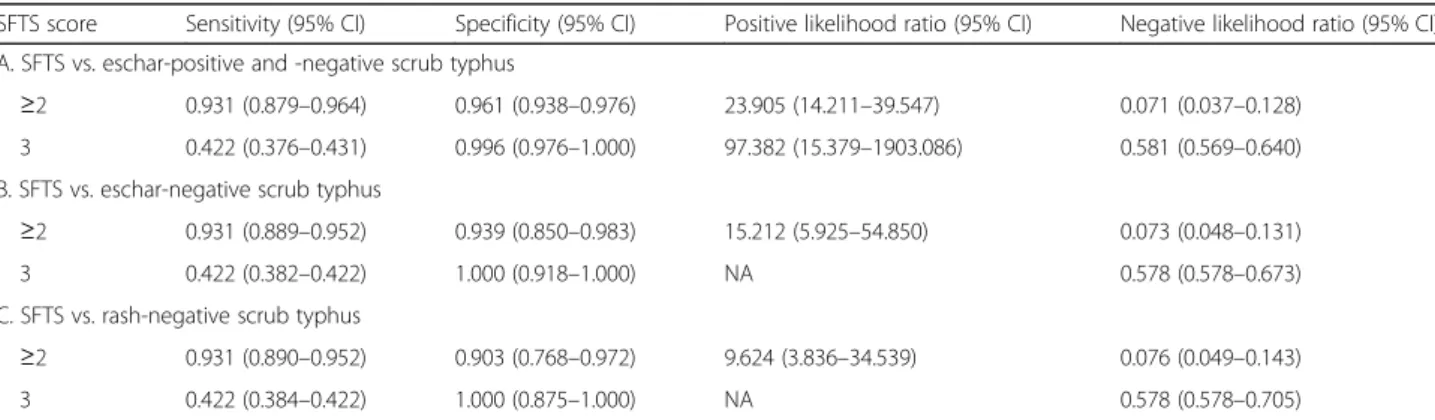
< 1 mg/dL); the total score ranged from 0 to 3. On the ROC curve obtained for this model, the optimal cut-off was ≥2. A score ≥ 2 had a sensitivity of 93.1% (95% confi- dence interval [CI], 87.9–96.4%) and a specificity of 96.1%
(95% CI, 93.8–97.6%) for SFTS, with an ROC area under the curve of 0.972 (95% CI, 0.952–0.990) (Table 6). In the eschar-negative scrub typhus subgroup, a score ≥ 2 had a sensitivity of 93.1% (95% CI, 88.9–95.2%) and a specificity of 93.9% (95% CI, 85.0–98.3%) for SFTS. In the subgroup of rash-negative scrub typhus, a score ≥ 2 had a sensitivity of 93.1% (95% CI, 89.0–95.2%) and a specificity of 90.3%
(95% CI, 76.8 –97.2%) for SFTS (Table 6).
Discussion
A considerable number of SFTS cases occur during the
epidemic season of scrub typhus in South Korea. In 2017,
86.7% (9132/10,528) of scrub typhus and 48.1% (131/272)
of SFTS cases were officially reported to occur from Sep-
tember to November. Although the case-fatality rate of
scrub typhus is low with antibiotic treatment, severe scrub
typhus remains an unresolved issue [18, 19]. The concur-
rent presence of the typical eschar with a compatible cli-
nical manifestation makes the clinical diagnosis of scrub
typhus obvious [20], but the poor sensitivity of diagnostic assays for scrub typhus and the presence of eschar-nega- tive scrub typhus make the diagnosis uncertain in some patients. Given this diagnostic uncertainty combined with the potential of severe clinical form, a clinical prediction tool will be very useful to narrow the differential diagnosis for those requiring further urgent investigation [14].
Our prediction tool used 3 variables; leukopenia (WBC count < 4000/mm
3), thrombocytopenia (platelet count < 80,000/mm
3) and low CRP (< 1 mg/dL), which could be obtained from routine basic laboratory blood tests and are readily applicable in primary care settings.
The cut-off level of thrombocytopenia as platelet count of 80,000/mm
3was determined by considering the distribution of platelet counts among the study subjects and the kinetics of initially persistent thrombocytopenia in SFTS [5]. One study previously proposed a similar scoring system using 4 variables: altered mental status, leukopenia, prolonged activated partial thromboplastin
time and normal C-reactive protein [21]. In our study,
‘altered mental status’ was not a significant factor to be incorporated into the prediction analysis. We objec- tively assessed mental status using the Glasgow coma scale, which is one of the basic tools used in critical care. We did not include the coagulation panels in the initial comparison because we do not routinely check coagulation panels during the investigation of possible scrub typhus cases.
A larger sample size might have led to different re- sults. Our study also included a relatively larger num- ber of eschar-negative scrub typhus cases, which causes diagnostic challenges and requires a clinical decision to guide further diagnostic evaluations. We adjusted the comparison with the duration from the onset of illness to the initial presentation because the kinetics of clin- ical variables are closely time-dependent. The predic- tion tool worked similarly in all subgroups of scrub typhus (Table 6). The data collection from multiple Table 1 Baseline characteristics of the subjects (n = 362)
Variable Scrub typhus (n = 255) SFTS P
value
cEschar-positive P
value
aEschar-negative P value
bn = 205 n = 50 n = 107
Age, years (median, IQR) 60 (49 –71) < 0.001 64.5 (51.2 –70.7) 0.004 71 (61 –78) < 0.001
Male gender, n (%) 81 (39.5) 0.044 22 (44.0) 0.387 55 (51.4) 0.054
Comorbidity
Diabetes mellitus 19 (9.3) 0.027 5 (10.0) 0.200 19/106 (17.9) 0.023
Hypertension 44 (21.5) 0.030 13 (26.0) 0.395 35 (32.7) 0.039
CVA 7 (3.4) 1.000 1 (2.0) 1.000 4 (3.7) 0.771
Congestive heart failure 9 (4.4) 0.342 2 (4.0) 0.593 2 (1.9) 0.254
Chronic liver disease 7 (3.4) 0.272 5 (10.0) 0.013 1 (0.9) 0.119
Asthma/COPD 7 (3.4) 0.583 1 (2.0) 0.665 5 (4.7) 0.474
Solid tumor 6 (2.9) 0.098 2 (4.0) 0.098 0 0.111
None 126 (61.5) 0.121 26 (52.0) 0.969 56 (52.3) 0.202
Seasonal occurrence, n (%)
Spring-summer (Mar-Aug) 0 < 0.001 0 < 0.001 67 (62.6) < 0.001
Autumn (Sep-Dec) 205 (100) 50 (100) 40 (37.4)
Geographical location
Western area 167 (81.5) < 0.001 42 (84.0) < 0.001 36/104 (34.6) < 0.001
Eastern area 38 (18.5) 8 (16.0) 68/104 (65.4)
Duration, mean (±SD), days
From onset of illness to admission 6.63 (3.918) < 0.001 6.38 (5.103) 0.006 4.39 (3.66)) < 0.001
Hospital stay 6.26 (11.927) < 0.001 6.18 (3.757) < 0.001 12.07 (9.57) < 0.001
Mortality, in-hospital 1 (0.5) < 0.001 0 < 0.001 43 (40.2) < 0.001
Abbreviations: SFTS severe fever with thrombocytopenia syndrome, IQR interquartile range, SD standard deviation, CVA cerebrovascular accident, COPD chronic obstructive lung disease
aP value when compared to SFTS
bP value when compared to SFTS
cP value when compared to all scrub typhus
hospitals in major endemic areas of the two diseases might strengthen the generalizability of our study.
In a binary comparison of clinical parameters, scrub typhus tended to present with more subjective manifes- tations, whereas SFTS showed more laboratory abnor- malities. In a subgroup comparison of eschar-negative scrub typhus with SFTS, skin rash and headache were significantly indicative of scrub typhus. As the skin rash in scrub typhus is a characteristic maculopapular type that is clinically distinct from that of SFTS [20], the presence of a maculopapular skin rash itself has diagnos- tic value if the clinician is sufficiently experienced. How- ever, approximately 10% of patients with scrub typhus are reported to lack the typical skin rash [22, 23]. There- fore, scrub typhus without eschar and a skin rash poses a further diagnostic challenge. In our study, 12.2% of eschar-positive and 22.0% of eschar-negative scrub typhus patients had no skin rash. The rate of eschar- negative scrub typhus has been reported to be approxi- mately 10%, although a skilled physician may observe a different rate [22, 23].
Reports regarding co-infection of both scrub typhus and SFTS are limited. In a recent report, 23.0% of patients clinically suspected of scrub typhus were SFTS-positive [24]. There was a case report of co-infection diagnosed by PCR [25]. In another report, however, none of the 38 patients with scrub typhus were SFTS-positive, whereas one of 21 patients with SFTS was serologically suggestive of scrub typhus. Thus, the clinical evidence on the
possibility of co-infection is not solid, and further moni- toring is necessary. From an ecological perspective, given the vectors of the two diseases, co-infection is not likely.
Although the epidemic seasons overlap and there is a risk of simultaneously acquiring the two diseases during out- door activity, they do not share vectors, and the ecologies of their vectors differs. Phenotypically, scrub typhus is highly prevalent in the rice field areas of western and southwestern South Korea [26], whereas the incidence of SFTS is low in this area, consistent with the low SFTSV infection rate in ticks. Conversely, the incidence of SFTS is high in the eastern and southeastern mountainous area of South Korea [5, 27].
The causative agent of scrub typhus, O. tsutsugamushi, is transmitted by trombiculid chigger mites. The causative Trombiculidae have a nationwide distribution in South Korea [28, 29]. Although the larvae of trombiculid mites can parasitize most animals, rodents and some other small mammals are their primary hosts [30, 31]. The prevalence and abundance of chigger mites on small mammals are much higher in cultivated flatland landscapes [32]. Mean- while, Haemaphysalis longicornis is the predominant vector for SFTSV, but other tick species such as H. flava, Amblyomma testudinarium and Ixodes nipponensis can also carry SFTSV in South Korea [27, 33, 34]. H.
longicornis is able to transmit SFTSV via both transo- varial and transstadial modes [35]. H. longicornis is widely distributed in Australia, New Zealand, Korea, Japan and China [36]. Larger mammals, such as rabbits, badgers, Table 2 Clinical symptom and sign at the initial presentation (n = 362)
Variable Scrub typhus (n = 255) SFTS P
value
cEschar-positive P
value
aEschar-negative P
value
bn = 107
n = 205 n = 50
Fever 205 (100) < 0.001 50 (100) 0.017 95/106 (89.6) < 0.001
Headache 179 (87.3) < 0.001 30 (60.0) < 0.001 26/105 (24.8) < 0.001
Myalgia 172 (83.9) < 0.001 29 (58.0) 0.794 58/104 (55.8) < 0.001
Conjunctival injection 78 (38.0) < 0.001 6 (12.0) 0.215 6/98 (6.1) < 0.001
Sore throat 74 (36.1) < 0.001 11 (22.0) 0.005 7/105 (6.7) < 0.001
Cough 76 (37.1) < 0.001 15 (30.0) 0.004 12/105 (11.4) < 0.001
Dyspnea 47 (22.9) 0.004 9 (18.0) 0.138 10/104 (9.6) 0.006
Nausea/vomiting 81 (39.5) 0.463 14 (28.0) 0.370 37/105 (35.2) 0.718
Abdominal pain 58 (28.3) 0.019 9 (18.0) 0.778 17/105 (16.2) 0.033
Arthralgia 62 (30.2) < 0.001 4 (8.0) 0.429 5/104 (4.8) < 0.001
Skin rash 180 (87.8) < 0.001 39 (78.0) < 0.001 6/105 (5.7) < 0.001
Presence of bite wound 205 (100) < 0.001 0 (0) < 0.001 30/106 (28.3) < 0.001
Altered mentality 11 (5.4) < 0.001 5 (10.0) 0.012 29/104 (27.9) < 0.001
Shock (MAP < 65 mmHg) 15 (7.3) 0.085 4 (8.0) 0.427 14/105 (13.3) 0.079
Abbreviations: SFTS severe fever with thrombocytopenia syndrome, MAP mean arterial pressure
aP value when compared to SFTS
bP value when compared to SFTS
cP value when compared to all scrub typhus
deer and wild boars, rather than rodents, have been sug- gested to be the principal hosts for this tick species [36].
The differences between the main hosts and habitats of the vectors makes their simultaneous exposure less likely.
However, the enhanced intrusion of wild animals into cul- tivated farmland areas and serologic evidence of SFTSV
infection in domestic animals may indicate the exposure risk to dual vectors [37, 38]. The case-fatality trend of scrub typhus also deserves to be mentioned for indirect evidence of co-infection. Although there are approxi- mately 10,000 cases of scrub typhus annually in South Korea, the recent annual mortality was stable in the range Table 3 Laboratory findings at the initial presentation (n = 362)
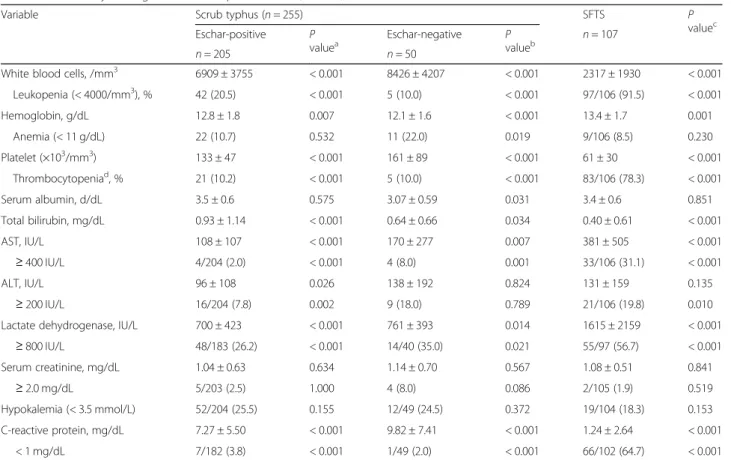
Variable Scrub typhus (n = 255) SFTS P
value
cEschar-positive P
value
aEschar-negative P value
bn = 107
n = 205 n = 50
White blood cells, /mm
36909 ± 3755 < 0.001 8426 ± 4207 < 0.001 2317 ± 1930 < 0.001
Leukopenia (< 4000/mm
3), % 42 (20.5) < 0.001 5 (10.0) < 0.001 97/106 (91.5) < 0.001
Hemoglobin, g/dL 12.8 ± 1.8 0.007 12.1 ± 1.6 < 0.001 13.4 ± 1.7 0.001
Anemia (< 11 g/dL) 22 (10.7) 0.532 11 (22.0) 0.019 9/106 (8.5) 0.230
Platelet (×10
3/mm
3) 133 ± 47 < 0.001 161 ± 89 < 0.001 61 ± 30 < 0.001
Thrombocytopenia
d, % 21 (10.2) < 0.001 5 (10.0) < 0.001 83/106 (78.3) < 0.001
Serum albumin, d/dL 3.5 ± 0.6 0.575 3.07 ± 0.59 0.031 3.4 ± 0.6 0.851
Total bilirubin, mg/dL 0.93 ± 1.14 < 0.001 0.64 ± 0.66 0.034 0.40 ± 0.61 < 0.001
AST, IU/L 108 ± 107 < 0.001 170 ± 277 0.007 381 ± 505 < 0.001
≥ 400 IU/L 4/204 (2.0) < 0.001 4 (8.0) 0.001 33/106 (31.1) < 0.001
ALT, IU/L 96 ± 108 0.026 138 ± 192 0.824 131 ± 159 0.135
≥ 200 IU/L 16/204 (7.8) 0.002 9 (18.0) 0.789 21/106 (19.8) 0.010
Lactate dehydrogenase, IU/L 700 ± 423 < 0.001 761 ± 393 0.014 1615 ± 2159 < 0.001
≥ 800 IU/L 48/183 (26.2) < 0.001 14/40 (35.0) 0.021 55/97 (56.7) < 0.001
Serum creatinine, mg/dL 1.04 ± 0.63 0.634 1.14 ± 0.70 0.567 1.08 ± 0.51 0.841
≥ 2.0 mg/dL 5/203 (2.5) 1.000 4 (8.0) 0.086 2/105 (1.9) 0.519
Hypokalemia (< 3.5 mmol/L) 52/204 (25.5) 0.155 12/49 (24.5) 0.372 19/104 (18.3) 0.153
C-reactive protein, mg/dL 7.27 ± 5.50 < 0.001 9.82 ± 7.41 < 0.001 1.24 ± 2.64 < 0.001
< 1 mg/dL 7/182 (3.8) < 0.001 1/49 (2.0) < 0.001 66/102 (64.7) < 0.001
Abbreviations: SFTS severe fever with thrombocytopenia syndrome, AST aspartate aminotransferase, ALT alanine aminotransferase
aP value when compared to SFTS
bP value when compared to SFTS
cP value when compared to all scrub typhus
dThrombocytopenia, < 80 × 103/mm3
Table 4 Major complications during the clinical course (n = 362)
Variable Scrub typhus (n = 255) SFTS P
value
cEschar-positive P
value
aEschar-negative P value
bn = 107
n = 205 n = 50
CNS involvement
Altered mentality (GCS < 15) 14/199 (7.0) < 0.001 8 (16.0) < 0.001 62/105 (59.0) < 0.001
Seizure 0/199 (0) < 0.001 0 (0) 0.002 17/105 (16.2) < 0.001
Lung involvement
Mechanical ventilation 5 (2.4) < 0.001 2 (4.0) < 0.001 34/106 (32.1) < 0.001
Renal involvement
Acute kidney injury 5 (2.4) < 0.001 5 (10.0) 0.080 23 (21.5) < 0.001
Abbreviations: SFTS severe fever with thrombocytopenia syndrome, CNS central nervous system, GCS Glasgow coma scale
aP value when compared to SFTS
bP value when compared to SFTS
cP value when compared to all scrub typhus
of 11–13 cases (http://www.cdc.go.kr/npt). This suggests that co-infection is rare, considering the high mortality and rising incidence of SFTS.
The magnitude of the prevalence of SFTS and the need for intense differential vigilance in the community must be further investigated. The seroprevalence for SFTSV antibodies in South Korea were reported to be 2.7 to 7.7%
in rural area and 1.9% in urban areas in small-scale studies [39, 40]. SFTS is already endemic throughout South Korea, and the rapidly increasing trend of its incidence is obvious [5]. The severity and poor prognosis of SFTS de- mand accurate initial clinical triage. Human granulocytic anaplasmosis and human monocytotrophic ehrlichiosis which have similar clinical presentations are also import- ant differential diseases. Hence, we suggest a clinical deci- sion algorithm based on our findings as follows. In an atypical febrile disease during the epidemic season of scrub typhus, the empirical administration of doxycycline is desirable. Positive findings of eschar or a maculopapular
skin rash on physical examination are strongly suggestive of scrub typhus and are an indication for maintaining doxycycline treatment. If there is no eschar or maculopa- pular rash, the calculation of the prediction score in our study may guide the degree of suspicion for SFTS. Until confirming the presence of SFTS, continuing doxycycline may be clinically useful to cover eschar- and skin rash- negative scrub typhus, anaplasmosis, and ehrlichiosis, which all respond to doxycycline.
This study has several limitations. First, the prediction scoring tool was evaluated only in comparison with scrub typhus. Further performance assessment in general febrile patients may be useful to demonstrate the utility of this tool for clinically identifying SFTS. Second, although the SFTS data were primarily obtained from intensive clinical care settings, the retrospective nature of data collection might lead to a bias in contrast to the prospective collec- tion of scrub typhus cases. In addition, only viremic SFTS cases were included, which might exclude mild cases.
Table 6 Diagnostic performance of the SFTS prediction scoring system
SFTS score Sensitivity (95% CI) Specificity (95% CI) Positive likelihood ratio (95% CI) Negative likelihood ratio (95% CI) A. SFTS vs. eschar-positive and -negative scrub typhus
≥2 0.931 (0.879 –0.964) 0.961 (0.938 –0.976) 23.905 (14.211 –39.547) 0.071 (0.037 –0.128)
3 0.422 (0.376 –0.431) 0.996 (0.976 –1.000) 97.382 (15.379 –1903.086) 0.581 (0.569 –0.640) B. SFTS vs. eschar-negative scrub typhus
≥2 0.931 (0.889 –0.952) 0.939 (0.850 –0.983) 15.212 (5.925 –54.850) 0.073 (0.048 –0.131)
3 0.422 (0.382 –0.422) 1.000 (0.918 –1.000) NA 0.578 (0.578 –0.673)
C. SFTS vs. rash-negative scrub typhus
≥2 0.931 (0.890 –0.952) 0.903 (0.768 –0.972) 9.624 (3.836 –34.539) 0.076 (0.049 –0.143)
3 0.422 (0.384 –0.422) 1.000 (0.875 –1.000) NA 0.578 (0.578 –0.705)
Abbreviations: SFTS severe fever with thrombocytopenia syndrome, CI confidence interval, NA not available
Table 5 Multivariate analysis for the predictive factors of SFTS compared to those of eschar-positive and negative scrub typhus
Variables Odds ratio (95% CI) P value
Age 1.03 (0.98 –1.09) 0.145
Male gender 2.09 (0.58 –7.44) 0.255
Diabetes mellitus 2.53 (0.36 –17.43) 0.345
Hypertension 0.64 (0.12 –3.28) 0.593
Duration, from onset of illness to initial visit 0.80 (0.68 –0.94) 0.008
Altered mentality 2.31 (0.27 –19.33) 0.439
Shock 1.72 (0.23 –12.68) 0.592
Leukopenia (WBC < 4000/mm
3) 30.13 (6.08 –149.22) < 0.001
Thrombocytopenia (< 80,000/mm
3) 19.73 (4.60 –84.58) < 0.001
AST ≥400 IU/L 8.33 (0.39 –176.07) 0.173
ALT ≥200 IU/L 0.36 (0.03 –4.07) 0.411
LDH ≥800 IU/L 3.05 (0.65 –14.15) 0.154
C-reactive protein < 1 mg/dL 67.46 (14.29 –318.30) < 0.001
Abbreviations: SFTS severe fever with thrombocytopenia syndrome, CI confidence interval, WBC white blood cell, AST aspartate aminotransferase, ALT alanine aminotransferase,LDH lactate dehydrogenase
However, our concentration on moderate to severe cases might have greater clinical impacts in practice.
Third, we did not evaluate other parameters such as ac- tivated partial thromboplastin time or ferritin, which have also been suggested as useful markers for SFTS, because the previous scrub typhus studies did not in- clude those variables [21, 41]. However, these labora- tory variables are not readily available at a point of care or are not necessary for usual clinical practices in the primary care settings where the confirmatory assays for both diseases are not available. Lastly, we used several cohorts of different time points. As there is no evidence that the clinical features of scrub typhus and SFTS have changed respectively, and we have used a same meth- odology, mixing of cohorts might not lead to the sig- nificant inhomogeneity.
Conclusions
We suggested a clinical prediction scoring tool for SFTS in comparison with scrub typhus that consists of 3 variables:
leukopenia (WBC count < 4000/mm
3), thrombocytopenia (platelet count < 80,000/mm
3) and low CRP (< 1 mg/dL). It is a simple and readily applicable tool that can be used in primary care settings. It will be useful for differentiating between SFTS and eschar- and skin rash-negative scrub typhus. We also showed an in-depth comparison of SFTS and scrub typhus to better understand the clinical features of both diseases. This tool may also be used to screen out SFTS in areas where SFTS has not yet been reported but is geographically capable of existing because of proximity to the endemic countries.
Abbreviations
ALT: Alanine transaminase; AST: Aspartate transaminase; CI: Confidence interval; CRP: C-reactive protein; IFA: Indirect immunofluorescence antibody assay; KCDC: Korea Centers for Disease Control and Prevention; LDH: Lactate dehydrogenase; OR: Odds ratio; PCR: Polymerase chain reaction; RT: Reverse transcription; SFTS: Severe fever with thrombocytopenia syndrome;
SFTSV: SFTS virus Acknowledgements
We thank our collaborators for collecting the data: Jacob Lee (Hallym University Medical Center, Seoul), Eun Hee Song (GangNeung Asan Hospital, GangNeung), Ki-Ho Park (Kyung Hee University Hospital, Seoul), Joon Young Song (Korea University Guro Hospital, Seoul), Dae Won Park (Korea University Ansan Hospital), Young Kyung Yoon (Korea University Anam Hospital), Hyun Hee Kwon (Daegu Catholic University Medical Center, Daegu), Cheol-In Kang (Samsung Medical Center, Seoul), Yu Mi Wi (Samsung Changwon Hospital, Changwon), Seong-Heon Wie (St. Vincent ’s Hospital, Suwon), Sang Hoon Han (Severance Hospital, Seoul), Yong Kyun Cho (Gachon University Gil Medical Center, Incheon), Jin-Soo Lee (Inha University Hospital, Incheon), Yoon Hee Jun (Cheju Halla Hospital, Jeju), Min Hee Lim (Changwon Fatima Hospital, Changwon), Kyung-Wook Hong (Hallym University Sacred Heart Hospital, Chuncheon), Moon-Hyun Chung (Hanmaeum Hospital, Jeju), Jae Myung Kang (Sunlin Hospital, Pohang) and Sung min Kiem (Inje University Paik Hospital, Busan), all from the Republic of Korea.
Funding
This study was supported by grants from the Korea Centers for Disease Control and Prevention (2015-E24001 –00,
http://www.cdc.go.kr; MdO), theSeoul National University Hospital Research Fund (04 –2009-610,
snuh.org;SWP) and the Seoul Metropolitan Government Seoul National University Boramae Medical Center (03 –2018-14,
http://www.brmh.org; SWP). Thefunders had no role in study design, data collection and analysis, decision to publish, or preparation of the manuscript.
Availability of data and materials
The datasets used and/or analyzed during this study are available from the corresponding author on reasonable request.
Authors ’ contributions
SWP and MdO designed and had funding sources for the study. CSL, JHK, IGB, CSM, YGK, BNK, JHL, SYR, HCJ, JH, JBJ, YJ, HHC, YKK, JHH, YSK, HWJ, KHS, WBP and ESK substantially contributed to the acquisition, analysis and interpretation of data. SWP, JHK and MdO have drafted the work and finalized the manuscript. All authors critically read, revised and approved the final version of the manuscript, took part sufficiently in the work to take public responsibility for all aspects of the work in ensuring that questions related to the accuracy or integrity of any part of the work were appropriately investigated and resolved.
Ethics approval and consent to participate
This study was approved by the institutional review board of Boramae Medical Center (10 –2018-68), which waived the need of obtaining consent from the patients. Personal information was de-identified before data retrieval and the anonymized data were processed by different analyzers. All clinical investigations were conducted according to the principles expressed in the Declaration of Helsinki.
Consent for publication Not applicable.
Competing interests
The authors declare that they have no competing interests.
Publisher ’s Note
Springer Nature remains neutral with regard to jurisdictional claims in published maps and institutional affiliations.
Author details
1
Department of Internal Medicine, Seoul National University College of Medicine, 103 Daehak-ro, Jongno-gu, Seoul 03080, the Republic of Korea.
2
Department of Internal Medicine, Boramae Medical Center, Seoul, Republic of Korea.
3Department of Internal Medicine, Chonbuk National University Medical School, Jeonju, Republic of Korea.
4Department of Internal Medicine, Gyeongsang National University School of Medicine, Jinju, Republic of Korea.
5
Department of Internal Medicine, Inje University College of Medicine, Busan, Republic of Korea.
6Department of Internal Medicine, Wonkwang University School of Medicine, Iksan, Republic of Korea.
7Department of Internal Medicine, Keimyung University Dongsan Medical Center, Daegu, Republic of Korea.
8Department of Infectious Diseases, Chonnam National University Medical School, Gwangju, Republic of Korea.
9Department of Internal Medicine, Yeungnam University College of Medicine, Daegu, Republic of Korea.
10Department of Internal Medicine, Ulsan University Hospital, University of Ulsan College of Medicine, Ulsan, Republic of Korea.
11
Department of Internal Medicine, Hallym University Sacred Heart Hospital, Anyang, Republic of Korea.
12Department of Internal Medicine, School of Medicine, Kyungpook National University Kyungpook National University Hospital, Daegu, Republic of Korea.
13Department of Internal Medicine, Yonsei University Wonju College of Medicine, Wonju, Republic of Korea.
14
Department of Internal Medicine, Chungnam National University School of
Medicine, Daejeon, Republic of Korea.
15Department of Internal Medicine,
Chungbuk National University College of Medicine, Cheongju, Republic of
Korea.
16Department of Internal Medicine, Seoul National University Bundang
Hospital, Seongnam, Republic of Korea.
Received: 23 November 2018 Accepted: 1 February 2019
References
1. Yu XJ, Liang MF, Zhang SY, Liu Y, Li JD, Sun YL, et al. Fever with thrombocytopenia associated with a novel bunyavirus in China. N Engl J Med. 2011;364(16):1523 –32.
2. Kim KH, Yi J, Kim G, Choi SJ, Jun KI, Kim NH, et al. Severe fever with thrombocytopenia syndrome, South Korea, 2012. Emerg Infect Dis. 2013;
19(11):1892 –4.
3. Takahashi T, Maeda K, Suzuki T, Ishido A, Shigeoka T, Tominaga T, et al. The first identification and retrospective study of severe fever with
thrombocytopenia syndrome in Japan. J Infect Dis. 2014;209(6):816 –27.
4. Jia B, Yan X, Chen Y, Wang G, Liu Y, Xu B, et al. A scoring model for predicting prognosis of patients with severe fever with thrombocytopenia syndrome. PLoS Negl Trop Dis. 2017;11(9):e0005909.
5. Choi SJ, Park SW, Bae IG, Kim SH, Ryu SY, Kim HA, et al. Severe fever with thrombocytopenia syndrome in South Korea, 2013-2015. PLoS Negl Trop Dis. 2016;10(12):e0005264.
6. Gokuden M, Fukushi S, Saijo M, Nakadouzono F, Iwamoto Y, Yamamoto M, et al. Low seroprevalence of severe fever with thrombocytopenia syndrome virus antibodies in individuals living in an endemic area in Japan. Jpn J Infect Dis. 2018;71(3):225 –8.
7. Reece LM, Beasley DW, Milligan GN, Sarathy VV, Barrett AD. Current status of severe fever with thrombocytopenia syndrome vaccine development. Curr Opin Virol. 2018;29:72 –8.
8. World Health Organization. 2017 Annual review of diseases prioritized under the research and development blueprint, 2017.
http://www.who.int/blueprint/what/research-development/2017-Prioritization-Long-Report.
pdf?ua=1. Accessed 30 Oct 2018.