EXPERIMENTAL and MOLECULAR MEDICINE, Vol. 32, No. 4, 193-196, December 2000
Alternative splicing in 5’-untranslational region of STK-15 gene, encoding centrosome associated kinase, in breast cancer cell lines
Sei One Shin
1, Kwan Ho Lee
2, Jung Hye Kim
3, Suk-Hwan Baek
3, Jong-Wook Park
4,
Edward W Gabrielson
5and Taeg Kyu Kwon
4,61
Department of Therapeutic Radiology and Oncology,
College of Medicine, Yeungnam University, Taegu 705-717, Korea
2
Department of Internal Medicine, College of Medicine, Yeungnam University, Taegu 705-717, Korea
3
Department of Biochemistry and Molecular Biology,
College of Medicine, Yeungnam University, Taegu 705-717, Korea
4
Department of Immunology, School of Medicine, Keimyung University, Taegu 700-712, Korea
5
Department of Pathology, Johns Hopkins University School of Medicine, Baltimore, MD 21224, USA
6
Corresponding author: Tel, +82-53-250-7846;
Fax, +82-53-255-1398; E-mail, [email protected] Accepted 14 November 2000
Abbreviations: STK15, serine/threonine kinase 15; RT-PCR, reverse transcription-polymerase chain reaction; bp, base pair
Abstract
Centrosomes maintain genomic stability by estab- lishing the bipolar spindles during cell division and execute accurate segregation of chromosomes dur- ing mitosis. In this study, we have demonstrated that there are three forms of STK-15 gene in breast can- cer cell lines. Alternative splice positions are located in 5’-untranslated region of STK15 gene. The results of in vitro translation experiments revealed that the alternative splicing in the 5’-untranslated region of STK15 had no effect on protein translation. The dif- ferential expression patterns of these alternatively spliced STK15 in breast cell lines and primary tumors therefore suggest that STK15 gene tran- scription may be differentially regulated or stabilized in these cells.
Keywords: STK15, centrosome, breast cancer, alterna- tive splicing, serine/threonine kinase, cell cycle
Introduction
Clear segregation of chromosomes during mitosis is an
important regulatory event in the progression of cell cycle.
A bipolar spindle composed of two centrosomes and a dynamic array of mircotubule assemblies and disassem- blies carries out the chromosome segregation (Elledge, 1998; Hardwick, 1998). The functions of centrosome and spindle may be regulated by phosphorylation, since phosphorylation of centrosomal proteins in Drosophila and vertebrates were found to influence microtuble nucleation and dynamics at the centrosomes (Kellogg et al., 1995; Taagepera et al., 1995). Two related kinases, Ipl1 and aurora, were identified in budding yeast and flies respectively and mutational inactivation of these kinases cause missegregation of chromosome and dis- turbance of centrosome separation (Francisco et al., 1994; Glover et al., 1995).
STK15 (also known as BTAK and aurora 2) encodes a centrosome associated kinase and was identified in chromosome 20q13, a region amplified in a number of human breast cancer cell lines (Sen et al., 1997;
Bischoff et al., 1998). Expression of the STK15 protein and its kinase activity are regulated during cell cycle and both are at a highest levels during the G
2/M phase (Kimura et al., 1997; Yanai et al., 1997; Bischoff et al., 1998; Kimura et al., 1999). Recently, Zhou et al. (1998) reported that overexpression of STK15 induced centro- some amplification, chromosomal instability, transformation in mouse NIH 3T3 cells. Deregulation of the centro- somes was implicated in chromosomal abnormalities (Fukasawa et al., 1996). Therefore, the abnormal num- bers of centrosomes were observed in a variety of human cancers (Lingle et al., 1998).
In this study, we have found the alternative splicing in the 5’-untranslated region of STK15 gene in breast cancer cell lines. The alternative splicing of transcripts in the 5’-untranslated region does not affect protein trans- lation. Interestingly, STK15 transcripts exhibit differential alternative splicing patterns in breast cancer cell lines and primary tumors. These results suggest that the ex- pression levels of STK15 transcripts may be differenti- ally regulated or stabilized in cell lines and primary tumors.
Materials and Methods
Cell culture and patient samples
The human cell lines, MDA231, MDA468, MDA361,
MDA436 and MCF7 were obtained from the ATCC
194 Exp. Mol. Med. Vol. 32(4), 193-196, 2000
(Rockville, MD). The culture medium used throughout these experiments was Dulbecco’s modified Eagle’s medium containing 10% FCS and 100 µg/ml gentami- cin. For the study, informed consents were obtained from those patients with confirmed diagnoses of breast tumor at the Johns Hopkins Bayview Hospital.
Reverse transcription-PCR (RT-PCR)
Total RNA was extracted from breast cancer cell lines and primary tumor samples using the Ambion Totally RNA kit (Ambion Inc., Austin, TX) according to the manufac- turer’s specifications. Two micrograms of total RNA was reverse-transcribed (RT-Superscript, Gibco BRL, Gaithers- burg, MD) with random hexamers to generate cDNA.
Two microliters of each cDNA mixture was amplified using 1 unit Taq polymerase and the following oligo- nucleotide primers: 5’-CCTGTAGGATACTGCTTGTTA- CT-3’ (upstream primer) and 5’-TATGGCAGCCCTGG- CTCAAGGATT-3’ (downstream primer). PCR was per- formed for 30 cycles under the conditions of annealing at 57
oC (45 s), extension at 72
oC (1 min), and denatur- ing at 94
oC (45 s) using a Perkin-Elmer thermocycler.
One-tenth of each reaction mixture was electrophoresed through a 1% agarose gel.
DNA sequence analysis
Amplified cDNA fragments were isolated from gel slices using a Qiagen gel extraction kit and subcloned into the TOPO vector. Sequencing was performed by the dideoxy- nucleotide chain termination procedure using a double stranded plasmid DNA with a Sequenase kit version 2.0 (Gibco BRL, Gaithersburg, MD).
In vitro translation
In vitro translation products of alternatively spliced STK15 were generated with a TNT-coupled reticulocyte lysate system (Promega, Madison, WI) in the presence of 40
µCi [35S]methionine (1000 Ci/mmol; Amersham, Arlington Heights, IL) following the manufacturers instructions. In vitro translated proteins were separated in a 10% SDS- polyacrylamide gel. The gel was fixed in 50% methanol/
10% acetic acid, incubated in 1 M sodium salicylate (pH 6.0) for 30 min, and exposed to an X-ray film.
RT- PCR and Southern blot analysis
RT-PCT products were separated in a 1.5% agarose gel and transferred to a GeneScreen Plus membranes (Bio- technology Systems, Boston, MA). The membrane was incubated in Church buffer at 62
oC with [α-
32P]dCTP- labeled full size of STK15 cDNA. The probe was labeled using the random primer method.
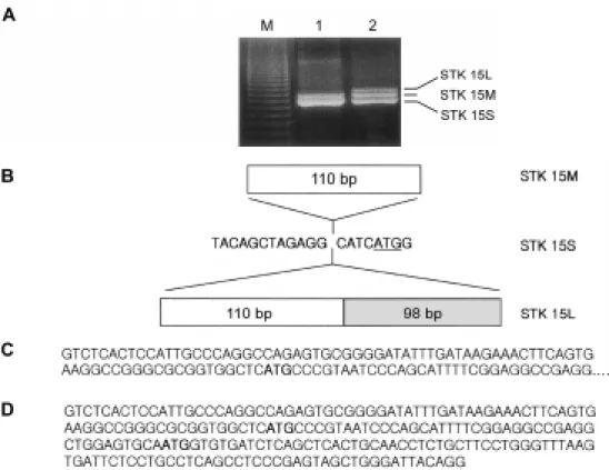
Figure 1. Alternative splicing generates three distinct mRNA transcripts. Primary tumor RNA samples and MDA 231 breast cancer RNA samples were incubated with reverse transcriptase and the resultant DNA was amplified by PCR as described under Materials and Methods. (A) Primary breast tumor (lane 1) and human MDA 231 (lane 2) RT-PCR yielded two and three sizes of cDNA (1295, 1305 and 1503 bp), respectively. (B) Schematic representation of the alternative splicing forms of STK15. (C) DNA sequence of 110 bp insert in 5’-untranslated region of the STK15M gene. (D) DNA sequence of 208 bp insert in 5’-untranslated region of the STK15L gene. The underline and bold letters indicate the translation start codon.
Alternative splicing of STK15 gene 195
Results and Discussion
Alternative splicing of STK15 in breast cancer cell lines
Recently, STK15 was found to encode a cell cycle regulating serine/threonine kinases that appears to be amplified and overexpressed in human breast cancers and colorectal cancers (Sen et al., 1997; Bischoff et al., 1998). Zhou et al. (1998) reported that overexpression of STK15 induced centrosome amplification and transfor- mation in mouse NIH 3T3 cells. To determine whether chromosomal instability was linked to STK15 expression levels in breast cancer cells, we first cloned STK15 cDNA by RT-PCR using specific primers. These primers correspond to regions outside the STK15 coding region.
RT-PCR using RNA of MDA231 human breast cancer cells yielded three distinct cDNA transcripts consisting of 1295, 1305 and 1503 bp. To further characterize the three MDA231 STK15 transcripts, the three RT-PCR products were subcloned into the TOPO vector and sequenced. Sequence analysis revealed that the shor- test cDNA (referred to as STK15S) was identical to the STK15 mRNA (Genbank AF011468) reported in the literature (Zhou et al., 1998). The middle sized STK15 transcript (referred to as STK15M) and the largest STK15 (referred as STK15L) had a 110 bp and 208 bp insertion in 5’-untranslated region, respectively. These data sug- gest that the STK 15 gene was alternatively spliced in the 5’-untranslated region in the MDA 231 breast cancer cells.
Alternatively spliced transcripts do not effect on protein translation
Interestingly, the sequence differences among the three distinct STK15 transcripts were in the 5’-untranslated region which is the region critical for transcriptional and translation control. The alternative transcripts of STK15M and STK15L contain one or two additional translation start codons (ATG), respectively, in the 5’-untranslated region. To determine whether the 110 bp and 208 bp insertions in 5’-untranslated region had an effect on STK15 protein translation, each transcript subclone was transcribed and translated in vitro using the TNT-cou- pled reticulocyte system. The in vitro translated [
35S]
methionine-labeled STK15 proteins were then analyzed by SDS-PAGE. As shown in Figure 2, alternatively splic- ed STK15 transcripts yielded the same sized protein and showed no difference in translational efficiency.
These results suggest that the differences in the STK15 transcripts do not affect the protein code, although the transcripts maybe are generated by differential promoter usage and thus possibly differentially expressed in human tissues or cell type.
Alternatively spliced transcript patterns in breast cancer cell lines and primary tumors
In order to get an overview of the pattern of expression of the STK15 mRNA transcript, we analyzed different breast cancer cell lines, primary tumors and normal cells using RT-PCR. Single stranded cDNAs were synthesiz- ed from total RNA from various breast cancer cell lines, primary tumors and normal cells. The RT-PCR experi- ment used an upstream primer corresponding to posi- tion 215-237 of the STK15 cDNA sequence and a down- stream primer corresponding to position 448-470 of the cDNA sequence. Results showed differential expression of STK15 alternatively spliced transcripts among the tested samples. To confirm the specificity of the PCR products, the PCR products were separated in an agarose gel and transferred by Southern blot to a mem- brane that was then hybridized with the cDNA probes.
As shown in Figure 3, breast cancer cell lines had at least three alternatively spliced transcripts, while primary tumors contained two transcript forms. Interestingly, these results suggested that the STK15 gene exhibits alternative splicing patterns in cell lines and primary tumors that are distinctive.
In conclusion, our data indicate the existence of at least three STK15 transcript forms. Their expression level may be differentially regulated or stabilized in different cell lines and primary tumors.
Acknowledgements
This research was supported by the Yeungnam Univer- sity Medical Center Research grants in 1999.
Figure 2. In vitro translation of the alternatively spliced STK15 gene.
Alternatively spliced STK15-L, STK15-M and STK15-S were in vitro translated using the TNT-coupled reticulocyte system. The translation products were separated in a 10% SDS-polyacrylamide gel. The gel was fixed and exposed to X-ray film for 1 day.
Figure 3. Alternatively spliced transcript patterns in breast cancer cell lines and primary tumors. Total RNA was isolated from 5 human breast cancer cell lines (lane 1, MCF7; lane 2, MDA231; lane 3, MDA361; lane 4, MDA468; lane 5, MDA436), 6 primary tumor samples (lane 6-11) and normal cell (N). RT-PCR analysis was performed and the PCR products were hybridized to full length of cDNA probe.
196 Exp. Mol. Med. Vol. 32(4), 193-196, 2000
References
Bischoff, J. R., Anderson, L., Zhu, Y., Mossie, K., Ng, L., Souza, B., Schryver, B., Flanagan, P., Clairvoyant, F., Ginther, C., Chan, C. S., Novotny, M., Slamon, D. J. and Plowman, G.
D. (1998) A homologue of Drosophila aurora kinase is oncogenic and amplified in human colorectal cancers. EMBO J. 17: 3052-3065
Elledge, S. J. (1998) Mitotic arrest: Mad2 prevents sleepy from waking up the APC. Science 279: 999-1000
Francisco, L., Wang, W. and Chan, C. S. (1994) Type 1 protein phosphatase acts in opposition to IpL1 protein kinase in regulating yeast chromosome segregation. Mol. Cell Biol.
14: 4731-4740
Fukasawa, K., Choi, T., Kuriyama, R., Rulong, S. and Vande Woude, G. F. (1996) Abnormal centrosome amplification in the absence of p53. Science 271: 1744-1747
Glover, D. M., Leibowitz, M. H., McLean, D. A. and Parry, H.
(1995) Mutations in aurora prevent centrosome separation leading to the formation of monopolar spindles. Cell 81: 95- 105
Hardwick, K. G. (1998) The spindle checkpoint. Trends Genet.
14: 1-4
Kellogg, D. R., Oegema, K., Raff, J., Schneider, K. and Alberts, B. M. (1995) CP60: a microtubule-associated protein that is localized to the centrosome in a cell cycle-specific manner. Mol. Biol. Cell 6: 1673-1684
Kimura, M., Kotani, S., Hattori, T., Sumi,N., Yoshioka, T., Todokoro, K.and Okano, Y. (1997) Cell cycle-dependent expression and spindle pole localization of a novel human protein kinase, Aik, related to Aurora of Drosophila and yeast Ipl1. J. Biol. Chem. 272: 13766-13771
Kimura, M., Matsuda, Y., Yoshioka, T. and Okano, Y. (1999) Cell cycle-dependent expression and centrosome localization of a third human aurora/Ipl1-related protein kinase, AIK3. J.
Biol. Chem. 274: 7334-7340
Lingle, W. L., Lutz, W. H., Ingle, J. N., Maihle, N. J. and Salisbury, J. L. (1998) Centrosome hypertrophy in human breast tumors: implications for genomic stability and cell polarity. Proc. Natl. Acad. Sci. U. S. A .95: 2950-2955 Sen, S., Zhou, H. and White, R. A. (1997) A putative serine/
threonine kinase encoding gene BTAK on chromosome 20q13 is amplified and overexpressed in human breast cancer cell lines. Oncogene 14: 2195-2200
Taagepera, S., Campbell, M. S. and Gorbsky, G. J. (1995) Cell-cycle-regulated localization of tyrosine and threonine phosphoepitopes at the kinetochores of mitotic chromosomes.
Exp. Cell Res. 221: 249-260
Yanai, A., Arama,E., Kilfin, G. and Motro, B. (1997) ayk1, a novel mammalian gene related to Drosophila aurora centro- some separation kinase, is specifically expressed during meiosis. Oncogene 14: 2943-2950
Zhou, H., Kuang, J., Zhong, L., Kuo, W. L., Gray, J. W., Sahin, A., Brinkley, B. R. and Sen, S. (1998) Tumour amplified kinase STK15/BTAK induces centrosome amplification, aneuploidy and transformation. Nat. Genet. 20: 189-193