Korean J Environ Agric (2011) Online ISSN: 2233-4173 Vol. 30, No. 4, pp. 459-465 http://dx.doi.org/10.5338/KJEA.2011.30.4.459 Print ISSN: 1225-3537
Antibacterial Effects of Extracts of Thuja Orientalis cv Aurea Nana Cones against Food-spoilage and Food-borne Pathogens
Xiao Nan Yang,
1Cher-Won Hwang,
2Gi-Seok Kwon
3and Sun Chul Kang
1*1
Department of Biotechnology, Daegu University, Gyeongsan, Gyeongbuk 712-714, Republic of Korea
2
School of Global Leadership, Handong Global University, Pohang, 791-708, Republic of Korea
3
Dept. of Bioresource Sciences, Andong National University, Andong 760-749, Republic of Korea
Received: 18 October 2011 / Accepted: 24 December 2011
ⓒ 2011 The Korean Society of Environmental Agriculture
*Prof. Sun Chul Kang Corresponding author,
Department of Biotechnology, College of Engineering, Daegu University, Kyoungsan, Kyoungbook 712-714, Republic of Korea
Phone: +82-53-850-6553; Fax: +82-53-850-6559;
E-mail: [email protected]; Phone: +82-53-850-6553;
Fax: +82-53-850-6559
459
Abstract
BACKGROUND: Nowadays, Chemical antiseptics have become great problems for health and environmental, so that developing of new substitutes for chemical antiseptics is more and more important. Natural product is a kind of environment-friendly additive that could be used as antiseptic in food industry. Thuja orientalis cv Aurea Nana is a gymnospermous plant of the family Cupressaceae, native to northwestern China and widely naturalised elsewhere in Korea and Japan. This study was aimed to investigate the antibacterial potential of various organic extracts from T. orientalis cones against some food-borne and food-spoilage bacteria.
METHODS AND RESULTS: Hexane extract (HE), chloroform extract (CE), ethyl acetate extract (EAE) and methanol extract (ME) were obtained from female cones of T. orientalis. The antibacterial activities of various extracts were tested by standard agar diffusion and minimum inhibitory concentrations (MICs) against five gram-positive and six gram-negative bacteria. Cell viability and morphology change of L. monocytogenes ATCC 10943 treated with
hexane extract were also observed. The various extracts displayed remarkable antibacterial effects against all the gram-positive bacteria but did not show any effect against the gram-negative bacteria. Hexane extract has the highest inhibitory effect on cell viability of L. monocytogenes ATCC 10943. SEM observation also demonstrated the damaging effect of the hexane extract on the morphology of L. monocytogenes ATCC 10943 at the minimum inhibitory concentration.
CONCLUSION(s): The tested gram-positive bacteria were significantly inhibited by organic extracts of T.
orientalis cone. Hexane extract was the most potent against Listeria monocytogenes ATCC 10943, as evidenced by the lowest MIC level and the complete inhibition of cell viability within shortest exposure time, along with SEM observation.
Key Words: Antibacterial activity, Food-borne and Food-spoilage pathogens, Thuja orientalis cv Aurea Nana
Introduction
Food-borne diseases encompass a wide spectrum of
illnesses and are a growing public health problem
worldwide. In industrialized countries, 30% of populations
are affected by food-borne diseases annually, and the
problem is likely to be even more widespread in
developing countries. In spite of modern improvements in
slaughter hygiene and food production techniques, food
safety is an increasingly important public health issue
(WHO, 2002). Mostly, the food industries are using chemical preservatives to prevent the growth of food-borne and food-spoiling microbes. Some natural products have also been used as antiseptics to reduce the amount of the chemicals added to food or to substitute them. Natural plant extracts are of growing interest to both of food industry and researchers because their antibacterial, antifungal, and antioxidant properties provide a great potential as an alternative to the chemical food preservatives (Deba et al ., 2008).
Thuja orientalis (= Platycladus orientalis (L.)) is a gymnospermous plant of the family Cupressaceae, which is native to northwestern China and widely distributes in Northeast Asian countries including Korea and Japan. T. orientalis cv Aurea Nana is a dwarf plant with yellow-green leaves that compactly branch, which widely used as an ornamental plant in South Korea. It has been reported that the leaves of T. orientalis were used in the treatment of various inflammatory diseases (Kim et al ., 2010). In Chinese herbal medicine, the leaves have been used for treatments of gout, rheumatism, diarrhea and chronic tracheitis (Zhu et al ., 2004). The plant also exhibits anti-plasmodial (Asili et al ., 2004), fungi toxic (Guleria et al ., 2008) and molluscicidal (Singh and Singh, 2009) effects, and improves impaired memory acquisition (Nishiyama et al ., 1995). There are many reports on such phytochemical and biological studies of this plant leaves all over the world. However, other parts of this plant have not been adequately studied for their medicinal or industrial use. Therefore, this study was carried out to investigate in vitro antibacterial activity of seed cone extracts of T. orientalis .
Materials and methods
Plant material
5 kg of female cones of T. orientalis were collected from local area of Kyungsan, Kyungpook, South Korea in October, 2010. The collected plant was identified to species level by morphological features.
A voucher specimen (DU-TO396) was deposited at Daegu University for further reference. The cones were washed, dried and ground.
Samples extraction
The ground cones (150 g) of T. orientalis were extracted with 500 ml of four different organic solvents including hexane, chloroform, ethyl acetate and methanol for 7
days at room temperature, respectively. And then, the solvents were evaporated by vacuum rotary evaporator (EYELA N1000, Japan) to get the hexane extract (HE), chloroform extract (CE), ethyl acetate extract (EAE) and methanol extract (ME). The extracts were maintained at 4°C until further use. Solvents (analytical grade) for extraction were obtained from a commercial source (SAMCHUN PURE CHEMICAL CO., LTD, Korea).
Test microorganisms (food-spoilage and food-borne pathogens)
Eleven food-spoilage and food-borne pathogens used in this study include five gram-positive bacteria ( Bacillus cereus KCTC 14042, Bacillus subtilis ATCC 6633, Listeria monocytogenes ATCC 10943, Staphylococcus aureus Wild Type, Staphylococcus aureus ATCC 6538) and six gram-negative bacteria ( Escherichia coli 0157 KCTC 14034, Escherichia coli ATCC 10536, Escherichia coli ATCC 8739, Salmonella enteritidis KCTC 12243, Salmonella typhimurium Wild Type, Pseudomonas aeruginose ATCC 15522). Fresh cultures were prepared by transferring a loop of cells from each stock culture to a new tube containing Luria-Bertani (LB) broth medium. The cultures were grown at 37°C for 24 h and maintained on LB agar medium at 4°C.
Assay for antibacterial potential
Standard agar diffusion method was used for antibacterial assay (Murray et al., 1995). Organic extracts were dissolved in DMSO. Fresh bacterial cultures were prepared by transferring a loop of cells from the stock culture to a flask containing LB medium, and incubated at 37°C for 24 h. The cultures were diluted with LB broth to achieve an optical density of 10
7CFU/ml for the test organisms at 600 nm by UV/Vis Spectrophotometer Optizen 2120 UV.
0.1 ml of each standardized bacterial inoculum (10
7CFU/ml) was poured into Petri plates containing 20 ml of LB agar medium, uniformly spread, and allowed to dry for 5 min. Sterile filter paper discs (6 mm diameter, Waterman No.1) were impregnated with 1 mg/disc of HE, CE, EAE and ME, respectively, and placed on the inoculated agar plates.
After the plates were kept at room temperature for 30
min to allow the extracts to diffuse into the agar, they
were incubated at 37°C for 24 h. Antibacterial activity
of the extracts was evaluated by measuring the
diameter of inhibition zone developing around the
discs.
Determination of minimum inhibitory concentrations The minimum inhibitory concentrations (MICs) of extracts were tested by the method described by Chandrasekaran and Venkatesalu (2004). Each extract was incorporated into LB broth medium in a tube and adjusted to a final concentration in the range of 0 to 1500 μg/ml. 10 μl of standardized suspension of each test organism (10
7CFU/ml) was transferred to the tubes, and incubated at 37°C for 24 h. MIC was determined as the lowest concentration (μg/ml) of each extract where no visible growth of test organisms occur.
Effect of hexane extract on cell viability of Listeria monocytogenes
Hexane extract at the MIC in 10 ml of LB broth was added into the tubes containing bacterial suspension (approximately 10
7CFU/ml) of L.
monocytogenes ATCC in LB broth medium. After incubation at 37°C, 10 μl of samples for viable cell counts were removed at 30-min intervals up to 240 min. Each sample was diluted appropriately with sterile water, and 50 μl of the diluted suspension was spread on the surface of LB agar. Colonies that formed on each plate were counted after 24 h of
incubation at 37°C. The control which inoculated without hexane extract was treated at same experimental condition as mentioned above.
Scanning electron microscopy analysis
The method of sample preparation for scanning electron microscopy (SEM) was modified from that of Kockro et, al (2000). Bacterial cells of L. monocytogenes ATCC 10943 which were treated with and without the MIC level of hexane extract for 4 h were washed three times using 50 mM phosphate buffer solution (PBS, pH 7.3), and centrifuged at 4000 g. The supernatant was removed and the centrifuged cells were suspended in a new PBS. A thin smear of the suspension was coated on a glass slide (4 mm
2) and fixed for 3 h in 2.5% (v/v) glutaraldehyde (Electron Microscopy Science, Washington, USA). After the fixation, the bacterial cells on each glass slide were rinsed, dehydrated using a series of graded ethanol (concentrations ranging from 50% to 100%), and dried with CO
2. The dried cells were coated with gold in a sputter coater (Hitachi, Japan). Samples were observed under a Scanning electron microscope (Hitachi-S4300, Japan).
Microorganism Diameter of inhibition zones A (mm)
HE CE EAE ME
Bacillus cereus
KCTC 14042 22.8 ± 0.9 a 19.7 ± 0.7 a 17.8 ± 0.7 a 26.2 ± 0.8 aBacillus subtilis
ATCC 6633 10.2 ± 0.4 e 12.2 ± 0.8 d 12.8 ± 0.4 c 13.2 ± 0.5 dListeria monocytogenes
ATCC 10943 11.4 ± 0.6 d 12.7 ± 0.8 d 18.5 ± 0.8 a 16.3 ± 0.6 cStaphylococcus aureus
Wild Type 17.5 ± 0.8 b 15.0 ± 0.5 c 15.9 ± 0.8 b 16.0 ± 0.6 cStaphylococcus aureus
ATCC 6538 15.1 ± 0.4 c 16.3 ± 0.3 b 17.2 ± 0.7 a 18.0 ± 0.4 bEscherichia coli
0157 KCTC 14034 nd B nd nd ndEscherichia coli
ATCC 10536 nd nd nd ndEscherichia coli
ATCC 8739 nd nd nd ndSalmonella enteritidis
KCTC 12243 nd nd nd ndSalmonella typhimurium
Wild Type nd nd nd ndPseudomonas aeruginosa
ATCC 15522 nd nd nd ndA Mean ± SD. Results are based on triplicate tests. Different letters next to the values indicate significant differences at
p
< 0.05. (Tested concentration of extracts: 1 mg/disc). HE: hexane extract, CE: chloroform extract, EAE: ethyl acetate extract, ME: methanol extract.B nd, no inhibition detected.
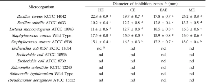
Table 1. Diameter of inhibition zones (mm) produced by cone extracts of
T. orientalis
on the test pathogens.Microorganism MICs A
HE CE EAE ME
Bacillus cereus
KCTC 14042 10 75 500 500Bacillus subtilis
ATCC 6633 10 75 75 100Listeria monocytogenes
ATCC 10943 25 100 750 500Staphylococcus aureus
Wild Type 50 750 500 500Staphylococcus aureus
ATCC 6538 10 750 750 500Escherichia coli
0157 KCTC 14034 nd B nd nd ndEscherichia coli
ATCC 10536 nd nd nd ndEscherichia coli
ATCC 8739 nd nd nd ndSalmonella enteritidis
KCTC 12243 nd nd nd ndSalmonella typhimurium
Wild Type nd nd nd ndPseudomonas aeruginosa
ATCC 15522 nd nd nd ndA MICs, Minimum inhibitory concentrations (values in μg/ml). HE: hexane extract, CE: chloroform extract, EAE: ethyl acetate extract, ME: methanol extract.
B nd, no inhibition detected.
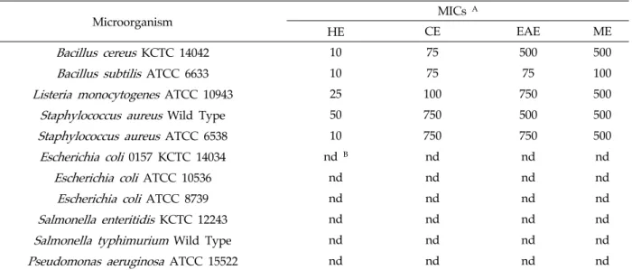
Table 2. Minimum inhibitory concentrations of cone extracts of
T. orientalis
against the growth of food-spoilage and food-borne pathogens.Fig. 1. Effects of extracts of
T. orientalis
cones (at MIC levels) on the viability of theL. monocytogenes
ATCC 10943. Control cells were not treated with the extracts.Bars indicate standard errors. HE: hexane extract, CE:
chloroform extract, EAE: ethyl acetate extract, ME:
methanol extract.
Results
In vitro antibacterial activity
In vitro antibacterial activities of T. orientalis extracts were determined by the presence/absence and the diameter of inhibition zone (Table 1). All extracts were found to have antibacterial effects against all gram-positive bacteria tested, but none of the gram-negative bacteria. The diameters of inhibition zones of HE, CE, EAE and ME against the gram-positive bacteria were in the range of 10.2-22.8 mm, 12.2-19.7 mm, 12.8-18.5 mm and 13.2-26.2 mm, respectively. For all of the extracts, the largest inhibition zone was developed against B. cereus KCTC 14042 (17.8-26.2 mm). Among all the extract tested, ME exhibited the highest antibacterial activities against B. cereus KCTC 14042, B. subtilis ATCC 6633 and S. aureus ATCC 6538. Its inhibitory effect was also high against L. monocytogenes ATCC 10943 and S. aureus Wild Type.
Minimum inhibitory concentrations
The minimum inhibitory concentrations of various extracts against the gram-positive bacteria tested were in the range of 10-750 μg/ml (Table 2). The HE showed the most potent inhibitory capacity with the MICs in the range of 10-50 μg/ml. The CE was more potent to B. cereus KCTC 14042, B. subtilis ATCC 6633 and L. monocytogenes ATCC 10943 compared to EAE and ME. However, the inhibitory effects against
S. aureus Wild Type and S. aureus ATCC 6538 were stronger than vice versa when treated with EAE and ME. The EAE and ME also showed potential inhibitory effects against all the test gram-positive bacteria with the MICs in the range of 75-750 μg/ml and 100-500 μg/ml, respectively. None of the extracts inhibited the growth of the five gram-negative bacteria tested.
Effects of extracts on cell viability of bacterium Reduced viability of L. monocytogenes ATCC 10943 was observed at MIC of all extracts tested (Fig.
1). After 150 min exposure, all the extracts revealed
complete inhibition of CFU number against L.
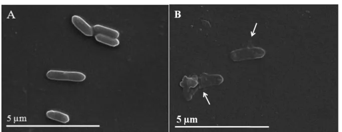
Fig. 2. Effect of hexane extract of
T. orientalis
cones on the morphology ofL. monocytogenes
ATCC 10943. A:cells without treatment showing a regular, smooth surface; B: cells lysis (arrows) caused by hexane extract inoculation at the MIC level (25 μg/ml).