ISSN 1225-6552, eISSN 2287-7630 http://dx.doi.org/10.7853/kjvs.2016.39.1.35
< Original Article >
Veterinary Service
Available online at http://kjves.org
*Corresponding author: Gil-Jae Cho, Tel. +82-53-950-5978, Fax. +82-53-950-5974, E-mail. [email protected]
경북지역 재래산양의 세균성, 바이러스성 설사병 병원체 검출률 조사
손준형
1ㆍ도재철
2ㆍ조길재
3*
경상북도가축위생시험소1, 경상북도가축위생시험소 동부지소2, 경북대학교 수의과대학3
Detection ratio of bacterial and viral pathogens of diarrhea from Korean indigenous goat feces in Gyeongbuk province
Jun-Hyung Sohn
1, Jae-Cheul Do
2, Gil-Jae Cho
3*
1Gyeongbuk Veterinary Service Laboratory, Daegu 41405, Korea
2East-Branch, Gyeongbuk Veterinary Service Laboratory, Gyeongju 38101, Korea
3College of Veterinary Medicine, Kyeongbuk National University, Daegu 41566, Korea (Received 8 January 2016; revised 4 February 2016; accepted 17 March 2016)
Abstract
The purpose of this study was to survey on infection status of pathogens of diarrhea from Korean in- digenous goat. A total of 800 fecal samples was collected from 50 farms from January to October 2015 and was tested by automatic biochemical machine and polymerase chain reaction (PCR). The overall detection ratio of bacterial pathogens was 22.4% and viral pathogens was 16.3%, respectively. The de- tection ratio of Escherichia coli (E. coli), Salmonella spp., bovine viral diarrhea virus (BVDV), rotavirus and coronavirus were 21.5%, 0.9%, 7.6%, 5.6% and 3.0%, respectively. In the rates of mixed detection, single was 78.2%, double 8.4%, triple 11.6% and quadruple 1.8% in each sample and 38%, 12%, 16%, 20% in each farm, respectively.
Key words : Korean indigenous goat, Diarrhea, E. coli, BVDV, Rotavirus
서 론
재래산양은 식용, 약용으로 오랫동안 우리나라에서 사육되어 왔으며 산악지형에도 잘 적응하여 방목사 육에 용이하다. 최근에는 소규모 부업 사육형태를 벗 어나 대규모 집단사육으로 변화함에 따라 여러 가지 질병에 대한 문제점이 드러나고 있으며, 이에 인해 효과적인 방역대책의 수립이 요구되고 있다. 그러나 오랜 사육역사에 비하여 재래산양의 질병에 대한 연 구는 세균 및 기생충성 질병에 대한 몇몇 보고와 포 괄적인 연구동향 보고가 있으나, 다른 가축들에 비해 그 수가 상당히 적으며 특히 경북지역에 대한 조사는 극히 미진한 실정이다. 기존 연구에 따르면 재래산양
에서 발생하는 질병 중 약 78%가 소화기 및 호흡기 질병으로 사육농장에 큰 손실을 입히고 있으며, 소화 기 질병 중 설사는 어린 동물에서 폐사의 주요 원인 으로 알려져 있다. 일반적으로 다른 가축들에 비해 질병과 환경에 대한 저항력이 강하지만 재래산양에 서 발생하는 질병 중 설사병이 약 40%에 이를 정도 로 많은 부분을 차지하고 있고, 설사병이 발생할 경 우 어린 일령의 폐사와 증체량 감소로 인하여 농가에 피해를 주고 있다. 설사는 기생충, 세균, 바이러스 등 의 병원체와 사육온도, 사료 등 환경적인 요인으로 인해 발생하고 있으며 이 중 E. coli, Salmonella spp., rotavirus, coronavirus, BVDV 등이 주요 세균성, 바이 러스성 설사병 병원체로 알려져 있다(Cho 등, 2008;
Choe 등, 2012; Heo 등, 1999; Kim, 2000).
따라서 본 연구는 경북지역에서 사육중인 재래산
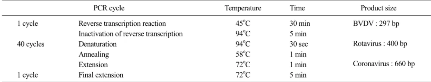
Table 1. PCR program and product size of viral diarrhea pathogens in goat
PCR cycle Temperature Time Product size
1 cycle Reverse transcription reaction 45oC 30 min BVDV : 297 bp
Rotavirus : 400 bp
Coronavirus : 660 bp Inactivation of reverse transcription 94oC 5 min
40 cycles Denaturation 94oC 30 sec
Annealing 58oC 1 min
Extension 72oC 1 min
1 cycle Final extension 72oC 5 min
Fig. 1. Gel electrophoresis of viral pathogen-specific gene by PCR.
Lane M: 1kb size marker. Lane 1, 2, 5, 6: triple detected (BVDV, rota- virus, coronavirus), Lane 3: single detected (BVDV), Lane 4: neg- ative, Lane 7: positive control strain.
양 설사병의 세균성 및 바이러스성 병원체 검출률을 조사하고 이를 바탕으로 질병발생 시 농가피해 감소 를 위한 효과적 방역대책의 기초 자료로 활용하고자 한다.
재료 및 방법
공시재료
2015년 1월에서 10월까지 10개월 동안 경북지역 18개 시ㆍ군 50 농장에 대하여 사육규모, 소독시설 유무등에 대한 사전조사를 완료한 재래산양 사육농 장에서 농장별 16점의 염소분변을 채취하였다. 사육 규모별 검사 대상농장은 50 두 미만(24 농장), 51∼
100 두(21 농장), 101 두 이상(5 농장)로 확인하였으 며, 채취한 분변은 24시간이내에 실험에 사용하였고 실험을 실시할 때까지 4oC에서 냉장ㆍ보관하였다.
검사방법
Automatic biochemical machine을 이용한 세균성 병원 체 동정: 분변을 blood agar plate 상에서 37oC 24시간 배양한 후 E. coli와 Salmonella spp. 등 병원체로 의심 되는 집락을 제조사에서 제공한 매뉴얼에 따라 0.45%
saline에 주입하여 0.6 McF로 탁도를 조절한 후 VITEK2 GN test kit, VITEK2 GN test kit와 VITEK2 Compact (Biomerieux, FRANCE) 자동화 장비를 사용하여 확인 하였다.
PCR을 이용한 바이러스성 병원체 동정: 바이러스성 병원체 동정은 PCR법을 사용하여 확인하였는데 이 를 위한 유전자 추출은 Genomic DNA & Viral RNA extraction kit와 자동화장비(Malcom, JAPAN)를 사용 하여 실시하였다. 각 질병에 대한 병원체 동정은 Bovine Coronavirus/rotavirus/diarrhea disease virus Detection kit
(iNtRON, KOREA)를 사용하여 제조사의 매뉴얼에 따 라 PCR을 실시한 후 ethidium bromide (EtBr)가 첨가 된 1.5% agarose gel 상에서 BVDV 297 bp, rotavirus 400 bp, coronavirus 660 bp에서 각각 특이밴드를 확인 하였다(Table 1, Fig. 1).
검사결과 분석
검사결과는 사육규모별로 구분하여 병원체 검출률 을 확인하였고, 병원체 검출정도에 따라 1종류 병원 체의 단독검출과 2종류 이상의 혼합검출 여부를 나 누어 살펴보았다.
결과 및 고찰
경북지역 50개 농장에서 채취한 800점 중 43 농장, 225점에서 1개 이상의 설사병 병원체가 검출되어 검
Table 2. Detection ratio of bacterial and viral pathogens in goats feces
Total Bacterial pathogens E. coli Salmonella spp. Viral pathogens BVDV Rotavirus Coronavirus
No of positive 309 179 172 7 130 45 24 61
Positive ratio (%) 38.6 22.4 21.5 0.9 16.3 5.6 3.0 7.6
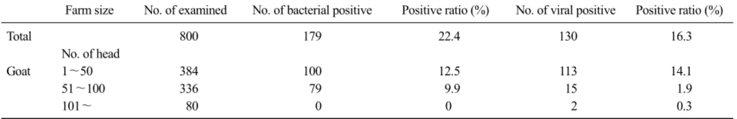
Table 3. Detection ratio of pathogens in goats according to farm sizes
Farm size No. of examined No. of bacterial positive Positive ratio (%) No. of viral positive Positive ratio (%)
Total 800 179 22.4 130 16.3
No. of head
Goat 1∼50 384 100 12.5 113 14.1
51∼100 336 79 9.9 15 1.9
101∼ 80 0 0 2 0.3
Table 4. Detected patterns in each farms
Farm size No. of detected farms (%)
No. of single detected (%)
No. of double detected (%)
No. of triple detected (%)
No. of quadruple detected (%)
Total 43 (86.0) 19 (38.0) 6 (12.0) 8 (16.0) 10 (20.0)
No. of head
Goat 1∼50 23 (46.0) 5 (10.0) 3 (6.0) 5 (10.0) 10 (20.0)
51∼100 18 (36.0) 12 (24.0) 3 (6.0) 3 (6.0) 0 (0)
101∼ 2 (4.0) 2 (4.0) 0 (0) 0 (0) 0 (0)
사호수 대비 86.0% (43/50), 검사시료 대비 28.1%
(225/800)의 양성률을 나타내었다. 세균성 설사병 병 원체는 22.4% (179/800)였으며, 바이러스성 설사병 병 원체는 16.3% (130/800)로 나타났다. 세부적으로는 E.
coli, salmonella가 각각 21.5% (172/800), 0.9% (7/800)로 나타났고 BVDV, rotavirus, coronavirus는 각각 7.6%
(61/800), 5.6% (45/800), 3.0% (24/800)의 양성률을 보 였다(Table 2). 사육규모별 검출률을 살펴본 결과 50 두 이하의 소규모 농장에서는 세균성 병원체와 바이 러스성 병원체가 각각 12.5% (100/800), 14.1%
(113/800), 51∼100 두를 사육하는 농장에서는 9.9%
(79/800), 1.9% (15/800), 101 두 이상을 사육하는 농장 에서는 0% (0/800), 0.3% (2/800)로 확인되었다. 또한, 세균과 바이러스성 병원체 모두 농장 출입 소독시설 이 없고 자체 소독장비가 미비한 50 두 미만 사육농 장에서 가장 높은 검출률을 보였다(Table 3).
병원체별로 기존 연구들과 비교해 본 결과 E. coli 와 Salmonella spp.는 각각 2.4%, 18.8%의 결과를 나 타낸 Bosilevac 등(2015)의 연구에 비해 E. coli는 그 비율이 상당히 높게 나왔고 Salmonella spp.는 낮은 검출률을 보였다. BVDV와 rotavirus는 각각 21.1%, 28.6%의 감염률을 보인 Fulton 등(1982), Kaminjolo 등
(1994)의 결과에 비해 낮았으며, coronavirus는 3%
(24/800)의 검출률을 보여 1%의 보인 Yang 등(2008) 의 연구에 비해 높게 나타났다. 농장별 병원체 검출 형태로 살펴본 결과 1종의 단독검출농장은 38.0%
(19/50), 2종 복합검출 12.0% (6/50), 3종 복합검출 16.0%
(8/50), 4종이상의 복합검출 농장이 20.0% (10/50)으로 확인되었으며 특히 4종류 이상의 병원체가 검출된 10 개 농장은 모두 50 두 미만을 사육하는 소규모 농장 이었다(Table 4). 이같은 결과로 볼 때 국내 재래산양 사육농장 특히 부업형태의 소규모 농장에 대한 정기 적인 검사와 방역교육이 실시되어야 함을 알 수 있었 고, 재래산양의 설사병과 관련하여 방역정책 수립, 사양관리 및 소독실태조사, 농가지도 등 재래산양 질 병관리방안이 조속히 마련되어야 할 것으로 판단된다.
결 론
경북지역 50호의 농장에서 채취한 800점의 분변 시료를 대상으로 실시한 재래산양 설사병 병원체 조 사 결과 전체 86.0% (43/50)의 농장에서 1개 이상의 병원체가 검출되어 대부분의 농장이 설사병에 노출
되어 있음을 확인할 수 있었다. 설사병 병원체의 검 출률은 38.6% (309/800)였으며 세균성 22.4% (179/800), 바이러스성 16.3% (130/800)로 확인되었다. 병원체별 검출률은 E. coli, Salmonella spp., BVDV, rotavirus, coronavirus가 각각 21.5% (172/800), 0.9% (7/800), 7.6% (61/800), 5.6% (45/800), 3.0% (24/800) 로 확인 되었다. 농장의 사육규모별로 분석해본 결과 세균성, 바이러스성 병원체 모두에서 50 두 이하의 농장에서 검출률이 가장 높은 것으로 조사되어 소규모 농장이 질병에 특히 취약함을 알 수 있었다.
REFERENCES
Bosilevac JM, Gassem MA, Al Sheddy IA, Almaiman SA, Al-Mohizea IS, Alowaimer A, Koohmaraie M. 2015.
Prevalence of Escherichia coli O157:H7 and Salmonella in camels, cattle, goats, and sheep harvested for meat in Riyadh. J Food Prot 78: 89-96.
Broaddus CC, Holyoak GR, Dawson L, Step DL, Funk RA, Kapil S. 2007. Transmission of bovine viral diarrhea virus to adult goats from persistently infected cattle. J Vet Diagn Invest 19: 545-548.
Cho KH, Lee JH, Kim DJ, Kim SJ, Kwon OD, Kwak DM. 2008.
Infection rate of parasites from feces of Korean in- digenous goats in northern areas of Gyeongbuk province.
Korean J Vet Serv 31: 357-362.
Choe CY, Kang DW, Cho CY, Jung BY, Son JK, Hur TY, Jung YH, Kang SJ, Do YJ, Ryu IS, Kin UH, Park YS, Son DS. 2012. A Survey of disease occurrence in Korean black goats. J Vet Clin 29: 160-164.
Cortés C, de la Fuente R, Contreras A, Sánchez A, Corrales JC, Martínez S, Orden JA. 2006. A survey of Salmonella spp. and Campylobacter spp. in dairy goat faeces and bulk tank milk in the Murcia region of Spain. Ir Vet J 59: 391-393.
Czopowicz M, Kaba J, Schirrmeier H, Bagnicka E, Szaluś- Jordanow O, Nowicki M, Witkowski L, Frymus T. 2011.
Serological evidence for BVDV-1 infection in goats in Poland. Acta Vet Hung 59: 399-404.
Da Costa Mendes VM, De Beer MC, Els HJ, Goosen GH, Theron J, Steele AD. 1994. Rotavirus in Saanen goats. J S Afr Vet Assoc 65: 132-133.
Duffy L, Barlow R, Fegan N, Vanderlinde P. 2009. Prevalence and serotypes of Salmonella associated with goats at two Australian abattoirs. Lett Appl Microbiol 48: 193-197.
Fulton RW, Downing MM, Hagstad HV. 1982. Prevalence of bo- vine herpesvirus-1, bovine oral diarrhea, parainfluenza-3, bovine adenoviruses-3 and -7, and goat respiratory syn- cytial viral antibodies in goats. Am J Vet Res 43:
1454-1457.
Hariharan H, López A, Conboy G, Coles M, Muirhead T. 2007.
Isolation of Escherichia fergusonii from the feces and internal organs of a goat with diarrhea. 2007. Can Vet J 48: 630-631.
Heo JH, Jung MH, Cho MH, Ahn DW, Lee SS. 1999. A survey on the actual management and the prevalence of interal parasite in the Korean indigenous goats of southern Kyoungnam area. Korean J Vet Serv 22: 71-77.
Hertig C, Pauli U, Zanoni R, Peterhans E. 1991. Detection of bo- vine viral diarrhea (BVD) virus using the polymerase chain reaction. Vet Microbiol 26: 65-76.
Kaminjolo JS, Adesiyun AA. 1994. Rotavirus infection in calves, piglets, lambs and goat kids in Trinidad. Br Vet J 150:
293-299.
Kim TJ. 2000. Study on digestive and respiratory viral pathogens of Korean native black goat:1. Establishment of serum bank for epidemiological study;2. Isolation of viral pathogens from digestive and respiratory tracts;3.
Development of vaccine to the isolated virus. Konkuk University press, Seoul.
McAuley CM, McMillan K, Moore SC, Fegan N, Fox EM. 2014.
Prevalence and characterization of foodborne pathogens from Australian dairy farm environments. J Dairy Sci.
97: 7402-7412.
Mishra N, Rajukumar K, Tiwari A, Nema RK, Behera SP, Satav JS, Dubey SC. 2009. Prevalence of Bovine viral diar- rhoea virus (BVDV) antibodies among sheep and goats in India. Trop Anim Health Prod 41: 1231-1239.
Molla W, Molla B, Alemayehu D, Muckle A, Cole L, Wilkie E.
2006. Occurrence and antimicrobial resistance of Salmonella serovars in apparently healthy slaughtered sheep and goats of central Ethiopia. Trop Anim Health Prod 38: 455-462.
Omatsu T, Tsuchiaka S, Hirata T, Shiroma Y, Okazaki S, Katayama Y, Oba M, Nishiura N, Sassa Y, Furuya T, Nagai M, Ochiai H, Tamaki S, Mizutani T. 2014.
Detection of enterovirus genome sequence from diarrheal feces of goat. Virus Genes 48: 550-552.
Pao S, Patel D, Kalantari A, Tritschler JP, Wildeus S, Sayre BL.
2005. Detection of Salmonella strains and Escherichia coli O157:H7 in feces of small ruminants and their iso- lation with various media. Appl Environ Microbiol 71:
2158-2161.
Papp H, Malik YS, Farkas SL, Jakab F, Martella V, Bányai K.
2014. Rotavirus strains in neglected animal species in- cluding lambs, goats and camelids. Virusdisease 25:
215-222.
Pratelli A, Martella V, Tempesta M, Buonavoglia C. 1999.
Characterization by polymerase chain reaction of ru- minant rotaviruses isolated in Italy. New Microbiol 22:
105-109.
Roug A, Byrne BA, Conrad PA, Miller WA. 2013. Zoonotic fecal pathogens and antimicrobial resistance in county fair animals. 2013. Comp Immunol Microbiol Infect Dis 36:
303-308.
Sharpee RL, Mebus CA, Bass EP. 1979. Characterization of a calf diarrheal coronavirus. Am J Vet Res 37: 1031-1041.
Stadler HP, Strasser M, Meier P, Tontis A, Hofmann M, Ruggli N, Peterbans E. 1996. PCR: a look behind the scenes at bovine viral diarrhea virus. Schweiz Arch Tierheilkd 138: 63-66.
Stirling J, Griffith M, Dooley JS, Goldsmith CE, Loughrey A, Lowery CJ, McClurg R, McCorry K, McDowell D, McMahon A, Millar BC, Rao J, Rooney PJ, Snelling WJ, Matsuda M, Moore JE. 2008. Zoonoses associated with petting farms and open zoos. Vector Borne Zoonotic Dis 8: 85-92.
Stone GG, Chengappa MM, Oberst RD, Gabbert NH, McVey S,
Hennessy KJ, Muenzenberger M, Staats J. 1993.
Application of polymerase chain reaction for the correla- tion of Salmonella serovars recovered from greyhound feces with their diet. J Vet Diagn Invest 5: 378-385.
Verbeek A, Tijssen P. 1991. Sequence analysis of the turkey en- teric coronavirus nucleocapsid and membrane protein genes: a close genomic relationship with bovine coro- navirus. J Gen Virol 72: 1659-1666.
Yang DK, Hwang IJ, Kim BH, Kweon CH, Lee KW, Kang MI, Lee CS, Cho KO. 2008. Serosurveillance of viral dis- eases in Korean native goats (Capra hircus). J Vet Med Sci 70: 977-979.