Prognostic Factors and Characteristics of Pancreatic Neuroendocrine Tumors: Single Center Experience
Tak Geun Oh,
1Moon Jae Chung,
1Jeong Yeop Park,
1Seung Min Bang,
1Seung Woo Park,
1Jae Bok Chung,
1and Si Young Song
1,21Division of Gastroenterology, Department of Internal Medicine and Yonsei Institute of Gastroenterology,
2Brain Korea 21 Project for Medical Science, Yonsei University College of Medicine, Seoul, Korea.
Received: September 8, 2011 Revised: November 8, 2011 Accepted: November 9, 2011
Corresponding author: Dr. Si Young Song, Division of Gastroenterology,
Department of Internal Medicine, Yonsei University College of Medicine, 50 Yonsei-ro, Seodaemun-gu, Seoul 120-752, Korea.
Tel: 82-2-2228-1957, Fax: 82-2-2227-7900 E-mail: [email protected]
∙ The authors have no financial conflicts of interest.
© Copyright:
Yonsei University College of Medicine 2012 This is an Open Access article distributed under the terms of the Creative Commons Attribution Non- Commercial License (http://creativecommons.org/
licenses/by-nc/3.0) which permits unrestricted non- commercial use, distribution, and reproduction in any medium, provided the original work is properly cited.
Purpose: Pancreatic neuroendocrine tumors (PNET) are a rare subgroup of tumors.
For PNETs, the predictive factors for survival and prognosis are not well known.
The purpose of our study was to evaluate the predictive factors for survival and dis- ease progression in PNETs. Materials and Methods: We retrospectively analyzed 37 patients who were diagnosed with PNET at Severance Hospital between No- vember 2005 and March 2010. Prognostic factors for survival and disease progres- sion were evaluated using the Kaplan-Meier method. Results: The mean age of the patients was 50.0±15.0 years. Eight cases (21.6%) were described as functioning tumors and 29 cases (78.4%) as non-functioning tumors. In univariate analysis of clinical factors, patients with liver metastasis (p=0.002), without resection of prima- ry tumors (p=0.002), or American Joint Committee on Cancer/Union for Interna- tional Cancer Control (AJCC/UICC) stage III/IV (p=0.002) were more likely to demonstrate shorter overall survival (OS). Patients with bile duct or pancreatic duct invasion (p=0.031), sized-lesions larger than 20 mm (p=0.036), liver metastasis (p=0.020), distant metastasis (p=0.005), lymph node metastasis (p=0.009) or with- out resection of primary tumors (p=0.020) were more likely to demonstrate shorter progression-free survival (PFS). In multivariate analysis of clinical factors, bile duct or pancreatic duct invasion [p=0.010, hazard ratio (HR)=95.046] and tumor loca- tion (non-head of pancreas) (p=0.036, HR=7.381) were confirmed as independent factors for predicting shorter PFS. Conclusion: Patients with liver metastasis or without resection of primary tumors were more likely to demonstrate shorter OS.
Patients with bile duct or pancreatic duct invasion or tumors located at body or tail of pancreas were more likely to demonstrate shorter PFS.
Key Words: Pancreatic neuroendocrine tumor, prognostic factor, liver metastasis, bile duct invasion, pancreatic duct invasion, location of tumor
INTRODUCTION
Pancreatic neuroendocrine tumors (PNETs), one of the rarest neoplasms, occur in fewer than one in 100000 people per year and represent 1-2% of all pancreatic tu-
The clinical and laboratory data of patients were obtained from a retrospectively enrolled database of the patients. As candidate predictive factors for survival or disease progres- sion, clinicopathological parameters, including patient gen- der, age, tumor size, location, endocrine function, duct inva- sion, resection of primary tumor, distant metastasis, lymph node metastasis, were investigated from a database of the enrolled patients. Diagnosis of PNET was confirmed by pa- thologists via immunohistochemical staining (chromogranin A, synaptophysin), of a surgical specimen or biopsy sample.
Data analysis and statistical considerations
The primary end points were overall survival (OS) and pro- gression-free survival (PFS). OS was calculated from the date of diagnosis until death from any cause or the patient’s last visit to the hospital. PFS in cases of resected tumors was calculated from the date of operation until the date of recur- rence or the day of the last radiological evaluation (comput- ed tomography or magnetic resonance imaging). PFS in cas- es of unresected tumors was calculated from the date of diagnosis until the date of radiological evaluation, demon- stration of tumor size increase, or the day of the last radio- logical evaluation.
For univariate analysis, OS and PFS were calculated using Kaplan-Meier methods. For multivariate analysis, OS and PFS were calculated using the Cox regression method with a 95% confidence interval. All analyses were performed with the SPSS statistical program (version 18.0; SPSS Inc., Seoul, Korea). A p-value of less than 0.05 was considered statistical- ly significant.
RESULTS
Baseline characteristics of patients
The baseline demographic and clinical characteristics are summarized in Table 1. The patient population included 17 men and 20 women, and the mean age of the patients was 50.0±15.0 years. The most common clinical symptom was hypoglycemia (62.5%) in functioning tumors, but in non- functioning tumors, 69.0% had no specific symptoms. Tu- mor size ranged from 6 to 100 mm (average 28.08±19.29).
Eight cases (21.6%) were listed as functioning tumors and 29 cases (78.4%) as non-functioning tumors. Five patients (14%) had bile duct or pancreatic duct invasion, confirmed by imaging study (4 patients) or pathologic findings (1 pa- tient) (Fig. 1). A total of 13 patients (35.1%) had distant mors.1 PNETs arise in all ages with a peak incidence be-
tween 30 and 60 years.1 Their incidence is thought to be in- creasing over the past 20 years.2 During the 1980s and 1990s, the term “carcinoid” was used, but this term was confusing for pathologists and clinicians. Since 2000, the terms “neuroendocrine tumor” and “neuroendocrine carci- noma” have been introduced to describe neuroendocrine tu- mors of the gastroentero-pancreatic system.3
PNETs can be classified as functional or non-functional tu- mors based on symptoms and endocrinologic laboratory tests.4 Functional PNETs secrete biologically active peptides, such as insulin, gastrin, glucagon, somatostatin, and vasoac- tive intestinal polypeptides.4 Most functional tumors cause glycemic symptoms, such as hypoglycemia, but most non- functional tumors are found by chance. PNETs can be spo- radic or may be part of genetic syndrome, such as multiple endocrine neoplasia type 1 syndrome, von Hippel-Lindau disease, neurofibromatosis type 1, and tuberous sclerosis.3
Approximately 60% of patients with PNET have been re- ported to have liver metastasis at presentation. However, the slow growth pattern of PNET along with improvement in the methods of pre- and intra-operative tumor localiza- tion, more aggressive treatments, including surgical inter- ventions, have lead to better outcomes.3 Even though pa- tients with PNET often have liver metastasis, 5-year survival can exceed 80% with liver resection or resection of the pri- mary tumor and multimodal medical therapy.5 On the other hand, a much poorer 5-year survival of 29% has been re- ported among groups in which primary tumors were not re- sected.6
There have been some reports concerning the prognostic factors for predicting survival and disease progression of PNETs. Functional status, primary mass size, resectability, lymph node metastasis, distant metastasis, and location of tumor have been reported as prognostic factors.7-9 Up to now, prognostic factors have been inconclusive, because there are no consistent prognostic factors.
The aim of our study was to evaluate the prognostic factors for predicting survival and disease progression in PNETs.
MATERIALS AND METHODS
Patients
Thirty seven patients diagnosed with PNET at Severance Hospital, Yonsei University, in Seoul, South Korea, between November 2005 and March 2010, were enrolled in this study.
Control (AJCC/UICC) staging, there were 21 (56.8%), 6 (16.2%), 0 (0%), 10 (27.0%) cases of stages I, II, III, and IV tumors, respectively. According to European Neuroen- docrine Tumors Society (ENETS) staging, there were 16 (43.2%), 6 (16.2%), 5 (13.5%), 10 (27.0%) cases of stages metastasis. Among them, 10 patients (27%) had liver me-
tastasis. Twelve patients (32%) were positive for lymph node metastasis. There were 25 patients (68%) with tumors located at the head of the pancreas. According to American Joint Committee on Cancer/Union for International Cancer Table 1. Demographic and Clinical Characteristics of the Study Population
Characteristics (n=37) Total (%)
Age (mean±SD) 50±15
Gender
Male 17 (45.9)
Female 20 (54.1)
Symptom
Functioning group (n=8)
Asymptomatic 2 (25.0)
Hypoglycemia 5 (62.5)
Galactorrhea 1 (12.5)
Non-functioning group (n=29)
Asymptomatic 20 (69.0)
Abdominal pain 4 (13.8)
Weight loss 2 (6.9)
Jaundice 1 (3.4)
Diarrhea 1 (3.4)
Constipation 1 (3.4)
Size (mm) (mean±SD) 28.08±19.29
Location
Head 25 (68)
Body 7 (18.9)
Tail 5 (13.5)
Distant metastasis 13 (35.1)
Site of metastasis
Liver 10 (27)
Peritoneum 1 (2.7)
Stomach 1 (2.7)
Adrenal gland 1 (2.7)
Ductal invasion 5 (14)
Bile duct invasion 2 (5.4)
Pancreatic duct invasion 3 (8.1)
Multicentricity 4 (10.8)
Metastasis to LN 12 (32)
AJCC/UICC stage
I 21 (56.8)
II 6 (16.2)
III 0 (0)
IV 10 (27)
ENETS stage
I 16 (43.2)
II 6 (16.2)
III 5 (13.5)
IV 10 (27)
AJCC/UICC, American Joint Committee on Cancer/Union for International Cancer Control; ENETS, European Neuroendocrine Tumors Society.
Fig. 1. Evidence of bile or pancreatic duct invasion, confirmed by cholangi- ography (A), CT (B), magnetic resonance cholangio pancreatography (C), and pathologic findings (D).
A
B
C
D
tumor without resection (p=0.002), with liver metastasis (p=0.002), or AJCC/UICC stage III/IV (p=0.002) were more likely to demonstrate shorter OS (Table 2). Without liver metastasis, the 5-year survival rate was 96.3%, but it was 60% for the group with liver metastasis (Fig. 2A). If the primary tumors were resected, the 5-year survival rate was 96.3%, where as it was 60% for the group in which primary tumors were not resected (Fig. 2B).
Prognostic factors related to progression-free survival (PFS)
As of March 2010, 9 patients (24.3%) showed disease pro- gression. The cumulative progression free survival was 89.2%
at 1 year and 81.1% at 2 years. Median progression free sur- vival could not be calculated because patients who experi- enced disease progression did not amount to 50% of all pa- tients. Univariate analysis of clinical factors showed that patients with bile duct or pancreatic duct invasion (p=0.031), tumor size larger than 20 mm (p=0.036), liver metastasis (p=0.020), distant metastasis (p=0.005), lymph node metasta- I, II, III, and IV tumors. The mean follow-up period was
23.3±16.6 months (Table 1).
Prognostic factors related to overall survival (OS) As of March 2010, 5 patients (13.5%) died. The cumulative survival rate was 94.6% at 1 year and 91.9% at 2 years.
Univariate analysis of clinical factors showed that primary Table 2. Univariate Analysis of Clinical Factors Associated with Overall Survival
Factor OS p value
Age (%) 0.623
>50 44.935
<50 47.698
Gender (%) 0.785
Male 46.951
Female 45.835
Size (%) 0.318
≥20 mm 42.745
<20 mm 51.219
Location (%) 0.621
Head 45.811
Non-head 49.327
Function of tumor 0.136
Function NA*
Non-function NA*
Ductal invasion 0.691
Yes NA*
No NA*
Treatment of primary tumor 0.002
Resection 53.091
No resection 30.575
Liver metastasis 0.002
Yes 30.575
No 53.091
Distant metastasis except liver 0.325
Yes 30.000
No 48.331
Lymph node metastasis 0.071
Yes 51.290
No 35.257
AJCC/UICC stage 0.002
I/II 53.091
III/IV 30.575
ENETS stage 0.050
I/II 52.524
III/IV 38.149
OS, overall survival; NA, not applicable; AJCC/UICC, American Joint Com- mittee on Cancer/Union for International Cancer Control; ENETS, European Neuroendocrine Tumors Society.
*OS was not available because survival rate was 100% in the group with functioning tumors and the group without duct invasion.
Fig. 2. (A) Disease-specific survival comparing patients with liver metasta- sis and those without liver metastasis (p=0.002, univariate analysis). (B) Disease-specific survival comparing patients who underwent definitive re- section of the primary tumor and those who did not (p=0.002, univariate analysis).
0.0
0.0 0.2
0.2 0.4
0.4 0.6
0.6 0.8
0.8 1.0
1.0
Cumulative survivalCumulative survival
0
0 10
10 20
20 30
30 40
40 50
50 60
60 Overall survival (months)
Overall survival (months) With liver metastasis
Without liver metastasis
With liver metastasis-censored Without liver metastasis-censored
Resectable Unresectable
Resectable-censored Unresectable-censored
A
B
Treatment modality
Surgical resection was performed in 27 patients. Among them, R0 resection was performed in 24 patients, all of which were still alive without recurrence. Surgical methods consisted of pylorus preserving pancreatoduodenectomy (n=7), distal pancreatectomy (n=9), enucleation (n=8), cen- tral pancreatectomy (n=1), total pancreatectomy (n=1), or wedge resection (n=1). The other treatment options that were performed included concurrent neoadjuvant chemora- diotherapy (n=3), palliative chemotherapy (n=6), transarte- rial chemoembolization (n=2), transarterial chemoinfusion (n=1), or somatostatin analog (n=3).
DISCUSSION
Although 64.3% of gastroentero-pancreatic endocrine tu- mors present metastasis at diagnosis, the 5-year survival is up to 77.5%.10 For pancreatic sites, poor differentiation and distant extra-hepatic metastasis have been reported as ma- jor negative prognostic factors.10 Han, et al.7 reported that large PNETs, regardless of their functional status, were more likely to be associated with malignancy and a predic- tor of worse survival. Paik, et al.8 demonstrated that resec- tion of primary tumors in patients with PNET was associat- ed with improved survival regardless of tumor stage.Kang, et al.9 reported that non-functioning tumors were more like- ly to show recurrence. In our report, patients with liver me- tastasis or without resection of primary tumors had shorter overall survival. Patients with bile duct or pancreatic duct invasion or tumors located in the body or tail of the pancre- as were more likely to demonstrate shorter PFS. We as- sumed that tumors located at the body or tail of the pancre- as are less likely to present symptoms, so that tumors are found at advanced stages and are more likely to show dis- ease progression. But, in our data, there was no definite dif- ference in tumor stages between the two groups [ENETS stage III, IV: 40% (head) vs. 41.7% (non-head)].
As for diagnostic tools of PNETs, Chromogranin A ap- pears to be the most useful serum marker for the diagnosis, staging and monitoring thereof.11 Chromogranin A is gain- ing acceptance as a serum marker for neuroendocrine tu- mors.12 In our report, almost all patients were diagnosed by positive chromogranin A staining, but serum tests for chro- mogranin A was not performed. EUS is of high value for localizing primary lesions, and EUS-guided FNA can accu- rately diagnose and predict prognoses based on cytopatho- sis (p=0.009), without resection of primary tumor (p=0.020),
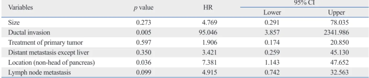
AJCC/UICC stage III/IV (p=0.020), or ENETS stage III/IV stage (p=0.009) were more likely to demonstrate shorter PFS (Table 3). Multivariate analysis for prognostic factors demon- strated that bile duct or pancreatic duct invasion [p=0.010, hazard ratio (HR)=95.046], and tumor location (non-head portion of pancreas) (p=0.036, HR=7.381) were significant factors for predicting shorter PFS (Table 4) (Fig. 3).
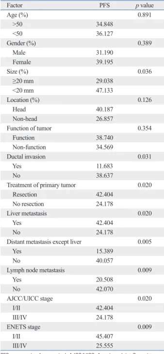
Table 3. Univariate Analysis of Clinical Factors Associated with Progression Free Survival
Factor PFS p value
Age (%) 0.891
>50 34.848
<50 36.127
Gender (%) 0.389
Male 31.190
Female 39.195
Size (%) 0.036
≥20 mm 29.038
<20 mm 47.133
Location (%) 0.126
Head 40.187
Non-head 26.857
Function of tumor 0.354
Function 38.740
Non-function 34.569
Ductal invasion 0.031
Yes 11.683
No 38.637
Treatment of primary tumor 0.020
Resection 42.404
No resection 24.178
Liver metastasis 0.020
Yes 42.404
No 24.178
Distant metastasis except liver 0.005
Yes 15.389
No 40.057
Lymph node metastasis 0.009
Yes 20.508
No 42.070
AJCC/UICC stage 0.020
I/II 42.404
III/IV 24.178
ENETS stage 0.009
I/II 45.407
III/IV 25.555
PFS, progression free survival; AJCC/UICC, American Joint Committee on Cancer/Union for International Cancer Control; ENETS, European Neuro- endocrine Tumors Society.
intermediate grade), and 3 (neuroendocrine carcinoma) in an attempt to predict natural history from pathology re- ports.4 In 2006, Rindi, et al.15 introduced a four stage TNM classification for PNETs, which has subsequently been ad- opted by the ENETS. In 2010, the new AJCC/UICC TNM staging for PNETs was proposed distinguishing between localized tumors (stage I), locally advanced resectable tu- mors (stage II), locally advanced unresectable tumors (stage III), and distantly metastasized tumors (stage IV).16 In our study, we were able classify PNETs by AJCC/UICC TNM staging, but we could not classify PNETs based on histo- logic factors (WHO classification) due to insufficient data.
From now on, PNETs need to be diagnosed based on both AJCC/UICC TMN staging and histologic findings (tumor differentiation, Ki-67 index, mitotic rate) through detailed data collection.
In contrast to our study, some articles reported that a small and pathologically benign nature did not predict a good prognosis in PNETs, so curative resection should be considered initially, even in cases of incidental PNETs.8 Even in patients with PNET metastatic lesions, resection of primary tumors should be considered for reasonable opera- tive candidates.17 In particular, resection of primary PNETs logic examination with immunocytochemistry. Somatosta-
tin receptor scintigraphy is a very sensitive procedure for diagnosing gastrinomas but not insulinomas. Computed to- mography, ultrasonography and magnetic resonance imag- ing are primarily useful for visualizing metastasis, which are able to predict prognoses.13 Cholangiography can be useful for evaluating bile duct invasion. Cholangiography was performed and was helpful in recognizing bile or pan- creatic duct invasion, which was confirmed as a significant prognostic factor in our study. Although EUS-guided FNA was performed, it was not helpful in predicting prognoses, because most of patients’ data did not include Ki-67 index and mitotic rate. Further evaluation, such as mitoses mea- surement and Ki-67 labeling, will be needed for predicting more reliable prognoses.
Previous classification systems make a distinction be- tween low and high-grade malignant NETs, but could not differentiate further prognosis. In contrast, according to tu- mor stage and Ki-67 index, new TNM classification can dif- ferentiate prognosis significantly.14 Also, according to size, mitoses, invasiveness, and Ki-67 labeling, recent WHO classification classifies PNETs into grades 1 (neuroendo- crine neoplasm, low grade), 2 (neuroendocrine neoplasm,
Fig. 3. (A) Disease-specific recurrence or progression comparing patients with duct invasion and those without duct invasion [p=0.010, HR=95.046 (3.857- 2341.986), multivariate analysis]. (B) Disease-specific recurrence or progression comparing patients with tumors in the head of pancreas and those not within the head of the pancreas [p=0.036, HR=7.381 (1.143-47.652), multivariate analysis].
0.0 0.0
0.2 0.2
0.4 0.4
0.6 0.6
0.8 0.8
1.0 1.0
Cumulative survival Cumulative survival
0 10 20 30 40 50 60 0 10 20 30 40 50 60
Progression-free survival (months) Progression-free survival (months) Without duct invasion
With duct invasion
Without duct invasion-censored With duct invasion-censored
Head of pancreas Non-head of pancreas
Head of pancreas-censored Non-head of pancreas-censored
A B
Table 4. Multivariate Analysis of Risk Factors for Progression Free Survival
Variables p value HR 95% CI
Lower Upper
Size 0.273 4.769 0.291 78.035
Ductal invasion 0.005 95.046 3.857 2341.986
Treatment of primary tumor 0.597 1.906 0.174 20.850
Distant metastasis except liver 0.350 3.421 0.259 45.130
Location (non-head of pancreas) 0.036 7.381 1.143 47.652
Lymph node metastasis 0.099 4.915 0.742 32.563
HR, hazard ratio; CI, confidence interval.
In conclusion, patients with bile duct or pancreatic duct invasion or tumors located at non-head portions of the pan- creas or without resection of primary tumor should be mon- itored carefully. But, because PNETs is a rare subgroup of tumor, we need more time and enough histopathologic data such as mitotic count and Ki-67 index for reliable prognos- tic prediction.
REFERENCES
1. Oberg K, Eriksson B. Endocrine tumours of the pancreas. Best Pract Res Clin Gastroenterol 2005;19:753-81.
2. Eriksson B, Oberg K. Neuroendocrine tumours of the pancreas.
Br J Surg 2000;87:129-31.
3. Ong SL, Garcea G, Pollard CA, Furness PN, Steward WP, Rajesh A, et al. A fuller understanding of pancreatic neuroendocrine tu- mours combined with aggressive management improves outcome.
Pancreatology 2009;9:583-600.
4. Ehehalt F, Saeger HD, Schmidt CM, Grützmann R. Neuroendo- crine tumors of the pancreas. Oncologist 2009;14:456-67.
5. Norton JA, Warren RS, Kelly MG, Zuraek MB, Jensen RT. Ag- gressive surgery for metastatic liver neuroendocrine tumors. Sur- gery 2003;134:1057-63.
6. Chen H, Hardacre JM, Uzar A, Cameron JL, Choti MA. Isolated liver metastases from neuroendocrine tumors: does resection pro- long survival? J Am Coll Surg 1998;187:88-92.
7. Han JH, Kim MH, Moon SH, Park SJ, Park do H, Lee SS, et al.
[Clinical characteristics and malignant predictive factors of pan- creatic neuroendocrine tumors]. Korean J Gastroenterol 2009;53:
98-105.
8. Paik WH, Yoon YB, Lee SH, Park JK, Woo SM, Yang KY, et al.
[Pancreatic endocrine tumors: clinical manifestations and predic- tive factors associated with survival]. Korean J Gastroenterol 2008;52:171-8.
9. Kang TW, Lee KT, Ryu MK, Moon W, Lee SS, Lee SY, et al.
[Clinical features of neuroendocrine tumor of the pancreas: single center study]. Korean J Gastroenterol 2006;48:112-8.
10. Panzuto F, Nasoni S, Falconi M, Corleto VD, Capurso G, Cassetta S, et al. Prognostic factors and survival in endocrine tumor pa- tients: comparison between gastrointestinal and pancreatic local- ization. Endocr Relat Cancer 2005;12:1083-92.
11. Metz DC, Jensen RT. Gastrointestinal neuroendocrine tumors:
pancreatic endocrine tumors. Gastroenterology 2008;135:1469- 92.
12. Nobels FR, Kwekkeboom DJ, Coopmans W, Schoenmakers CH, Lindemans J, De Herder WW, et al. Chromogranin A as serum marker for neuroendocrine neoplasia: comparison with neuron- specific enolase and the alpha-subunit of glycoprotein hormones. J Clin Endocrinol Metab 1997;82:2622-8.
13. Zimmer T, Stölzel U, Bäder M, Koppenhagen K, Hamm B, Buhr H, et al. Endoscopic ultrasonography and somatostatin receptor scintigraphy in the preoperative localisation of insulinomas and gastrinomas. Gut 1996;39:562-8.
14. Pape UF, Jann H, Müller-Nordhorn J, Bockelbrink A, Berndt U, Willich SN, et al. Prognostic relevance of a novel TNM classifica- tion system for upper gastroenteropancreatic neuroendocrine tu-
should be given to patients with treatable hepatic metasta- sis. Aggressive surgical resection for select individuals with PNETs can be performed safely and may improve both symptomatic disease and overall survival. Prognostic indi- ces such as tumor differentiation and the ability to achieve R0/R1 resection have been prognostic factors in PNETs and should be considered when planning aggressive surgi- cal management.18 Surgical strategy for PNET depends on the size and location of the tumor as well as the risk of ma- lignancy. Selection of the proper surgical method is impor- tant to preventing postoperative complications and recur- rence. Radical resection including primary and metastatic lesion may improve survival of malignant PNETs.19 Only patients without distant metastasis underwent surgical re- section in our study. Through analyzing prognostic factors, aggressive surgical resection can be considered even in cas- es of distant metastasis.
Transarterial chemoembolization, peptide receptor radio- nuclide therapy (PRRT), systemic chemotherapy, and ra- diotherapy can also be treatment options. PRRT uses soma- tostatin analogs to convey radioactivity within the tumor itself through somatostatin receptors. New biological agents and somatostatin tagged radionuclides are also under investigation20 as antiproliferative agents capable of stabi- lizing tumor growth in patients with metastatic PNETs.21 Advances in therapy and cooperative multicenter studies are needed to improve diagnosis, treatment and survival of patients with PNETs.22 Various treatment options besides surgery were tried in our study. Concurrent chemoradiother- apy was performed preoperatively in patients with lymph node metastasis or vascular invasion. Palliative chemother- apy (Etoposide, cisplatin) was performed in patients with distant metastasis. Transarterial chemoembolization was performed in patients with liver metastasis where surgical resection was not feasible. Somatostatin analogs were tried in patients with carcinoid syndromes such as flushing, diar- rhea, and abdominal pain.
Prognostic factors were inconclusively reported in previ- ous reports. We analyzed prognostic factors for predicting survival and disease progression in order to predict preoper- ative prognostic factors and postoperative results. From our report, resection of primary tumors can improve survival, and pancreatic or bile duct invasion, tumor location (body or tail of pancreas) were poor prognostic factors for disease progression. However, our study has some limitations, in- cluding a very wide confidence interval, due to small num- ber of patients, and insufficient data for WHO classification.
19. Liu H, Zhang SZ, Wu YL, Fang HQ, Li JT, Sheng HW, et al. Di- agnosis and surgical treatment of pancreatic endocrine tumors in 36 patients: a single-center report. Chin Med J (Engl) 2007;120:
1487-90.
20. Kaemmerer D, Prasad V, Daffner W, Hörsch D, Klöppel G, Hom- mann M, et al. Neoadjuvant peptide receptor radionuclide therapy for an inoperable neuroendocrine pancreatic tumor. World J Gas- troenterol 2009;15:5867-70.
21. Strosberg J, Kvols L. Antiproliferative effect of somatostatin ana- logs in gastroenteropancreatic neuroendocrine tumors. World J Gastroenterol 2010;16:2963-70.
22. Massironi S, Sciola V, Peracchi M, Ciafardini C, Spampatti MP, Conte D. Neuroendocrine tumors of the gastro-entero-pancreatic system. World J Gastroenterol 2008;14:5377-84.
mors. Cancer 2008;113:256-65.
15. Rindi G, Klöppel G, Alhman H, Caplin M, Couvelard A, de Herd- er WW, et al. TNM staging of foregut (neuro) endocrine tumors: a consensus proposal including a grading system. Virchows Arch 2006;449:395-401.
16. Edge SB, Compton CC. The American Joint Committee on Can- cer: the 7th edition of the AJCC cancer staging manual and the fu- ture of TNM. Ann Surg Oncol 2010;17:1471-4.
17. Hill JS, McPhee JT, McDade TP, Zhou Z, Sullivan ME, Whalen GF, et al. Pancreatic neuroendocrine tumors: the impact of surgi- cal resection on survival. Cancer 2009;115:741-51.
18. Hodul PJ, Strosberg JR, Kvols LK. Aggressive surgical resection in the management of pancreatic neuroendocrine tumors: when is it indicated? Cancer Control 2008;15:314-21.