Vol. 13, No. 3, September, 2006
저산소증-섬유근통 증후군의 병인 중 하나인가?
동국대학교 의과대학 경주병원 류마티스내과, 해부학교실*
김 성 호․문 일 수*
= Abstract =
Hypoxia-A Possibility in Fibromyalgia Syndrome Pathogenesis
Seong-Ho Kim, M.D., Il Soo Moon, Ph.D.*Departments of Internal Medicine and Anatomy*, College of Medicine, Dongguk University, Gyeongju Hospital, Gyeongju, Korea
Objective: We studied the expression of pain-related molecules such as substance P involved in chronic pain of fibromyalgia syndrome (FMS) patients using rat cortical cells in hypoxia.
Methods: We sacrificed pregnant Sprague-Dawley rat and got embryo. We cultured the cortical cells and compared the expression of pain-related molecules in 1st, 3rd, 5th day cortical cells exposed to hypoxia (37°C, 5% CO2, 98% N2) to control by immunohistochemistry. We measured the density at soma using softwear ‘Scion image'.
Results: The expression of substance P was increased in hypoxic cortical cell group than control (control mean: 49.9 vs. hypoxia 1st day: 75.4 (p<0.001), 3rd day: 65.6 (p<0.001), 5th day: 79.9 (p<0.001)). The expression of kainate receptor was increased in hypoxic cortical cell group than control (control mean: 58.4 vs. hypoxia 1st day : 64.9 (p<0.001), 3rd day: 63.3 (p<
0.001), 5th day: 62.9 (p<0.001)). The expression of N-methyl-D-aspartate receptor 2B was increased in hypoxic cortical cell group than control (control mean : 59.4 vs. hypoxia 1st day:
60.8 (p<0.001), 3rd day: 62.6 (p<0.001), 5th day: 67.1 (p<0.001)). But, the expression of calcitonin gene related peptide was decreased in hypoxic cortical cell group than control (control mean: 76.8 vs. hypoxia 1st day: 76.4 (p<0.001), 3rd day: 71.5 (p<0.001), 5th day: 61.3 (p<
0.001)).
Conclusion: Hypoxia during night could increase the expression of some pain-related molecules, which might be the cause of chronic pain in FMS patients.
ꠏꠏꠏꠏꠏꠏꠏꠏꠏꠏꠏꠏꠏꠏꠏꠏꠏꠏꠏꠏꠏꠏꠏꠏꠏꠏꠏꠏꠏꠏꠏꠏꠏꠏꠏꠏꠏꠏꠏꠏꠏꠏꠏꠏꠏꠏꠏꠏꠏꠏꠏꠏꠏꠏꠏꠏꠏꠏꠏꠏꠏꠏꠏꠏꠏꠏꠏꠏꠏꠏꠏꠏꠏꠏꠏꠏꠏꠏꠏꠏꠏꠏꠏꠏꠏꠏ Key Words: Fibromyalgia, Hypoxia, Substance P, Glutamate, Calcitonin gene related
peptide
ꠏꠏꠏꠏꠏꠏꠏꠏꠏꠏꠏꠏꠏꠏꠏꠏꠏꠏꠏꠏꠏꠏꠏꠏꠏꠏꠏꠏꠏꠏꠏꠏꠏꠏꠏꠏꠏꠏꠏꠏꠏꠏꠏꠏꠏꠏꠏꠏ
<접수일:2006년 3월 29일, 심사통과일:2006년 8월 2일>
※통신저자:김 성 호
경북 경주시 석장동 1090-1 동국대학교 경주병원 류마티스내과
Tel:054) 770-8563, Fax:054) 770-8378, E-mail:[email protected]
서 론
섬유근통 증후군은 원인 불명의 만성 통증 증후군 으로 광범위한 비관절성 근골격계 통증 및 전신적인 압통점들이 특징적이다 (1,2). 섬유근통 증후군의 유 병률은 외국의 경우 0.5∼5%로 보고되며 (3), 국내 유병률은 저자가 보고한 바에 의하면 2.2%이다 (4).
두통, 피곤감, 수면 장애, 과민성 대장 증후군, 감각 이상 등이 나타날 수 있고, 날씨 또는 스트레스 수 준의 변화에 따라 증상의 굴곡이 나타날 수 있다.
섬유근통 증후군의 병인은 알려져 있지 않지만 만성 통증의 신경생물학적 연구에 의하면 잘못된 통증 조 정, 즉 비정상적인 민감화와 ‘wind-up' 현상이 중요 한 역할을 한다고 알려졌다 (5,6). 다른 요인들로는 일부 섬유근통 증후군 환자들의 뇌척수액에서 통각 의 매개물인 substance P (SP)의 증가 (7), 변형된 sero- tonin 대사 (8) 및 수면 시 낮은 melatonin 분비 등 (9)이 보고되고 있다. 일반적으로 섬유근통 환자들 통증의 발단은 주로 근육이라는 의견이 지배적이다 (10-12). 또한 침범된 근육에서 산소 조직압이 감소 되어 있는 것이 보고되면서 (13) 섬유근통 환자들의 근육에서 발견되는 변화들이 근육의 저산소증때문일 것이라는 가능성들이 대두되었다 (14-16).
섬유근통 증후군 환자들에서 이런 근육의 저산소 증이 수면 중 빈번히 발생하는 경증의 저산소혈증 때문임을 시사하는 연구가 있다. 섬유근통 증후군 환자들에서 수면중 동맥혈의 산소 포화도가 떨어지 며 저산소 상태에 머무는 시간이 대조군보다 더 길 었다. 즉 밤동안 동맥혈의 산소 포화도가 90% 및 92% 미만인 시간이 대조군보다 더 많았다. 이런 변 화들이 섬유근통과 관련된 수면 무호흡 증후군으로 인할 가능성이 배제되어서 저자들은 수면중 경도의 저산소혈증이 섬유근통 증후군 근골격계 병태생리에 중요할 것이라고 주장한다 (17).
전신적 저산소혈증 시에는 적절한 대뇌 산소 운반 을 위해 대뇌 혈류의 조절이 있는 것으로 알려져 있 지만, 최근의 한 연구에 따르면 non-rapid eye move- ment (NREM) 수면중엔 저산소혈증 상태라도 대뇌 혈류가 감소한다고 밝혀졌다 (18). 따라서 수면중 저 산소혈증은 근육뿐만 아니라 중추신경계에도 저산소
상태를 유발할 것이다. 이를 근거로 저자들은 쥐의 대뇌 피질 세포를 이용하여 저산소 상태에서 섬유근 통 증후군 환자들의 가장 주된 증상인 만성 통증에 관여하는 SP와 glutamate 수용체 같은 통증 관련 물 질들의 발현을 보기 위해 연구를 하였다.
대상 및 방법 1. 실험대상
본 연구에 사용한 동물은 Sprague-Dawley계 흰쥐 로서 임신 19일째 배아의 대뇌신경세포를 사용하였다.
2. 방법
1) 대뇌피질세포 배양: 임신 19일째 Sprague-Dawley 어미쥐를 에테르로 마취시킨 후 배아가 든 자궁을 신속히 꺼낸 다음 clean-bench 안에서 대뇌를 적출하 여 Ca2+과 Mg2+이 없는 Hank balanced salt solution (HBSS)에 넣었다. 대뇌피질을 분리한 후 뇌막과 혈 관을 제거하고 잘게 조각낸 후 HBSS를 제거하고 0.25% trypsin으로 37oC waterbath에서 1분간 처리하 였다. 조직이 따라 나오지 않게 조심하면서 trypsin 용액을 제거한 뒤 HBSS로 조직을 5회 씻었다. 최종 적으로 해마 10개(즉, 뇌 5개) 당 약 1 mL의 HBSS 를 넣고, 불에 달궈 끝을 가늘게 한 Pasteur pipette으 로 통과시켜 세포를 분산시켰다. 분산된 세포가 있 는 상등액을 얻고 원심분리하여 상등액을 제거한 후 동일양의 Ca2+과 Mg2+이 포함된 HBSS에 현탁시켰 다. 배양은 poly-L-lysine으로 coating한 culture plate에 B27을 첨가한 plating neurobasal 배지(100 mL neuro- basal, 2 mL B27 supplement, 0.25 mL glutamax I, 0.1 mL 25 mM glutamate, 0.1 mL 25 mM 2-mercaptoe- thanol; Invitrogen Life Technology)에 2000 cells/mm2 의 밀도로 37oC humidified CO2 배양조에서 배양하 고 2∼3일 간격으로 배양액을 feeding neurobasal media (100 mL neurobasal, 2 mL B27 supplement, 0.25 mL glutamax I)로 1/3씩 교환하였다.
2) 저산소증 유도: Culture plate를 2% O2/5% CO2로 맞춘 Water Jacketed Incubator (Forma Scientific Inc.) 에 넣고 3시간 처리하였다. 저산소처리가 끝나면 cul- ture plate를 다시 normoxic, 37oC humidified CO2 배 양조에 옮기고 계속 배양하였다.
3) 면역세포화학 염색: 세포를 4oC D-PBS (0.1 g/L CaCl2, 0.2 g/L KCl, 0.2 g/L KH2PO4, 0.1 g/L/ MgCl2
-6H2O, 8.0 g/L NaCl, 2.16 g/L Na2HPO4 -7H2O)로 잠깐 씻은 후 -20oC methanol 500μl를 넣고 -20oC 냉동고에서 20분간 고정하였다. D-PBS로 잠깐 씻은 다음 4oC preblock solution [0.05% triton, 5% normal goat serum in h-PBS (20 mM NaPO4, pH 7.4, 450 mM NaCl)] 1 mL 넣고 밤새 4oC 냉장고에 넣어 pre- block한 후 일차 항체들[rabbit anti-substance P (1:
500; ImmunoStar, Inc., Hudson, WI, USA), rabbit anti- glutamate receptor subunits 2 and 3 monoclonal anti- body (1:100; Chemicon International, Inc., Temecula, CA, USA), mouse anti-glutamate receptor subunits 5, 6 and 7 monoclonal antibody (1:500; Chemicon), affinity- pure goat anti-N-methyl-D-aspartate (NMDA) ε4 poly- clonal antibody (1:100; Santa Cruz Biotechnology Inc., Santa Cruz, CA, USA), rabbit anti-NMDA receptor 2B polyclonal antibody (1:500) (19), rabbit anti-α-calci- tonin gene related peptide (α-CGRP) (1:500; Immu- noStar)]을 넣고 1∼2일 동안 4oC에서 반응시켰다. 반 응이 끝나면 preblock solution으로 15분간씩 실온에 서 2회 세척하고, 이차항체 Alexa Fluor 568-conju- gated goat anti-mouse IgG 혹은 Alexa Fluor 488-con- jugated goat anti-rabbit IgG (1:1,000; Molecular Pro- bes, Leiden, The Netherlands)를 넣고 4oC에서 밤새
반응시켰다. 반응이 끝나면 preblock solution으로 15 분간 1회, PBS로 15분간 2회 세척한 후 mounting하 였다.
4) 이미지 획득 및 표현 분석: 형광이미지는 I3 및 N2.1 filter system이 장착된 형광현미경(Leica DM IRE2 Imaging System, Wetzlar, Germany)으로 관찰하 고, Cooled CCD camera (Photometrics Inc., Germany) 를 이용하여 디지털 이미지를 획득하였다. 표현강도 를 측정하기 위하여 Adobe Photoshop 5.0 (Adobe Sys- tems Inc.) software를 이용하여 이미지를 흑백으로 전환하고, 반전시킨 후 Scion Image Beta 4.03 soft- ware로 intensity를 측정하였다. 통계분석은 2회 이상 반복실험하여 얻은 100∼200개 세포의 형광이미지를 one-sample t-test로 분석하였다.
결 과
다양한 통각수용체들 중 SP는 섬유근통 증후군 환자의 뇌척수액에서 대조군보다 증가되어 있다 (20).
따라서 SP는 섬유근통 증후군 환자들에서 특히 의 미 있는 통증 전달 물질이다. 저산소 환경에 노출된 사람의 중추신경계 SP 자료는 지금까지는 없어 이 의 발현을 보고자 하였다. 저산소에 노출된 대뇌피 질세포군에서 대조군보다 SP의 발현이 증가하였다 (대조군 평균: 49.9 vs. 저산소 1일째: 75.4 (p<0.001),
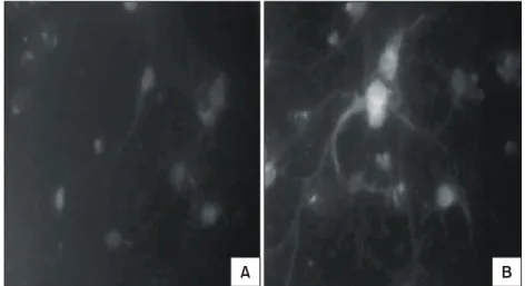
Fig. 1. More increased substance P expression in rat cortical cells after hypoxia (B) than before hypoxia (A) (×400).
A B
3일째: 65.6 (p<0.001), 5일째: 79.9 (p<0.001)) (그림 1).
Glutamate는 통각전달에 가장 의미 있는 물질 중 하나며, NMDA와 kainate는 그에 속하는 대표적인 물질이다. 최근 저자는 정상 대조군보다 섬유근통 증후군 환자들의 피부에서 NMDA 수용체의 발현이 증가되어 있음을 보고했다 (21). 따라서 이의 발현을 보고자 했다. 저산소에 노출된 대뇌피질세포군에서 대조군보다 kainate receptor의 발현이 증가하였다(대 조군 평균: 58.4 vs. 저산소 1일째: 64.9 (p<0.001), 3
일째: 63.3 (p<0.001), 5일째: 62.9 (p<0.001) (그림 2). 저산소에 노출된 대뇌피질세포군에서 대조군보 다 NMDA receptor 2B의 발현이 증가하였다(대조군 평균: 59.4 vs. 저산소 1일째: 60.8 (p<0.001), 3일째:
62.6 (p<0.001), 5일째: 67.1 (p<0.001) (그림 3).
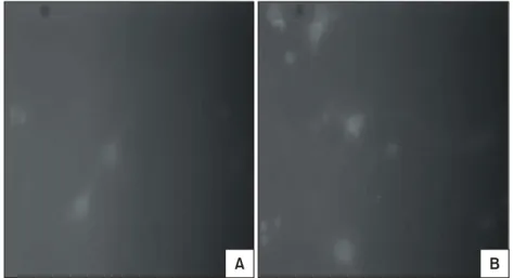
섬유근통 증후군의 뇌척수액에서 CGRP가 존재하 고 타키키닌(tachykinins)의 통각 활동도를 항진시킬 수 있기 때문에 CGRP가 섬유근통의 통증 발병기전 에 중요할 것이라는 연구가 있으며 (22), 저산소 손 Fig. 3. More increased N-methyl-D-aspartate receptor 2B expression in rat cortical cells after
hypoxia (B) than before hypoxia (A) (×400).
A B
Fig. 2. More increased kainate expression in rat cortical cells after hypoxia (B) than before hypoxia (A) (×400).
A B
상만으로 손상 전후의 CGRP 발현 차이를 본 연구 는 문헌상 찾을 수 없어 이를 보고자 하였다. 저산 소에 노출된 대뇌피질세포군에서 대조군보다 CGRP 의 발현이 감소하였다(대조군 평균: 76.8 vs. 저산소 1일째: 76.4 (p<0.001), 3일째: 71.5 (p<0.001), 5일 째: 61.3 (p<0.001) (그림 4).
고 찰
통각은 neuropeptide인 SP, CGRP 및 isolectin B4 등 을 발현하는 다양한 통각수용체들의 활성과 관련이 있다 (23). 이들의 감각뉴런은 주로 척수의 dorsal horn에서 끝나는데, 판(lamina) I, II가 주를 이루고 판 V도 일부 포함된다. 이 척수 부위엔 역시 통각 전달에 의미 있는 수용체를 발현하는 연접후 뉴런들 이 있다. 이런 수용체들엔 SP, neurokinin 1, neuro- kinin 2 및 glutamate 수용체들[NMDA, alpha-amino-3- hydroxy-5-methyl-4-isoxazolepropionic acid (AMPA), ka- inate, metabotropic]이 있다. 신체의 다른 조직들에 분 포하는 뉴런의 수용체 발현에 관하여 알려진 바가 거의 없다. 중추신경계에 SP, CGRP 및 glutamate 수 용체들이 존재하는 것은 이미 여러 문헌에서 알려져 있다 (24-26). 따라서 저자는 쥐의 대뇌세포를 배양 해서 이들 통각수용체들의 발현을 비교해 보았다.
쥐의 대뇌 피질이 저산소/저혈압 환경에 노출될
때 glutamate와 aspartate 같은 흥분성 아미노산의 방 출은 증가한다 (27,28). 특히 뉴런에서 NMDA 수용체 아형들(NMDA 2A, 2B, 2C, 2D)의 발현양은 상대적으 로 비슷하며 장시간 저산소에 노출되면(이들 연구에 선 10분) glutamate 수용체와 관련된 칼슘 유입이 제 한된다고 보고한다 (28). 저자들의 연구결과는 NMDA 2B와 kainate의 발현만이 증가되었다. 이런 차이는 첫째로 수용체 활동도를 보는 방법의 차이에 기인하 는 것이라고 추정해 본다. 세포내 칼슘 농도 상승으 로 수용체 활동도를 측정한 Bickler 등의 방법은 수 용체의 기능적 측면도 고려한 최신의 실시간 정량법 인데 반해 (28) 저자들의 면역세포화학 염색법은 비 특이적 결합 등을 간과할 수 없어 두 군 간의 정량 적 차이가 명확하지 않을 수 있다고 본다. 둘째로 저산소에 노출된 기간의 차이를 언급할 수 있겠다.
Bicker 등은 저산소 노출 시간을 100초 및 10분 두 군을 비교하였는데 (28), 저자들은 3시간 동안 저산 소에 노출시키고 1일, 3일, 5일째 관찰했기 때문에 수용체 발현 및 활동도에 영향을 줄 수 있는 또 다 른 요소일 수 있다고 생각된다.
SP는 섬유근통 증후군 환자의 뇌척수액에서 대조 군보다 증가되어 있다 (20). 따라서 SP는 섬유근통 증후군 환자들에서 특히 의미 있는 통증 전달 물질 이다. 저산소 환경에 노출된 사람의 중추신경계 SP 자료는 지금까지는 없으며, 쥐에서 만성으로 저산소 Fig. 4. More decreased calcitonin gene related peptide expression in rat cortical cells after
hypoxia (B) than before hypoxia (A) (×400).
A B
환경에 노출 시(10% O2 in nitrogen for 14 days) 측 정된 두 부위(ventrolateral medulla oblongata, striatum) 에서 노출 전후의 SP 발현 차이는 없었으며 뇌하수 체 전엽에서는 감소되었다는 보고가 있다 (29). 성인 고양이에서 인공환기법으로 저산소를 유발하면 nucleus tractus solitarii에서 SP-like immunoreactivity의 세포외 액 농도가 증가되어 있음을 보이고 SP가 저산소에 대한 중추반응의 매개체라는 주장도 있다 (30). 성인 토끼에서도 비슷한 방법으로 nucleus tractus solitarii 에서 SP-like immunoreactivity의 세포외액 농도가 증 가되어 있다는 보고가 있다 (31). 하지만 7∼8일된 신생쥐에서 저산소 허혈 뇌손상을 유발한 경우엔 줄 무늬체(striatum)에서 SP-like immunoreactivity의 농도가 반대측에 비해 반정도 감소되어 있다는 보고도 있어 신생아 저산소 허혈 후 뇌 신경펩티드 변화는 좀 더 복잡할 것임을 시사한다 (32). 저자들의 실험은 신경 세포에서의 발현을 측정한 것으로 세포외액에서 SP 농도를 측정한 것과는 차이가 있다. 이 펩티드가 개 별의 신경세포에서 기인함을 직접적으로 보여 주며 저자들의 문헌 고찰상 저산소 노출 후 신경세포 내 에서 SP의 발현이 증가됨을 보여 주는 첫 보고다.
뇌의 부위에 따른 발현 차이가 있을 수 있으나 (29) 뇌척수 세포외액이 아닌 뇌실질의 세포에서 저산소 노출 후 SP의 발현이 증가한 것은 기존의 보고와는 다른 새로운 점이다.
CGRP는 구심 신경 경로에서 SP와 같이 위치하는 신경펩티드로 주로 SP를 줄이는 펩티드분해효소를 경쟁적으로 억제한다 (33). 섬유근통 증후군의 뇌척 수액에서 CGRP는 정상 대조군보다 높았지만 통계 적으로 의미 있는 차이를 보이지 않는다 (34). 역시 섬유근통 증후군의 뇌척수액에서 CGRP의 존재를 증명하고 SP와 상관을 본 연구에서는 CGRP 수준은 SP 수준과 상관이 없으나 CGRP가 타키키닌(tachy- kinins)의 통각 활동도를 항진시킬 수 있기 때문에 CGRP가 섬유근통의 통증 발병기전에 중요할 것이 라 주장한다 (22). 저산소 손상만으로 손상 전후의 CGRP 발현 차이를 본 연구는 문헌상 찾을 수 없었 으며, 역시 영아 쥐에서 저산소 허혈 뇌손상 후 뇌 신경세포에서 CGRP의 면역활동도가 증가된 것을 보이고 영아 뇌에서 저산소 허혈 후 신경 복구에 CGRP가 관여할 가능성을 제시한 연구는 한편 있었
다 (35). 이들의 연구와 여러 조건의 차이가 있어서 인지 저자들의 실험은 저산소 노출 후 신경세포에서 CGRP의 발현이 감소되어 반대의 결과가 나왔다. 성 인 쥐를 사용한 점이며 허혈 손상은 없었던 점 등 여러 조건의 차이가 다양한 해석을 가능하게 하지만 저자들은 성급히 해석하기 이전에 새로운 발견이라 는 점을 우선은 강조하고 싶다.
섬유근통 환자들은 수면 중 저산소에 의하여 말초 나 중추에서 민감화(sensitization)가 일어날 수 있겠 다는 가정하에 저산소 상태의 신경세포에서 통증 관 련 수용체들의 변화를 본 것은 무척 의미 있는 일로 생각된다. 현재까지의 문헌 고찰로는 섬유근통 증후 군에서 중추신경계의 저산소 상태에 대한 직접적인 증거는 없는 실정이라 저산소 상태에서 쥐의 대뇌 피질 세포의 통증 관련 물질의 변화를 섬유근통 증 후군의 병인과 관련 짖기에는 다소 무리가 있다. 하 지만 섬유근통 증후군 환자들에서 대뇌의 국소적 혈 류량이 기본적으로 감소되어 있다는 사실과 (36,37) 수면 중 저산소 상태에 놓인다는 점은 (17) 간접적 인 증거라 생각되어 섬유근통 증후군의 병인 중 하 나의 가능성일 수도 있다고 생각한다. 현재 섬유근 통 증후군의 발병기전에 대한 주된 관심사가 비정상 적인 ‘중추성 민감화’ (central sensitization)라는 현상 에만 집중되어 있는데 (38), 이런 민감화란 현상을 자극하는 더 근본적인 원인들에 대한 관심도 가졌으 면 한다.
결 론
저산소 환경에 노출된 중추신경계 세포에서 일부 중요한 통증 관련 물질들의 발현이 증가됨을 알 수 있었다. 섬유근통 증후군 환자들에서 증명된 수면 중 저산소 환경은 통증 관련 물질들을 증가시킬 수 있겠으며, 이들 환자군에 특징적인 만성 통증의 한 원인이 될 것으로 생각된다.
REFERENCES
1) Wolfe F, Smythe HA, Yunus MB, Bennett RM, Bombardier C, Goldenberg DL, et al. The American College of Rheumatology criteria for the classifica- tion of fibromyalgia: report of the multicenter criteria
committee. Arthritis Rheum 1990;33:160-72.
2) Goldenberg DL. Fibromyalgia syndrome. JAMA 1987;257:2782-7.
3) Gran JT. The epidemiology of chronic generalized musculoskeletal pain. Best Pract Res Clin Rheumatol 2003;17:547-61.
4) 김성호, 배근량, 임현술. 한국의 두 지역사회에서 섬유 근통 증후군과 만성 광범위 통증의 유병률과 위험요인.
대한류마티스학회지 2006; 13: 18-25.
5) Okifuji A, Turk DC, Marcus DA. Comparison of generalized and localized hyperalgesia in patients with recurrent headache and fibromyalgia. Psychosom Med 1999;61:771-80.
6) Staud R, Vierck CJ, Cannon RL, Mauderli AP, Price DD. Abnormal sensitization and temporal summation of second pain (wind-up) in patients with fibromyal- gia syndrome. Pain 2001;91:165-75.
7) Schwarz MJ, Spath M, Muller-Bardorff H, Pongratz DE, Bondy B, Ackenheil M. Relationship of subs- tance P, 5-hydroxyindole acetic acid and tryptophan in serum of fibromyalgia patients. Neurosci Lett 1999;
259:196-8.
8) Offenbaecher M, Bondy B, de-Jonge S, Glatzeder K, Krger M, Schoeps P, et al. Possible association of fibromyalgia with a polymorphism in the serotonin transporter gene regulatory region. Arthritis Rheum 1999;42:2482-8.
9) Wikner J, Hirsch U, Wetterberg L, Rojdmark S. Fib- romyalgia: a syndrome associated with decreased noc- turnal melatonin secretion. Clin Endocrinol Oxf 1998;
49:179-83.
10) Rice JR. “Fibrositis” syndrome. Med Clin N Am 1986;70:455-68.
11) Bengtsson A, Henriksson KG, Jorfeldt L, Kagedal B, Lennmarken C, Lindstrom F. Primary fibromyalgia: a clinical and laboratory study of 55 patients. Scand J Rheumatol 1986;15:340-7.
12) Gee1 SE. The fibromyalgia syndrome: musculoske- letal pathophysiology. Sem Arthritis Rheum 1994;23:
347-53.
13) Lund N, Bengtsson A, Throborg P. Muscle tissue oxy- gen pressure in primary fibromyalgia. Scand J Rheu- matol 1986;15:165-73.
14) Yunus MB, Kalyan-Raman UP, Kalyan-Raman K, Masi AT. Pathologic changes in muscle in primary fibromyalgia syndrome. Am J Med 1986;81(Supple 3A):38-42.
15) Bengtsson A, Henriksson KG. The muscle in fibro- myalgia: a review of Swedish studies. J Rheumatol
1989;16(Supple 19):144-9.
16) Henriksson KG. Pathogenesis of fibromyalgia. J Mus- culoskel Pain 1993;1:3-16.
17) Lario BA, Valdivielso JLA, Lopez JA, Soteres CM, Banuelos JLV, Cabello AM. Fibromyalgia syndrome:
overnight falls in arterial oxygen saturation. Am J Med 1996;101:54-60.
18) Meadows GE, O'Driscoll DM, Simonds AK, Morrell MJ, Corfield DR. Cerebral blood flow response to isocapnic hypoxia during slow-wave sleep and wake- fulness. J Appl Physiol 2004;97:1343-8.
19) Moon IS, Apperson ML, Kennedy MB. The major tyrosine-phosphorylated protein in the postsynaptic density fraction is N-methyl-D-aspartate receptor subunit 2B. Proc Natl Acad Sci USA 1994;91:3954-8.
20) Russell IJ, Orr MD, Littman B, Vipraio GA, Al- boukrek D, Michalek JE, et al. Elevated cerebros- pinal fluid levels of substance P in patients with the fibromyalgia syndrome. Arthritis Rheum 1994;37:
1593-601.
21) Kim SH, Jang TJ, Moon IS. Increased expression of N-methyl-D-aspartate receptor subunit 2D in the skin of patients with fibromyalgia. J Rheumatol 2006;33:
785-8.
22) Vaeroy H, Sakurada T, Forre O, Kass E, Terenius L.
Modulation of pain in fibromyalgia (fibrositis syn- drome): cerebrospinal fluid (CSF) investigation of pain related neuropeptides with special reference to calcitonin gene related peptide (CGRP). J Rheumatol 1989;19(Supple):94-7.
23) Kitchener PD, Wilson P, Snow PJ. Selective labeling of primary sensory afferent terminals in lamina II of the dorsal horn by injection of Bandeiraea sim- plicifolia isolectin B4 into peripheral nerves. Neu- roscience 1993;54:545-51.
24) Nakanishi S. Molecular diversity of glutamate recep- tors and implications for brain function. Science 1992;
258:597-603.
25) Saria A. The tachykinin NK1 receptor in the brain:
pharmacology and putative functions. Eur J Phar- macol 1999;375:51-60.
26) Katafuchi T, Minamino N. Structure and biological properties of three calcitonin receptor-stimulating pe- ptides, novel members of the calcitonin gene-related peptide family. Peptides 2004;25:2039-45.
27) Phillis JW, Walter GA. Hypoxia/hypotension evoked release of glutamate and aspartate from the rat cere- bral cortex. Neurosci Lett 1989;106:147-51.
28) Bickler PE, Fahlman CS, Ferriero DM. Hypoxia
increases calcium flux through cortical neuron gluta- mate receptors via protein kinase C. Neurochem 2004;
88:878-84.
29) Poncet L, Denoroy L, Dalmaz Y, Pequignot JM, Jou- vet M. Alteration in central and peripheral substance P- and neuropeptide Y-like immunoreactivity after chronic hypoxia in the rat. Brain Res 1996;733:64- 72.
30) Lindefors N, Yamamoto Y, Pantaleo T, Lagercrantz H, Brodin E, Ungerstedt U. In vivo release of sub- stance P in the nucleus tractus solitarii increases du- ring hypoxia. Neurosci Lett 1986;69:94-7.
31) Srinvasan M, Goiny M, Pantaleo T, Lagercrantz H, Brodin E, Runold M, et al. Enhanced in vivo release of substance P in the nucleus tractus solitarii during hypoxia in the rabbit: role of peripheral input. Brain Res 1991;546:211-6.
32) Johnson M, Hanson GR, Gibb JW, Adair J, Filloux F. Effect of neonatal hypoxia-ischemia on nigro-stria- tal dopamine receptors and on striatal neuropeptide Y, dynorphin A and substance P concentrations in rats.
Brain Res Dev Brain Res 1994;83:109-18.
33) Tamatani M, Senba E, Tohyama M. Calcitonin gene-
related peptide- and substance P-containing primary afferent fibers in the dorsal column of the rat. Brain Res 1989;495:122-30.
34) Russell IJ. Advances in fibromyalgia: possible role for central neurochemicals. Am J Med Sci 1998;315:377- 84.
35) Dragunow M, Sirimanne E, Lawlor PA, Williams C, Gluckman P. Accumulation of calcitonin-gene related- peptide-like immunoreactivity after hypoxic- ischae- mic brain injury in the infant rat. Brain Res Mol Brain Res 1992;14:267-72.
36) Kwiatek R, Barnden L, Tedman R, Jarrett R, Chew J, Rowe C, et al. Regional cerebral blood flow in fibromyalgia. Arthritis Rheum 2000;43:2823-33.
37) Mountz JM, Bradley LA, Modell JG, Alexander RW, Triana-Alexander M, Aaron LA, et al. Fibromyalgia in women. Abnormalities of regional cerebral blood flow in the thalamus and the caudate nucleus are associated with low pain threshold levels. Arthritis Rheum 1995;38:926-38.
38) Price DD, Staud R. Neurobiology of fibromyalgia syn- drome. J Rheumatol 2005;32(Supple 75):22-8.