131 http://dx.doi.org/10.4196/kjpp.2012.16.2.131
ABBREVIATIONS: TNF, tumor necrosis factor; LPS, lipopolysac- charide; AMPK, AMP-activated protein kinase; PDE, phospho- diesterase; cAMP, cyclic AMP; AICAR, 5-Aminoimidazole-4-carbo- xamide 1-β-D-ribofuranoside; CTZ, cilostazol; PTX, pentoxifylline.
Received February 28, 2012, Revised March 15, 2012, Accepted April 3, 2012
Corresponding to: Jong Ryeol Eun, Department of Internal Medi- cine, School of Medicine, Yeungnam University, 317-1, Daemyung 5-dong, Daegu 705-717, Korea. (Tel) 82-53-620-3985, (Fax) 82-53- 654-8386, (E-mail) [email protected]
This is an Open Access article distributed under the terms of the Creative Commons Attribution Non-Commercial License (http://
creativecommons.org/licenses/by-nc/3.0) which permits unrestricted non-commercial use, distribution, and reproduction in any medium, provided the original work is properly cited.
Cilostazol Decreases Ethanol-Mediated TNFalpha Expression in RAW264.7 Murine Macrophage and in Liver from Binge Drinking Mice
Youn Ju Lee
1, and Jong Ryeol Eun
21
Department of Pharmacology, School of Medicine, Catholic University of Daegu, Daegu 705-718,
2Department of Internal Medicine, Yeungnam University College of medicine, Daegu 705-717, Korea
Alcoholic hepatitis is a leading cause of liver failure in which the increased production of tumor necrosis factor α (TNFα) plays a critical role in progression of alcoholic liver disease. In the present study, we investigated the effects of cilostazol, a selective inhibitor of type III phosphodiesterase on ethanol-mediated TNFα production in vitro and in vivo, and the effect of cilostazol was compared with that of pentoxifylline, which is currently used in clinical trial. RAW 264.7 murine macrophages were pretreated with ethanol in the presence or absence of cilostazol then, stimulated with lipo- polysacchride (LPS). Cilostazol significantly suppressed the level of LPS-stimulated TNFα mRNA and protein with a similar degree to that by pentoxifylline. Cilostazol increased the basal AMP- activated protein kinase (AMPK) activity as well as normalized the decreased AMPK by LPS. AICAR, an AMPK activator and db-cAMP also significantly decreased TNFα production in RAW264.7 cells, but cilostazol did not affect the levels of intracellular cAMP and reactive oxygen species (ROS) production. The in vivo effect of cilostazol was examined using ethanol binge drinking (6 g/kg) mice model. TNFα mRNA and protein decreased in liver from ethanol gavaged mice compared to that from control mice. Pre- treatment of mice with cilostazol or pentoxifylline further reduced the TNFα production in liver. These results demonstrated that cilostazol effectively decrease the ethanol-mediated TNFα production both in murine macrophage and in liver from binge drinking mice and AMPK may be responsible for the inhibition of TNFα production by cilostazol.
Key Words: Alcoholic hepatitis, AMPK, Cilostazol, Macrophage, Tumor necrosis factor α
INTRODUCTION
Alcohol consumption is one of the major causes of liver disease, which is a spectrum including simple alcoholic steatosis, alcoholic hepatitis and cirrhosis. Severe alcoholic hepatitis can be life-threatening and a common reason for hospitalization after heavy alcohol drinking. The progres- sion of alcoholic liver diseases is regulated by various fac- tors and the increased levels of pro-inflammatory factors are known to be important contributors. Heavy alcohol in- take causes increase of endotoxin/lipopolysaccharide (LPS) in the blood, which in turn activates Kupffer cells, the resi- dent hepatic macrophage, leading to the increased produc-
tion of several proinflammatory cytokines, such as TNFα and interleukin-6. In particular, TNFα has been shown to play a critical role in the progression of alcoholic liver dis- ease [1,2]. Previous animal studies have shown that rats treated with TNFα antibody and TNFα receptor I knock- out mice are resistant to alcohol-induced liver injury [3,4].
In addition, the treatment of antibiotics which reduce the level of blood endotoxin prevents the hepatic steatosis and inflammation in rats after exposure to alcohol [5].
A significant amount of studies, therefore, have focused on the regulation of TNFα production in liver exposed to alcohol to develop effective therapies for alcoholic hepatitis.
The expression of TNFα in LPS-stimulated Kupffer cell is
regulated by a number of signal molecules, including re-
active oxygen species (ROS) and cAMP [6,7]. Among the
regulators of TNFα expression, it has been clearly demon-
strated that the elevation of cyclic AMP (cAMP) suppresses
the expression of TNFα in macrophage [8,9]. In consistent
with these observations, phosphodiesterase (PDE) inhibi-
tors suppressed TNFα production in macrophage through
elevating intracellular cAMP [10-12].
holic hepatitis due to its safety compared to glucocorticoids [13]. However, the studies on its efficacy are limited and its effects are unclear in some clinical situations [14]. At this time, no other therapeutic modalities for severe alco- holic hepatitis are available. Therefore, more studies on pentoxifylline are needed as well as new pharmacological therapy is urgent.
Cilostazol is a selective PDE3 inhibitor, which is known to inhibit platelet aggregation by increase of intracellular cAMP. Thus, cilostazol is widely used for treatment of pe- ripheral vascular diseases [15,16]. In addition to its anti- platelet effect, recent studies have suggested its various pharmacologic effects including anti-inflammatory, anti- oxidant and anti-apoptotic effects via cAMP-dependent and -independent pathways [17-19]. Cilostazol has also shown a beneficial effect on liver steatosis in non-alcoholic fatty liver disease animal model [20].
In the present study, we have examined the effects of cilostazol on ethanol-mediated TNFα expression in in vitro and in vivo model using RAW264.7 murine macrophage and binge drinking mice and the effect of cilostazol was com- pared to that of pentoxifylline.
METHODS Materials
Cilostazol was donated by Otsuka Pharmaceuticals (Tokushima, Japan). Dulbecco’s modified eagle’s medium (DMEM), fetal bovine serum (FBS) and penicillin/strepto- mycin, and 5-(and-6)-carboxy-2’,7’-dichlorodihydrofluores- cein diacetate (carboxy-H2DCFDA) were purchased from Invitrogen (Carlsbad, CA, USA). LPS (Escherichia coli 0111:B4), db-cAMP, 5-Aminoimidazole-4-carboxamide 1-β- D-ribofuranoside (AICAR), pentoxifylline, ethanol, carbox- ymethylcellulose, 3-(4,5-dimethylthiazole-2yl)-2,5-diphenyl- 2H-tetrazolium bromide (MTT) and protease inhibitors (aprotinin, leupentin, pepstatin A) were obtained from Sigma-Aldrich (St. Louis, MO, USA). The phospho-AMPK antibody was from Cell Signaling Technology (Beverly, MA) and GAPDH antibody was from Santa Cruz Biotechnology (Santa Cruz, CA, USA).
Cell culture and treatment
The RAW264.7 murine macrophage was obtained from the Korean Cell Line bank (Seoul, Korea) and cultured in DMEM containing 10% FBS, 100 U/ml penicillin, 100 μg/
ml streptomycin and maintained at 37
oC in a humidified incubator with 5% CO
2atmosphere. For experiments, cells were plated at a density of 1×10
5/cm
2and treated with 25 mM ethanol for 24 h in the presence or absence of cilostazol and pentoxifylline [21,22]. Then, cells were stimulated with 50 ng/ml LPS.
Ethanol binge
Seven-week old male C57BL/6 (18∼22 g) mice were ob- tained from Central Lab. Animal Inc. (Seoul, Korea). Mice were housed in a specific pathogen-free animal care facility under a 12 h light/dark cycle and were allowed to free ac-
en groups: control, ethanol (6 g/kg body weight, p.o.), cil- ostazol (100 mg/kg/day, i.p.), cilostazol (50 and 100 mg/
kg/day, i.p.)+ethanol, pentoxifylline (50 and 100 mg/kg/
day, i.p.)+ethanol. Mice were administered cilostazol, pen- toxifylline, or vehicle (0.5% carboxyl methylcellulose) for 4 days before ethanol administration. Ethanol was diluted with sterile water (32% w/v) and was given orally 1 h after the last treatment with cilostazol or pentoxifylline. Mice were sacrificed 6 h after ethanol administration and liver was collected. The doses of cilostazol or pentoxifylline used in this study were selected by following previous studies reported by others [20,24-26]. The protocol for animal care and use was approved by Animal Care and Use Committee of Yeungnam University.
Cell viability
Cell viability was measured based on the conversion of water soluble tetrazolium MTT to water-insoluble blue for- mazan by viable cells. Cells were treated with various con- centrations of cilostazol or pentoxifylline in the presence or absence of 25 mM ethanol for 24 and 48 h. Then, cells were treated with MTT (5 mg/ml in PBS) and incubated for 4 h. The formation of formazan was dissolved in DMSO and the optical density was measured at 570/620 nm.
Protein extracts
Cells were lysed on ice in lysis buffer (20 mM HEPES (pH 7.5), 1 mM EDTA, 1 mM EGTA, 50 mM NaF, 2 mM MgCl
2, 150 mM NaCl, 10 mM KCl, 1% NP-40, 1 mM Na
3VO
4, 1 mM DTT, 1 mM benzamide, 1 mM PMSF, and protease inhibitors). Liver was homogenized in lysis buffer supplemented with 1% glycerol. After centrifugation at 13,000 g, the supernatant was taken and protein concen- tration was determined by Bradford reagent (Sigma, St.
Louis, MO).
Enzyme-linked immunosorbent assay (ELISA) of TNFα The levels of TNFalpha in cell and liver were determined using mouse TNFα ELISA kit (R&D Systems, Inc., Min- neapolis, MN). One hundred microliter of cell culture media or liver extract was used and the assay was performed ac- cording to protocol provided by manufacturer. The amount of TNFα production was expressed as pg/mg protein.
Semiquantative- and real time RT-PCR
Total RNA was extracted from RAW264.7 macrophage or
liver using Tri reagent (Sigma, St. Louis, MO). RNA was
reverse transcribed to cDNA from 1 μg of total RNA using
a High-Capacity cDNA Reverse Transcription Kit (Applied
Biosystems, Foster City, CA, USA). For semiquantitative
PCR, the following primers were used: TNFα (307 bp: for-
ward, 5'-GGCAGG-TCTACTTTGGAGTCATTGC-3'; rever-
se, 5'-ACATTCGAGGCTCCAGTGAATTCGG-3’); 18s rRNA
(209 bp: forward, 5'-CCCGGGGAGGTAGTGACGAAAAAT-
3'; reverse 5'-CGCCCGCTCCCAAGATCCAACTAC-3'). Quanti-
tative real-time PCR was performed using the Real-Time
PCR 7500 system and Power SYBR Green PCR master mix
(Applied Biosystems) according to the manufacturer’s
Fig. 1. The effects of cilostazol and pentoxifylline on viability of RAW264.7 cells. Cells were pretreated with cilo- stazol (0∼100 μM) or pentoxifylline (100 μM) for 1 h followed by treat- ment with 25 mM ethanol or vehicle control (culture media) for 24 h (A) and 48 h (B). Data represented as percentage of cell survival over the control cells are mean±SEM of four independent experiments. *p<0.05 vs. control (CTZ, cilostazol; PTX, pentoxifylline; EtOH, ethanol).
Fig. 2. The effects of cilostazol and pentoxifylline on LPS-stimu- lated TNFα expression in RAW264.7 cells exposed to ethanol. Cells were treated with 25 mM ethanol in presence of cilostazol (50 and 100 μM), pentoxifylline (100 μM) or DMSO (vehicle control) for 24 h. Then, cells were stimulated with 50 ng/ml LPS for 4 h. (A) The accumulation of TNFα in cell culture media was measured by ELISA and normalized by the amount of protein of each sample.
Data represented as percentage of TNFα production over DMSO group are mean±SEM of four independent experiments. (B) The level of TNFα mRNA was measured by RT-PCR. *p<0.05 vs.
DMSO group (CTZ, cilostazol; PTX, pentoxifylline).
instructions. The thermal cycling conditions were initial in- cubation at 95
oC for 10 minutes, followed by 45 cycles of 95
oC for 15 seconds, 55
oC for 20 seconds, and 72
oC for 35 seconds. Primers for mouse TNFα (71 bp: forward, 5'- CTATCTCCAGGTTCTCTTCAA-3'; reverse, 5'-GCAGAG- AGGAGGTTGACTTTC) and β-actin (121 bp: forward, 5'- TGGACAG-TGAGGCAAGGATAG-3'; reverse, 5'-TACTG- CCCTGGCTCCTAGCA-3') were designed using the Primer Express program (Applied Biosystem). The expression level of β-actin was used as an internal control.
Reactive oxygen species (ROS) measurement
To determine ROS generation, FACS analysis was per- formed. Cells were incubated with 50 μM carboxy- H2DCFDA (Invitrogen, Carlasbad, CA) for 40 min. Cells were washed with PBS and subjected to flow cytometry us- ing a Becton-Dickinson FACS Caliber and analyzed by Cell Quest software (Becton-Dickinson, San Jose, CA).
Intracellular cAMP measurement
The concentration of cAMP was measured using cAMP EIA kit (Cayman, Ann Arbor, MI) according to the manu- facturer’s protocols. Briefly, cells were lysed in 1 ml of lysis buffer and 50 μl of supernatant after centrifugation was used for assay and then the absorbance was measured at 405 nm.
Western blotting
Equal amounts of proteins from cell lysates were sepa- rated by 10% SDS-PAGE gel and transferred to nitro- cellulose membrane (Bio-Rad, Hercules, CA). The mem- brane was blocked with 5% non-fat dry milk in Tris- buf- fered saline. The blots were incubated with primary anti- body for phospho-AMPK (Cell signaling, Beverly, MA) and then, reacted with a peroxidase-conjugated secondary anti- body. The protein bands were detected using an enhanced chemiluminescence detection system (Millipore, Billerica, MA). The density of respective bands was analyzed by the LAS-3000 imaging system (Fuji film, Tokyo, Japan). The membrane was reprobed with anti-GAPDH antibody, which was used as loading control.
Statistics
Data are expressed as means±SEM. Statistical analyses
were made by the Student’s t-test to compare values be-
tween two groups or by one way ANOVA followed by
Tukey’s post hoc test to compare values among more than
three groups. A value of p<0.05 was considered significant.
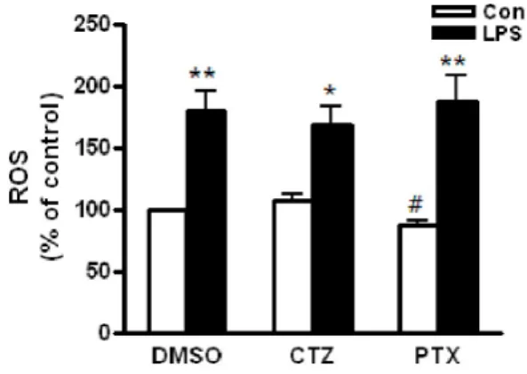
Fig. 3. The effects of cilostazol and pentoxifylline on LPS-induced ROS production in RAW264.7 cells exposed to ethanol. Cells were treated with 25 mM ethanol in presence of cilostazol (100 μM), pentoxifylline (100 μM) or DMSO for 24 h. Then, cells were stimulated with 50 ng/ml LPS for 4 h. After incubation of cells with 50 μM carboxy-H2DCFDA for 40 min, the production of ROS was measured by flow cytometry. Data represented as percentage increases over the DMSO control and are expressed as mean±SEM of three independent experiments. *p<0.05, **p<0.01 vs. control.
#