ABSTRACT
Vitamin D insufficiency is associated with obesity and its related metabolic diseases. Adipose tissues store and metabolize vitamin D and expression levels of vitamin D metabolizing enzymes are known to be altered in obesity. Sequestration of vitamin D in large amount of adipose tissues and low vitamin D metabolism may contribute to the vitamin D inadequacy in obesity. Vitamin D receptor is expressed in adipose tissues and vitamin D regulates multiple aspects of adipose biology including adipogenesis as well as metabolic and endocrine function of adipose tissues that can contribute to the high risk of metabolic diseases in vitamin D insufficiency. We will review current understanding of vitamin D regulation of adipose biology focusing on vitamin D modulation of adiposity and adipose tissue functions as well as the molecular mechanisms through which vitamin D regulates adipose biology. The effects of supplementation or maintenance of vitamin D on obesity and metabolic diseases are also discussed.
Keywords: Cholecalciferol; adipogenesis; adipose function; obesity; metabolic diseases
INTRODUCTION
Obesity, a state of excess accumulation of white adipose tissues (WATs), and its associated metabolic diseases are increasing worldwide. Adipose tissues play important roles in systemic metabolism by storing and releasing energy as well as acting as endocrine organ. Adipose tissues become dysfunctional in obesity, which is characterized by hypertrophied adipocytes, elevated inflammation, hypoxia and fibrosis and reduced angiogenesis [1]. Alterations in adipose derived factors, elevated levels of fatty acids (FAs) and proinflammatory cytokines along with low level of adiponectin from higher mass of dysfunctional adipose tissues, are thought to cause or exacerbate cardiometabolic diseases in obesity.
Adipose tissues are present in multiple locations throughout an organism and largely divided into intraabdominal visceral and subcutaneous depots. The major visceral depots are omental and epiploic fat and major subcutaneous depots are the upper-body abdominal subcutaneous and the lower-body femoral and gluteal fat in humans [2]. In addition to WATs, bioenergetically more active brown adipose tissue (BAT) as well as inducible brown-like
Review
Received: May 20, 2020 Revised: Jun 3, 2020 Accepted: Jul 16, 2020
§
Corresponding Author:
Mi-Jeong Lee
Department of Human Nutrition, Food and Animal Sciences, College of Tropical Agriculture and Human Resources, University of Hawaii at Manoa, 1955 East West Road, AgSci 314k, Honolulu, HI 96822, USA.
Tel. +1-808-956-9565 Fax. +1-808-956-4024 E-mail. [email protected]
©2020 The Korean Nutrition Society and the Korean Society of Community Nutrition This is an Open Access article distributed under the terms of the Creative Commons Attribution Non-Commercial License (https://
creativecommons.org/licenses/by-nc/4.0/) which permits unrestricted non-commercial use, distribution, and reproduction in any medium, provided the original work is properly cited.
ORCID iDs
Hataikarn Nimitphong
https://orcid.org/0000-0003-0151-1622 Eunmi Park
https://orcid.org/0000-0002-1911-4652 Mi-Jeong Lee
https://orcid.org/0000-0002-8171-7913 Conflict of Interest
The authors declare no potential conflicts of interests.
Author Contributions
Conceptualization: Lee MJ; Investigation:
Nimitphong H, Park E, Lee MJ; Writing -
Hataikarn Nimitphong 1 , Eunmi Park 2 , and Mi-Jeong Lee 3§
1
Division of Endocrinology and Metabolism, Department of Medicine, Faculty of Medicine, Ramathibodi Hospital, Mahidol University, Bangkok 10400, Thailand
2
Department of Food and Nutrition, Hannam University, Daejeon 34430, Korea
3
Department of Human Nutrition, Food and Animal Sciences, College of Tropical Agriculture and Human Resources, University of Hawaii, Honolulu, HI 96822, USA
Vitamin D regulation of adipogenesis
and adipose tissue functions
original draft: Nimitphong H, Park E, Lee MJ;
Writing - review & editing: Nimitphong H, Park E, Lee MJ.
adipocytes, beige or brite adipocytes, exist. Brown and beige/brite adipocytes contain more mitochondria and express uncoupling protein 1 (UCP1) and hence, have higher thermogenic capacity [3]. Each adipose depot has differential influence on systemic metabolism. Visceral adiposity, over and above total fat mass per se, is independently associated with metabolic complications, whereas fat accumulation in the lower-body is protective [2]. The amount of BAT or beige/brite fat is reduced in obesity and increasing their amount and activity could confer protection against obesity and its associated metabolic diseases [4].
In addition to its well-known effects on calcium homeostasis and bone metabolism, vitamin D exerts many other actions including proliferation and differentiation of cells and immunomodulatory functions [5]. In adipose tissues, vitamin D has been shown to affect adipocyte development and their metabolic and endocrine functions [6]. While obesity is associated with low vitamin D status and metabolic diseases, whether deficiency of vitamin D predisposes to obesity or vitamin D supplementation improves obesity and metabolic diseases is not clear. We will review vitamin D regulation of adipose biology including adipose tissue development and metabolic and endocrine properties with emphasis on molecular mechanisms that link low vitamin D status with obesity and metabolic diseases. We also discuss the effects of supplementation or maintenance of vitamin D on obesity and metabolic diseases.
VITAMIN D METABOLISM AND BIOLOGICAL FUNCTION
Vitamin D metabolism
Vitamin D is synthesized from 7-dehydrocholesterol in the skin (vitamin D
3) or ingested as food (both vitamin D
2and D
3forms). Ultraviolet B photons cause the photolysis of 7-dehydrocholesterol to previtamin D
3, which thermally isomerizes to vitamin D
3. Vitamin D (D represents D
2and/or D
3) is activated through 2 hydroxylation steps (Fig. 1). In the liver, vitamin D is hydroxylated to 25-hydroxyvitamin D [25(OH)D] by 25-hydroxylases (CYP2R1, CYP27A1, CYP3A4, CYP2J2) [7]. 25(OH)D is then activated to 1,25 dihydroxyvitamin D [1,25(OH)
2D] by 1α-hydroxylase (CYP27B1) in the kidneys. Vitamin D binding protein is the specific chaperone for vitamin D and its metabolites in the blood and then to the storage sites (adipose tissues and skeletal muscle) or target tissues (liver, kidneys or parathyroid gland) and cells (monocytes and macrophages). Both 25(OH)D and 1,25(OH)
2D are hydroxylated by 24-hyroxylase (CYP24A1) and degraded. Other tissues including adipose tissues also express 1α-hydroxylase and 25-hydroxylase and can activate vitamin D locally [8].
Biological functions of vitamin D
In addition to regulation of calcium homeostasis and bone metabolism, vitamin D regulates broad biological processes including proliferation, differentiation and maturation of cells, immune functions and cellular metabolism. We refer to other reviews for the roles of vitamin D and we will briefly describe the molecular details through which vitamin D regulates cellular actions.
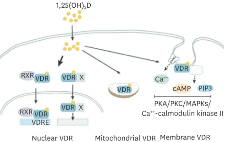
Most of biological actions of 1,25(OH)
2D
3are thought to be mediated through its nuclear
receptor, vitamin D receptor (VDR) (Fig. 2). Ligand bound VDR translocates into the nucleus
as a heterocomplex with retinoid X acid receptor and controls gene transcription by binding to
vitamin D response elements of genes [9]. Through interactions with other nuclear receptors
including nuclear factor kappa B (NF-κB), SP1 and STAT5 vitamin D controls transcription of
genes. 1,25(OH)
2D
3-VDR regulates hundreds of genes in cell- and tissue-specific manners [10].
VDR localizes in the caveolae structures of plasma membrane where it exerts rapid
membrane-initiated signaling responses that are referred as non-genomic actions of vitamin D [11] (Fig. 2). Once bound to vitamin D, VDR interacts with plasma membrane proteins including phospholipase A2, phosphatidylinositol-3 kinase and calcium transporters. These lead to generation of secondary messengers, Ca
++, cyclic adenosine monophosphate, and phosphatidylinositol 3,4,5 triphosphate (PIP3) and activation of downstream protein kinase A, protein kinase C, mitogen activated protein kinases (MAPKs) and Ca
++-calmodulin kinase II [11]. Through these signaling events, vitamin D is known to affect many cellular responses.
These signaling events can also lead to control of gene expression through modulation of transcriptional machinery.
VDR is present in mitochondria where 1,25(OH)
2D
3is known to negatively affect respiratory capacity in multiple cell types including platelets and keratinocytes [12,13]. Vitamin D can also inhibit cell respiration through the nuclear VDR-mediated suppression of transcription of mitochondrial respiratory chain complexes. Through regulation of mitochondrial
Vitamin D
2and D
3from diet and supplements
Vitamin D
Vitamin D
Vitamin D binding protein 25(OH)D
1,25(OH)
2D Liver
Kideny
25-hydroxylase(s) (CYP2R1, CYP27A1)
1α-hydroxylase (CYP27B1) 25-hydroxylase
(CYP24A1) UVB
7-deh ydr ochol ester ol → Vitamin D
3