Original Article
© 2013 The Korean Ophthalmological Society
This is an Open Access article distributed under the terms of the Creative Commons Attribution Non-Commercial License (http://creativecommons.org/licenses /by-nc/3.0/) which permits unrestricted non-commercial use, distribution, and reproduction in any medium, provided the original work is properly cited.
Ptosis Repair Using Preserved Fascia Lata with the Modified Direct Tarsal Fixation Technique
Ji Young Suh, Hee Bae Ahn
Department of Ophthalmology, Dong-A University Medical Center, Busan, Korea
Frontalis sling surgery is the treatment of choice for con- genital ptosis with poor levator function [1-3]. Autogenous fascia lata is the material of gold standard for this opera- tion, but it requires a second incision and cannot be used in very young children [4,5]. In these cases, allograft fascia lata may be the ideal substitute. Surgical techniques for frontalis sling surgery include pentagonal pattern, double rhomboid pattern, triangular pattern, and modified double
triangular pattern [6-10]. In 1990, Spoor and Kwitko [11]
invented a surgical technique of direct tarsal and frontalis fixation. With direct fixation, it is easier to create a lid crease and adjust the lid contour.
This paper describes the clinical results of frontalis sling operation using preserved fascia lata with modified direct tarsal fixation technique in congenital ptosis patients.
Materials and Methods
We conducted a retrospective study of all patients who underwent frontalis suspension surgery using preserved fascia lata with modified direct tarsal fixation for congeni- tal ptosis between March 2001 and December 2008. Pa- tients with blepharophimosis syndrome and Marcus-Gunn Purpose: To evaluate the clinical outcome of frontalis sling operation using preserved fascia lata with modified
direct tarsal fixation in congenital ptosis patients.
Methods: Forty-seven congenital ptosis patients (60 eyes) who underwent a frontalis sling operation using preserved fascia lata with modified direct tarsal fixation method between March 2001 and December 2008 with a mean follow-up time of 52 months (range, 26 to 122 months) were included in this study. The medical records were reviewed retrospectively.
Results: A retrospective chart review was conducted in patients who were diagnosed with congenital ptosis and underwent frontalis suspension surgery using preserved fascia lata with modified direct tarsal fixation from 2001 through 2008 at Dong-A University Hospital. The patients were 34 males and 14 females. The age of the patients ranged from 1 to 18 years with an average age of 4.51 years. At a mean follow-up of 60 months, good final results were achieved in 46 eyes (76.6%), fair in 8 eyes (13.3%), and poor in 6 eyes (10%). The poor results consisted of undercorrection of 1 eye and recurrence in 5 eyes. The accumulative survival rate was 87.2%, with all recurrences occurring within 12 months postoperatively.
Conclusions: Frontalis sling operation by preserved fascia lata with modified direct tarsal fixation appears to be an effective treatment for severe congenital ptosis, showing good long term results.
Key Words: Blepharoptosis, Congenital ptosis, Frontalis sling operation, Preserved fascia lata, Tarsal fixation
Received: August 21, 2012 Accepted: December 5, 2012
Corresponding Author: Hee Bae Ahn, MD. Department of Ophthalmolo- gy, Dong-A University Medical Center, #26 Daesingongwon-ro, Seo-gu, Busan 602-715, Korea. Tel: 82-51-240-5228, Fax: 82-51-254-1987, E-mail:
The abstract of this paper was presented on the 104th meeting of Korean Ophthalmological Society.
jaw winking synkynesis were excluded. Data collected in- cluded age, gender, history of prior surgery, comorbidity, preoperative and postoperative digital or Polaroid photo- graphs, ptosis assessment prior to surgery, post-surgical re- sults, and related complications.
All operations were performed by one surgeon (HBA) assisted by a resident or fellow. All patients were evaluated postoperatively by the same surgeon. All surgeries were performed under general anesthesia. A lid crease line was designed and drawn with a marking pen above the lash line. Another two stab incision sites were marked just above the eyebrow. The upper eyelid and suprabrow tissue were infiltrated with a mixture of lidocaine 2% and 1 : 100,000 epinephrine. The lid skin was incised at the lid crease, and dissection was propagated through the orbicu- laris muscle in order to expose some of the tarsal plate.
Two pieces of Tutoplast (Biodynamics International, Erlan- gen, Germany) a commercially available processed fascia lata allograft, were prepared. The ends of the two pieces of fascia lata were anchored to the upper tarsus with 6-0 ny- lon (Ethicon Inc., Somerville, NJ, USA) suture. Both ends of the anchoring suture were left uncut. A kelly was in- serted into the suprabrow incision, through the tissues and out through the lower lid crease. Then, grabbing the free end of the sutured fascia lata by the kelly, it was pulled through the side of the suprabrow incision. The remaining fascia lata was performed in the same manner. The orbital septum was penetrated at the arcus marginalis, creating a passage directed toward the eyelid. After adjusting the proper lid height, the remaining ends of the fascia lata were cut off. Then, the ends were fixed and buried in the suprabrow incision with 6-0 nylon (Ethicon Inc.) suture.
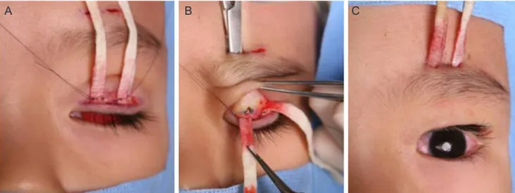
With the sutures left uncut, an eyelid fold was formed by fixing the skin incision to the tarsus. The lid crease was sutured with continuous technique, and the suprabrow in- cisions were closed with the vertical mattress technique using 6-0 plain gut (Fig. 1).
The patients were subdivided by the degree of ptosis.
Mild was defined as 2 mm (or less) drooping from its nor- mal level, moderate was 3 mm drooping, and severe was 4 mm (or more) drooping. Twelve eyes with moderate ptosis and 35 eyes with severe ptosis were included in the study.
Surgical outcome was graded on a three-point scale. Out- come was defined as good if margin reflex dis tance 1 (MRD1) was ≥3 mm, fair when MRD1 was 1.5 to 2.5 mm, and poor when MRD1 was ≤1 mm.
Results
A total of 47 patients (60 eyes) were included in the study. The average age of the patients was 4.5 ± 3.6 years (range, 1 to 18 years). Thirty-four of them (70.2%) were men. Thirty-two patients underwent unilateral surgery, and 14 had bilateral surgery. Eleven patients had epibleph- aron and underwent epiblepharon correction along with the ptosis correction surgery. Three patients had under- gone prior frontalis suspension with Supramid Extra II (S.
Jackson Inc., Alexandra, VA, USA). Demographics of the study population are summarized in Table 1. The mean follow-up period was 60 ± 21 months (range, 22 to 122 months).
Postoperatively, 46 (76.6%) of the operated lids were judged to have achieved good results, 8 eyes (13.3%) had fair lid results, and 6 eyes (10%) had poor results which in- cluded undercorrection (1 eye, 1.7%) and recurrence (5 eyes, 8.3%). One undercorrected case and 1 recurrent case underwent further surgery. Recurrence occurred after a mean of 3.7 ± 4.6 months; all were within 12 months post- operatively (1 week to 11 months). The cumulative survival rate was 87.2%, and the cumulative survival curve is shown in Fig. 2. Fig. 3. shows one patient before and after the surgery.
The correlation between the severity of the ptosis and surgical outcome was not statistically significant (p = 0.384, Mantel-Haenszel chi-square). The correlation be- tween the bilaterality of the ptosis and surgical outcome was also not statistically significant (p = 0.64, Pearson chi-
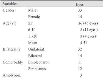
Table 1. Demographics of the study population
Variables Eyes
Gender Male 33
Female 14
Age (yr) ≥5 36 (45 eyes)
6-10 8 (11 eyes) 11-20 3 (4 eyes) Mean 4.51
Bilaterality Unilateral 32
Bilateral 14
Comorbidity Epiblepharon 11
Strabismus 12
Amblyopia 3
square test). Three patients had amblyopia, and all of them underwent unilateral surgeries. One of them was in the re- currence group. The correlation between amblyopia and recurrence was not statistically significant (p = 0.19, Pear- son chi-square test).
Eleven eyes (18.3%) had superficial punctate keratitis postoperatively, 12 eyes (20%) had poor lid contour (peak- ing in 2 eyes and temporal drooping in 10 eyes), but there were no cases of suture exposure, suture infection or pyo- genic granuloma.
Discussion
The treatment of choice for moderate to severe congeni- tal ptosis with minimal or no levator function is frontalis
sling operation [1-3]. Suspensory materials for the frontalis sling operation can be divided into three categories: syn- thetic, autograft fascia lata, and allograft fascia lata. Auto- graft fascia lata is generally considered to the best material [7,12-15] because it is not degraded and is believed to allow fibrovascular tissue ingrowth leading to biointegration without significant inflammation. Also, there is less chance of infection associated with this material. Autograft fascia lata has good long term results with maintenance of lid height [16,17]. However, Crawford [7] and Crawford [12]
recommended using it only in children older than 3 years because there is difficulty in harvesting this material when Fig. 1. (A) The ends of the two pieces of fascia lata were anchored to the upper tarsus with 6-0 nylon suture. (B) A Kelly was inserted into the suprabrow incision, grabbing the free end of the sutured fascia lata. (C) The ends of fascia lata were pulled through the sides of the suprabow incisions.
100
0.95
0.90
0.85
0 20 40 60
Time after operation (mon)
Probability of success
80 100 120
Fig. 2. The cumulative survival curve. The cumulative survival rate was 87.2%. Recurrence occurred after a mean of 3.7 ± 4.6 months.
Fig. 3. Pictures of one patient before (A) and after (B) the sur- gery. The degree of ptosis was moderate and surgical outcome was good.
A
B
A B C
the leg is too short, and insufficient amounts of fascia lata are obtained. Also, the process requires a long operation time, and there is a cosmetic problem of postoperative leg scarring [7,18]. For these reasons, the use of allograft fascia lata as an alternative material has been promoted. Being a synthetic material, it carries some risk of potential infec- tion, but it avoids the need for a second surgical site.
There have been several reports on the outcome of fron- talis sling operation with allograft, i.e., preserved fascia lata. Many of them reported a higher rate of late ptosis re- currence [14,15,19-21]. Our results with preserved fascia lata were very encouraging. The success rate was 86.6%, and all recurrence occurred within 12 months.
The commonly used surgical techniques for frontalis sling operation are rectangular pattern or rhomboid pat- tern. However, in our study, we used direct tarsal fixation technique. This technique was invented by Spoor and Kwitko [11] in 1990 and is superior with regard to adjust- ment of eyelid height and ensures firm anchoring of the sling material. Blephroplasty is easily performed at the completion of this fascia lata slinging procedure [11]. In the original technique, two fascia lata strips are folded in half, and the closed loops of each fascia lata are sutured to the tarsus. The suprabrow incision is large (3 to 4 cm) and is further enlarged until the frontalis muscle is exposed. In our study, the method was somewhat different. We also used two pieces of long fascia lata but did not fold them in half. The suprabrow incisions were small (3 to 4 mm) and were not enlarged. Moreover, we passed the fascia lata strips through the retroseptal area separately, making a two-tunneled space. We believe this modified method is better than the original technique because it requires smaller incisions, thus leaving smaller scars. Also, because the retroseptal dissection is smaller and two strips are sep- arately passed through the narrow tissue tunnel space, minimal trauma results to the adjacent tissue and incorpo- ration and biointegration are improved. This can possibly decrease the postoperative complications and recurrence.
The preserved fascia lata used in the operation showed a good safety profile with no infection or exposure over the follow-up period. Our study had a relatively long period of follow-up, with few complications and recurrence. The re- sults are quite encouraging.
We believe our report supports the long term stability of the procedure. Thereby, we conclude that frontalis sling operation by preserved fascia lata with modified direct tar-
sal fixation is a relatively simple procedure showing a good long term result and should be considered as a good surgi- cal solution for severe congenital ptosis.
Conflict of Interest
No potential conflict of interest relevant to this article was reported.
Acknowledgements
This work was supported by the Dong-A University Re- search Fund, Busan, Korea.
References
1. Beard C. Ptosis. 3rd ed. St. Louis: Mosby; 1981. p. 169-73.
2. Mustarde JC. Repair and reconstruction in the orbital re- gion. 2nd ed. Edinburgh: Churchill Livingstone; 1980. p.
325-8.
3. Fox SA. Congenital ptosis II. Frontalis sling. J Paediatr Ophthalmol 1966;3:25.
4. Song R, Song Y. Treatment of blepharoptosis: direct trans- plantation of the frontalis muscle to the upper eyelid. Clin Plast Surg 1982;9:45-8.
5. Crawford JS. Recent trends in ptosis surgery. Ann Ophthal- mol 1975;7:1263-7.
6. Fox SA. A new frontalis skin sling for ptosis. Am J Oph- thalmol 1968;65:359-62.
7. Crawford JS. Repair of ptosis using frontalis muscle and fascia lata. Trans Am Acad Ophthalmol Otolaryngol 1956;60:672-8.
8. Goldberger S, Conn H, Lemor M. Double rhomboid sili- cone rod frontalis suspension. Ophthal Plast Reconstr Surg 1991;7:48-53.
9. Seider N, Beiran I, Kaltreider SA. One medial triangular Tutoplast sling as a frontalis suspension for adult myogenic blepharoptosis. Acta Ophthalmol Scand 2006;84:121-3.
10. Mauriello JA Jr, Abdelsalam A. Effectiveness of homolo- gous cadaveric fascia lata and role of suture fixation to tar- sus in frontalis suspension. Ophthal Plast Reconstr Surg 1998;14:99-104.
11. Spoor TC, Kwitko GM. Blepharoptosis repair by fascia lata
suspension with direct tarsal and frontalis fixation. Am J Ophthalmol 1990;109:314-7.
12. Crawford JS. Repair of ptosis using frontalis muscle and fascia lata: a 20-year review. Ophthalmic Surg 1977;8:31- 40.
13. Crawford JS. Frontalis sling operation. J Pediatr Ophthal- mol Strabismus 1982;19:253-5.
14. Wagner RS, Mauriello JA Jr, Nelson LB, et al. Treatment of congenital ptosis with frontalis suspension: a compari- son of suspensory materials. Ophthalmology 1984;91:245-8.
15. Wasserman BN, Sprunger DT, Helveston EM. Comparison of materials used in frontalis suspension. Arch Ophthalmol 2001;119:687-91.
16. Kemp EG, James CR, Collin JR. Brow suspension in the management of ptosis: an analysis of over 100 cases. Trans Ophthalmol Soc U K 1986;105(Pt 1):84-7.
17. El-Toukhy E, Salaem M, El-Shewy T, et al. Mersilene mesh sling as an alternative to autogenous fascia lata in the man- agement of ptosis. Eye (Lond) 2001;15(Pt 2):178-82.
18. Leibovitch I, Leibovitch L, Dray JP. Long-term results of frontalis suspension using autogenous fascia lata for con- genital ptosis in children under 3 years of age. Am J Oph- thalmol 2003;136:866-71.
19. Beyer CK, Albert DM. The use and fate of fascia lata and sclera in ophthalmic plastic and reconstructive surgery.
Ophthalmology 1981;88:869-86.
20. Wilson ME, Johnson RW. Congenital ptosis: long-term re- sults of treatment using lyophilized fascia lata for frontalis suspensions. Ophthalmology 1991;98:1234-7.
21. Broughton WL, Matthews JG 2nd, Harris DJ Jr. Congenital ptosis: results of treatment using lyophilized fascia lata for frontalis suspensions. Ophthalmology 1982;89:1261-6.