© 2017 The Korean Ophthalmological Society
This is an Open Access article distributed under the terms of the Creative Commons Attribution Non-Commercial License (http://creativecommons.org/licenses /by-nc/3.0/) which permits unrestricted non-commercial use, distribution, and reproduction in any medium, provided the original work is properly cited.
Original Article
Neutrophil to Lymphocyte Ratio in Patients with Nonarteritic Anterior Ischemic Optic Neuropathy
Alime Gunes, Musa Yigit, Levent Tok, Ozlem Tok
Department of Ophthalmology, Süleyman Demirel University Research and Education Hospital, Isparta, Turkey
Purpose: To evaluate the neutrophil to lymphocyte ratio (NLR) in patients with nonarteritic anterior ischemic op- tic neuropathy (NAION).
Methods: We investigated 112 subjects comprising 56 patients with NAION and 56 healthy controls at Süley- man Demirel University. Complete blood count, demographic, and clinic data from NAION patients were eval- uated in this study. The NLR was calculated in all individuals and compared between the patient and control groups. Cut-off values were also determined. Then, the relationship between NLR and visual outcomes was investigated.
Results: The cut-off value for NLR was 1.64. NLR values were significantly higher in NAION patients than in healthy subjects (p < 0.001) and were directly correlated with erythrocyte sedimentation rate levels (r = 0.263, p = 0.006). Also, the NLR value was associated with visual outcomes. Receiver operator characteristic curve analysis revealed a 0.63 area under the curve (confidence interval, 53.7% to 74.1%), 85% sensitivity and 41%
specificity at the cut-off NLR value.
Conclusions: The NLR may be a biomarker with good sensitivity that is quick, cost effective and easily detect- ed in serum. It can be used in clinical practice to predict a NAION patient’s prognosis in terms of visual out- comes.
Key Words: Neutrophil to lymphocyte ratio, Optic neuropathy, Prognosis, Useful marker, Vision
Nonarteritic anterior ischemic optic neuropathy (NAION) is the most common cause of acute optic neu- ropathy in adults above 50 years of age [1]. It is an idio- pathic disease and most patients present with painless sud- den visual loss, optic disc swelling and visual field defects.
The patients generally have systemic vascular risk factors,
such as hypertension, diabetes mellitus, hypercholesterol- emia [2]. Other associations have been reported, including hyperhomocysteinemia, nocturnal hypotension, anemia, obstructive sleep apnea syndrome, and some coagulopa- thies [3].The diagnosis of NAION in most patients is clini- cal, considering age, presence of systemic vascular risk factors, pattern of visual loss, and appearance of the swol- len disc.
White blood cell count and its subtypes are well-known as classic inflammatory markers especially in cardiovascu- lar diseases [4]. In recent years, the blood neutrophil to lymphocyte ratio (NLR) has been identified as a potential-
Received: September 24, 2015 Accepted: December 14, 2015
Corresponding Author: Alime Gunes, MD. Department of Ophthalmol- ogy, Süleyman Demirel University Faculty of Medicine, Isparta 32260, Turkey. Tel: 90-5054828345, Fax: 90-2462112830, E-mail: dralimesefer@
hotmail.com
ly useful marker of clinical outcome in inflammatory dis- ease [5-11]. Also, neutrophil-mediated inflammation could play an important role in the pathogenesis of the ischemic disease of the optic nerve, and NLR may be used as a marker to diagnose and assess disease severity for NAION.
In the present study, we compared NLR values between NAION patients and healthy subjects. Also, we investigat- ed the relationship between NLR and visual outcomes. To the best of our knowledge, this is the first study to evaluate the NLR in patients with NAION.
Materials and Methods
In this study, we included patients diagnosed with NAION at the Department of Ophthalmology at Süleyman Demirel University between January 2011 and June 2014.
The study was conducted according to the Declaration of Helsinki and approved by the Süleyman Demirel Universi- ty, Department of Medical Sciences ethics committee. In- formed consent was obtained from each of the healthy subjects in the control group.
The study included 56 patients diagnosed with NAION and 56 healthy controls. The diagnosis of NAION was es- tablished according to the Ischemic Optic Neuropathy De- compression Trial criteria: sudden loss of vision within the previous 14 days, a relative afferent pupillary defect, optic disc edema, and visual field defects consistent with optic neuropathy. We differentiate NAION from ischemic optic neuropathy by the absence of complaints such as headache, weight loss, temporal artery tenderness, and jaw claudica- tion. All patients were treated with intravenous corticoste- roids (1 g/day methylprednisolone) for 3 days, followed by oral prednisone (1 mg per kilogram per day) for 11 days.
Patients were excluded if they had active infection, chronic inflammatory disease or autoimmune disease, glaucoma, ophthalmic or neurological disease, any other retinal pa- thology, ocular trauma or if they had undergone previous eye surgery (other than uneventful cataract surgery). Oph- thalmological examination included best-corrected visual acuity (BCVA [logarithm of the minimum angle of resolu- tion]), slit-lamp examination, and visual field test (Zeiss Humphrey Systems, Dublin, CA, USA). BCVA was re- corded at the initial onset of disease and after 6 months.
Complete blood count data were obtained for all sub- jects. Blood sampling was performed at the time of diag-
nosis with NAION in the patient group. The hospital in the present study had been employing an automation system since 2008. Furthermore, age, sex, visual acuity, laboratory values including leukocytes, neutrophils, lymphocytes, NLR, mean platelet volume (MPV), red blood cell distri- bution width (RDW), erythrocyte sedimentation rate (ESR), and C-reactive protein (CRP) parameters were re- corded for these patients. In addition, we calculated NLR in all individuals, compared NLR between the patient and control groups, and determined cut-off NLR values. Then, we investigated the relationship between NLR and visual outcomes.
Complete blood and inflammatory-marker counts Complete blood counts were performed by flow cytome- try (Beckman Coulter LH 780 Analyzer; Beckman Coulter, Miami, FL, USA). Calibration of the analyzer was performed twice a day by the control blood samples with low and high parameters. Peripheral blood was obtained at the time of diagnosis. Venous blood samples were drawn into EDTA tubes for the evaluation of hematological pa- rameters before treatment. Hematological parameters (leu- kocytes, neutrophils, lymphocytes, NLR, MPV, RDW, ESR, and CRP) were recorded. The NLR was calculated as follows: NLR = neutrophil count to lymphocyte count.
CRP was measured by the turbidimetric method (Toshiba ACCUTE TBA-40FR; Toshiba Medical Systems, Tokyo, Japan). ESR was measured by spectrophotometric assay (Alifax test-1 THL, 950 nm; Alifax, Polverara, Italy).
Statistical analysis
All statistical analyses were performed using the SPSS for Windows ver. 20.0 (IBM Corp., Armonk, NY, USA).
The Kolmogorov-Smirnov test was used to determine whether or not variables are normally distributed. Contin- uous variables were expressed as mean (standard devia- tion) or median according to distribution state. Categorical variables were expressed as numbers and percentage. The chi-square test was used to compare proportions in differ- ent groups. Student’s t-test or Mann-Whitney U-test was used to compare the two independent groups according to distribution state. The Kruskal-Wallis test was used for comparing more than two independent groups for non-nor- mal distributed variables. In cases where Kruskal-Wallis
test yielded a statistical significance, post hoc analysis was performed to identify the groups that showed differences by Bonferroni-corrected Mann-Whitney U-test. Receiv- er-operating characteristic curves were used to determine the cut-off values of NLR. The correlation coefficients and their significance between non-normally distributed vari- ables (NLR and other laboratory values: RDW, MPV, ESR, and CRP) were analyzed using the Spearman test. A p-val- ue less than 0.05 was considered significant.
Results
Fifty-six patients with NAION and 56 age- and sex- matched healthy subjects were included in this study. The mean age of patients with NAION and patients in the con- trol group were 54.7 ± 15.0 and 57.2 ± 13.8 years, respec- tively (p = 0.36). The percentage of women was 57.1% (n = 32) for patients, and 73.2% (n = 41) for the controls (p = 0.28). Demographic data, clinical characteristics, and blood parameters are given in Table 1. The initial and final BCVA (logarithm of the minimum angle of resolution) were significantly lower in the patient group (p < 0.001).
The patients with NAION had significantly higher neu- trophil count (p < 0.001), NLR (p = 0.006) and ESR (p <
0.001) than control group (Table 1). No difference was de- tected in the levels of lymphocyte, RDW, MPV, and CRP
between groups (p = 0.531, p = 0.893, p = 0.947, and p = 0.157, respectively). The NLR, and both the initial and final BCVA were negatively correlated (p = 0.014 and p = 0.04, respectively).
Table 1. Demographic data, clinical characteristics, and blood parameters of the study groups
Variable Patient group (n = 56) Control group (n = 56) p-value
Age (yr) 54.7 ± 15.0 57.2 ± 13.8 0.36
Sex, female 32 (57.1) 41 (73.2) 0.28
First BCVA (logMAR) 1.08 ± 0.93 - <0.001
Final BCVA (logMAR) 0.78 ± 84 - <0.001
Leucocyte (103/mL) 9.50 ± 3.99 6.78 ± 1.65 <0.001
Neutrophil (103/mL) 6.46 ± 3.91 4.13 ± 1.27 <0.001
Lymphocyte (103/mL) 2.25 ± 0.78 2.19 ± 0.75 0.531
NLR 3.62 ± 4.37 2.09 ± 0.96 <0.001
MPV (fl*) 8.28 ± 1.07 8.23 ± 0.86 0.947
RDW (fl*) 14.2 ± 1.69 14.2 ± 1.57 0.893
CRP (mg/dL) 6.16 ± 8.79 3.58 ± 2.44 0.157
ESR (mm/hr) 21.2 ± 16.1 11.4 ± 7.2 <0.001
Values are presented as mean ± standard deviation or number (%).
BCVA = best-corrected visual acuity; logMAR = logarithm of the minimum angle of resolution; NLR = neutrophil to lymphocyte ratio;
MPV = mean platelet volume; RDW = red blood cell distribution width; CRP = C-reactive protein; ESR = erythrocyte sedimentation rate.
*fl = (plateletcrit [%] / platelet count [×109/l]) × 105.
Sensitivity
0.2 0.4
1-Specificity
0.6 0.8 1.0
1.0
0.8
0.6
0.4
0.2
0.0
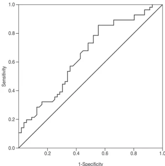
Fig. 1. Receiver operator characteristic curve showing specificity and sensitivity percentages of neutrophil to lymphocyte ratio in patients with nonarteritic anterior ischemic optic neuropathy.
Area under the curve 0.63, neutrophil to lymphocyte ratio cut-off value 1.64, sensitivity 85%, specificity 41%.
The cut-off value of NLR for predicting a NAION diag- nosis was 1.64. Receiver operator characteristic analysis revealed a 0.63 area under the curve (confidence interval, 53.7% to 74.1%), 85% sensitivity and 41% specificity with the cut-off of NLR (Fig. 1). The positive predictive value and negative predictive value for NLR were 60% and 71%, respectively.
The study group was divided into two groups according to NLR (high NLR [≥1.64] and low NLR [<1.64]). The ini- tial and final visual acuity were significantly worse in pa- tients with high NLR than in patients with low NLR (Table 2). In addition, the majority of patients with low NLR were female (75.0%).
There was a statistically significant positive correlation between ESR and NLR (r = 0.263, p = 0.006) (Fig. 2).
There was no correlation between NLR and MPV, RDW, and CRP (r = 0.061, p = 0.657; r = 0.061, p = 0.657; r = 0.199, p = 0.166, respectively).
Discussion
NLR is a simple, cost-effective and reliable indicator of inflammation. To our knowledge, this is the first study of NLR in NAION patients and the first report of a relation- ship between NAION and NLR, and a significant correla- tion between NLR and visual outcomes in NAION.
NAION is one of the most common causes of vision loss worldwide [12]. So far, there is no generally accepted, well-proven, effective treatment for NAION. The primary medications used in the treatment of NAION are cortico- steroids, but the mechanism by which steroids act in this disease is not known. Corticosteroid therapy has been sug- gested to be potentially beneficial in acute phase NAION through improvement of visual acuity and visual field [13].
NAION is known to be a consequence of acute ischemia of the optic nerve head, but the precise mechanism of the disease is unclear and presumably is multifactorial, includ- ing the development of a compartment syndrome [14], em- bolism, and vascular dysregulation [15]. Recently, early in- flammation components were determined in clinical NAION and its models [16-18]. NAION results in early cy- tokine mediated changes [19], and then sequential inflam- matory cellular activation and infiltration are observed [17].
As a new inflammation marker, NLR represents the bal- Table 2. The relationship between NLR, initial visual acuity, and final visual acuity in patients with NAION
High NLR* (n = 48) Low NLR† (n = 8) p-value
Age (yr) 55.0 ± 15.7 52.6 ± 10.9 0.67
Sex, female 26 (54.1) 6 (75.0) <0.001
CRP (mg/dL) 6.63 ± 9.28 2.76 ± 0.88 0.31
ESR (mm/hr) 22.1 ± 16.7 15.4 ± 11.0 0.31
Initial BCVA (logMAR) 1.17 ± 0.93 0.47 ± 0.70 0.014
Final BCVA (logMAR) 0.88 ± 0.87 0.31 ± 0.41 0.04
Values are presented as mean ± standard deviation or number (%).
NLR = neutrophil to lymphocyte ratio; NAION = nonarteritic anterior ischemic optic neuropathy; CRP = C-reactive protein; ESR = erythrocyte sedimentation rate; BCVA = best-corrected visual acuity; logMAR = logarithm of the minimum angle of resolution.
*NLR value of 1.64 or higher; †NLR value less than 1.64.
Neutrophil to lymphocyte ratio
20.00 40.00
r = 0.263, p = 0.006
60.00 80.00 100.00
Erythrocyte sedimentation rate 30.00
25.00 20.00 15.00 10.00 5.00
0.0
Fig. 2. Correlations between neutrophil to lymphocyte ratio and erythrocyte sedimentation rate (mm/hr) level. r = Spearman cor- relation coefficient; p < 0.05 was statistically significant.
ance between neutrophil and lymphocyte levels, which are indicators of systemic inflammation [5-11]. Unlike other inflammatory markers, NLR is simple to calculate, easily applicable in daily practice, and inexpensive. In addition, a high NLR has been found to be correlated with severity and poor prognosis of several diseases [5-11]. Also, the NLR has been assessed in several studies on eye disease [20,21].Ilhan et al. [20] found that patients with age-related macular degeneration have higher NLR compared with healthy subjects. Their study also showed that NLR cor- relates with disease severity. Karaca et al. [21] recently re- ported that the NLR is associated with progression of ker- atoconus. In our study, we found that patients with NAION have higher NLR and this value was correlated with visual results. According to our study, NLR may be helpful for diagnosing NAION and predicting the visual prognosis of NAION subjects.
In the previous study, a NLR result of 1.64 predicted the presence of NAION with 85% sensitivity and 41% specific- ity. The difference in initial and final visual acuity be- tween patients with high and low NLR was significant (p
= 0.014 and p = 0.04, respectively) (Table 2). According to our results, a high NLR value is associated with worse vi- sual results.
Dirican et al. [7] reported that there was a correlation between NLR and levels of ESR in patients with sarcoid- osis and the value of ESR increased significantly when the stage increased.In our study, we found higher level of ESR in NAION patients than in the control group, and there was a positive correlation between NLR and ESR values.
But, we did not find any difference between NAION pa- tients and the control group for the CRP value. According to these results, it is thought that the severity of disease may be more strongly associated with ESR than with CRP.
Our study had several limitations. This study had a ret- rospective design and evaluated a small patient group due to the rarity of the disease. In addition, usage of a single blood sample cannot be used to predict the persistence of NLR over time.
In conclusion, this study showed that NLR was signifi- cantly increased in patients with NAION compared with controls. We suggest that NLR can be used in clinical practice to predict prognosis for NAION patients in terms of visual outcomes.
Conflict of Interest
No potential conflict of interest relevant to this article was reported.
References
1. Hattenhauer MG, Leavitt JA, Hodge DO, et al. Incidence of nonarteritic anterior ischemic optic neuropathy. Am J Oph- thalmol 1997;123:103-7.
2. McCulley TJ, Lam BL, Feuer WJ. A comparison of risk factors for postoperative and spontaneous nonarteritic an- terior ischemic optic neuropathy. J Neuroophthalmol 2005;25:22-4.
3. Worrall BB, Moazami G, Odel JG, Behrens MM. Anterior ischemic optic neuropathy and activated protein C resis- tance: a case report and review of the literature. J Neu- roophthalmol 1997;17:162-5.
4. Arruda-Olson AM, Reeder GS, Bell MR, et al. Neutrophil- ia predicts death and heart failure after myocardial infarc- tion: a community-based study. Circ Cardiovasc Qual Outcomes 2009;2:656-62.
5. Azab B, Chainani V, Shah N, McGinn JT. Neutrophil-lym- phocyte ratio as a predictor of major adverse cardiac events among diabetic population: a 4-year follow-up study. Angi- ology 2013;64:456-65.
6. Ertas G, Sonmez O, Turfan M, et al. Neutrophil/lympho- cyte ratio is associated with thromboembolic stroke in pa- tients with non-valvular atrial fibrillation. J Neurol Sci 2013;324:49-52.
7. Dirican N, Anar C, Kaya S, et al. The clinical significance of hematologic parameters in patients with sarcoidosis.
Clin Respir J 2016;10:32-9.
8. Ahsen A, Ulu MS, Yuksel S, et al. As a new inflammatory marker for familial Mediterranean fever: neutrophil-to-lym- phocyte ratio. Inflammation 2013;36:1357-62.
9. Dirican A, Kucukzeybek BB, Alacacioglu A, et al. Do the derived neutrophil to lymphocyte ratio and the neutrophil to lymphocyte ratio predict prognosis in breast cancer? Int J Clin Oncol 2015;20:70-81.
10. Kao SC, Pavlakis N, Harvie R, et al. High blood neutro- phil-to-lymphocyte ratio is an indicator of poor prognosis in malignant mesothelioma patients undergoing systemic therapy. Clin Cancer Res 2010;16:5805-13.
11. Farah R, Khamisy-Farah R. Association of neutrophil to
lymphocyte ratio with presence and severity of gastritis due to Helicobacter pylori infection. J Clin Lab Anal 2014;28:219-23.
12. Hayreh SS, Zimmerman MB. Nonarteritic anterior isch- emic optic neuropathy: natural history of visual outcome.
Ophthalmology 2008;115:298-305.
13. Hayreh SS. Role of steroid therapy in nonarteritic anterior ischemic optic neuropathy. J Neuroophthalmol 2010;30:388-9.
14. Tesser RA, Niendorf ER, Levin LA. The morphology of an infarct in nonarteritic anterior ischemic optic neuropathy.
Ophthalmology 2003;110:2031-5.
15. Arnold AC. Pathogenesis of nonarteritic anterior ischemic optic neuropathy. J Neuroophthalmol 2003;23:157-63.
16. Zhang C, Guo Y, Miller NR, Bernstein SL. Optic nerve in- farction and post-ischemic inflammation in the rodent model of anterior ischemic optic neuropathy (rAION).
Brain Res 2009;1264:67-75.
17. Salgado C, Vilson F, Miller NR, Bernstein SL. Cellular in- flammation in nonarteritic anterior ischemic optic neuropathy and its primate model. Arch Ophthalmol 2011;129:1583-91.
18. Slater BJ, Vilson FL, Guo Y, et al. Optic nerve inflamma- tion and demyelination in a rodent model of nonarteritic anterior ischemic optic neuropathy. Invest Ophthalmol Vis Sci 2013;54:7952-61.
19. Goldenberg-Cohen N, Kramer M, Bahar I, et al. Elevated plasma levels of interleukin 8 in patients with acute anterior ischaemic optic neuropathy. Br J Ophthalmol 2004;88:1538- 40.
20. Ilhan N, Daglioglu MC, Ilhan O, et al. Assessment of neu- trophil/lymphocyte ratio in patients with age-related macu- lar degeneration. Ocul Immunol Inflamm 2015;23:287-90.
21. Karaca EE, Ozmen MC, Ekici F, et al. Neutrophil-to-lym- phocyte ratio may predict progression in patients with ker- atoconus. Cornea 2014;33:1168-73.