Articles published in Obstet Gynecol Sci are open-access, distributed under the terms of the Creative Commons Attribution Non-Commercial License (http://creativecommons.
org/licenses/by-nc/3.0/) which permits unrestricted non-commercial use, distribution, and reproduction in any medium, provided the original work is properly cited.
Copyright © 2015 Korean Society of Obstetrics and Gynecology
Case Report
Obstet Gynecol Sci 2015;58(4):327-330 http://dx.doi.org/10.5468/ogs.2015.58.4.327 pISSN 2287-8572 · eISSN 2287-8580
www.ogscience.org 327
Introduction
Tamoxifen has been widely used for adjuvant treatment of breast cancer, but several gynecological side effects after its use have been noted, including endometrial hyperplasia, endo- metrial polyps, endometrial carcinoma, and rare complications such as endometriosis, adenomyosis, and cervical polyps [1-3].
Polypoid endometriosis is a rare benign gynecological disease, and several reported cases were associated with the use of tamoxifen [2]. To the best of our knowledge, three cases of polypoid endometriosis have been reported in the Korean lit- erature, but in none of the cases, polypoid endometriosis was associated tamoxifen use. Herein, we report the first case of polypoid endometriosis associated with tamoxifen treatment in a breast cancer patient.
Case report
A 66-year-old woman, who had received left partial mastectomy due to ductal carcinoma in situ about 4 years ago and was tak- ing tamoxifen treatment until a recent date, was admitted for evaluation of an incidentally detected ovarian cyst. She had
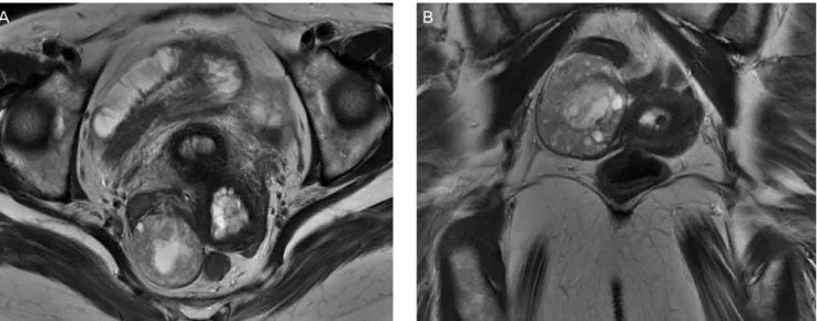
reached menopause 10 years ago and had no other medical history except that she was a carrier of hepatitis C virus. In the laboratory findings, liver enzymes showed an occasional increase but there was no specific problem, and CA-15-3 was within the normal range. Although the initial pelvic sonography in breast cancer work-up showed no abnormal findings, the second pelvic sonography performed 2 months before hysterectomy revealed thickened endometrium (9.7 mm), suggesting endometrial hy- perplasia, and a right ovarian cyst (41×36×35 mm). Pelvic mag- netic resonance imaging revealed multiple cystic structures in the endometrial cavity with mildly thickened endometrium and an approximately 4.1×3.4-cm-sized, well-defined, multicystic le-
Received: 2014.10.2. Revised: 2015.1.15. Accepted: 2015.2.6.
Corresponding author: Dong Won Kim
Department of Pathology, Soonchunhyang University Seoul Hospital, 59 Daesagwan-ro, Yongsan-gu, Seoul 140-743, Korea Tel: +82-2-709-9426 Fax: +82-2-709-9441
E-mail: [email protected]
Tamoxifen-associated polypoid endometriosis mimicking an ovarian neoplasm
In Ho Choi
1, So-Young Jin
1, Yoon Mi Jeen
1, Jeong Jae Lee
2, Dong Won Kim
1Departments of 1Pathology, 2Obstetrics and Gynecology, Soonchunhyang University Seoul Hospital, Soonchunhyang University College of Medicine, Seoul, Korea
Tamoxifen has been widely used for adjuvant treatment of breast cancer, but several gynecological side effects have been noted, including endometrial hyperplasia, polyp and carcinoma. Polypoid endometriosis is one of the extremely rare benign complications associated with tamoxifen therapy. A 66-year-old postmenopausal woman, who had received left partial mastectomy due to breast cancer (about 4 years ago) and was taking tamoxifen treatment, had an ovarian cyst on ultrasonography. Pelvic magnetic resonance imaging suggested tamoxifen-associated endometrial and ovarian changes, especially a 4.1×3.4-cm-sized, well-defined, multicystic mass in the right ovary. She received hysterectomy with bilateral salpingo-oophorectomy. Microscopically, the right paratubal mass showed endometrial glands and stroma, and immunohistochemical staining for CD10 confirmed the endometrial nature of the stroma. Three cases of polypoid endometriosis have been reported in the Korean literature, but in none of the cases, polypoid endometriosis was associated with tamoxifen use. Herein, we report the first case of polypoid endometriosis associated with tamoxifen treatment in Korea.
Keywords: Endometriosis; Polyps; Tamoxifen
www.ogscience.org 328
Vol. 58, No. 4, 2015
sion in the right ovary with peripheral enhancement, suggestive of tamoxifen-associated endometrial and ovarian changes (Fig.
1). Under the impression of endometrial hyperplasia and right ovarian cyst, she received hysterectomy with bilateral salpingo- oophorectomy. The right ovarian mass, which was detected by radiologic examination, was confirmed to be a right paratubal mass.
Grossly, a 4×3-cm-sized, well-circumscribed, polypoid mass was noted in the right paratubal area (Fig. 2A). Microscopically, it revealed a multilocular cystic structure comprising of cystically dilated endometrial glands and stroma together (Fig. 2B). No hy- perplastic feature or complexity of glands was noted. There were no areas of fibrosis, necrosis, and decidual change in the stroma.
The glands were lined by a single layer of columnar or cuboidal cells with no identifiable cellular atypia or mitosis (Fig. 2C). Both glandular epithelial cells and stromal cells were positive for estro- gen receptor (not shown in figure). Presence of CD10-positive stromal cells confirmed the endometrial nature (Fig. 2D). She was diagnosed as having an endometrial polyp with cervical endome- triosis and paratubal polypoid endometriosis and was discharged without any complications.
Discussion
Endometriosis is an estrogen-dependent disorder, and it is rare in postmenopausal women. There are a growing number of cases in the literature, which suggest that the tamoxifen
is an antagonist of the estrogen receptor in breast tissue but behaves as an agonist in the endometrium, and is associated with the development of endometriosis in post-menopausal woman with breast cancer [3-5].
Since Mostoufizadeh and Scully [6] first described polypoid endometriosis in 1980, it has been considered as a benign gynecologic lesion that develops in endometriosis. It usually simulates a malignant tumor during the operation and his- tologically resembles an endometrial polyp. According to the review of 24 cases of polypoid endometriosis by Parker et al. [7], polypoid endometriosis showed several clinico-pathologic dif- ferences when compared with typical endometriosis. It usually occurred in an older age group; 60% of patients with polypoid endometriosis in their study were more than 50 years of age as compared to <5% of patients with typical endometriosis.
The authors also mentioned that hormonal factors might play a role in its pathogenesis, including its association with tamoxi- fen treatment.
Pathologically, polypoid endometriosis should be differentiated from other neoplasms, such as adenofibroma and adenosarcoma due to the presence of benign glands. Indeed, focal or diffuse architectural atypia of the glandular epithelium can be observed in the above lesions. However, endometrioid adenofibroma usu- ally has an ovarian-type stroma in contrast to the endometrial- type stroma of polypoid endometriosis. Adenosarcoma can be distinguished from polypoid endometriosis because the former has projecting stromal papillae or broad polypoid fronds, atypia of stromal cells, and periglandular cellular stromal cuffs.
Fig. 1. (A,B) Pelvic dynamic magnetic resonance imaging shows multiple subendometrial cystic changes with mildly thickened endometrium and a 4.1×3.4-cm-sized, well-defined, multicystic lesion in the right adnexal portion.
A B
www.ogscience.org 329
In Ho Choi, et al. Tamoxifen treatment-associated polypoid endometriosis
In conclusion, three cases of polypoid endometriosis have been reported in Korea; in two cases, polypoid endometriosis origi- nated from the rectum mimicking rectal cancer, and in one case, it developed as a subserosal mass in the uterus [8-10]. However, in none of the cases, polypoid endometriosis was associated with tamoxifen use, and this is the first report of tamoxifen treatment- associated with polypoid endometriosis in Korea.
Conflict of interest
No potential conflict of interest relevant to this article was reported.
Acknowledgments
This work was supported by the Soonchunhyang University research fund.
References
1. McCluggage WG, Bryson C, Lamki H, Boyle DD. Benign, borderline, and malignant endometrioid neoplasia arising in endometriosis in association with tamoxifen therapy. Int J Gynecol Pathol 2000;19:276-9.
2. Schlesinger C, Silverberg SG. Tamoxifen-associated pol-
Fig. 2. (A) An approximately 4×3-cm-sized, polypoid mass is noted in the right paratubal area. (B) At lower magnification, it appears as a multilocular cystic mass, which consists of cystically dilated endometrial glands and stroma (H&E, ×12.5). (C) The glands are lined by a single layer of columnar or cuboidal cells with no cellular atypia or mitosis (H&E, ×400). (D) The stromal cells show positivity for CD10 (CD10, ×200).A
B
C D
www.ogscience.org 330