Received: May 29, 2018 Revised: June 25, 2018 Accepted: July 21, 2018
Address for Correspondence: Hyuk Jung, Department of Obstetrics and Gynecology, Chosun University College of Medicine, 365 Pilmun-daero, Dong-gu, Gwangju 61453, Korea
Tel: +82-62-220-3091, Fax: +82-62-232-2310, E-mail: [email protected]
Original Article
pISSN: 2288-6478, eISSN: 2288-6761 https://doi.org/10.6118/jmm.2018.24.2.81 Journal of Menopausal Medicine 2018;24:81-86
J MM
Introduction
Tamoxifen has been used as adjuvant therapy for breast cancer in postmenopausal women, but has been implicated in endometrial changes. Therefore, gynecologic surveillance of asymptomatic women is needed.1 Endometrial pathology has been identified in up to 35.5% of postmenopausal breast cancer tamoxifen-treated patients.2
Benign endometrial polyps are the most common pathol- ogy described in these patients, with an incidence of 8% to 36%.3 Some women develop recurrent polyps with an inci- dence of malignancy of up to 10.7%.4 There is therefore an
urgent need for a long-term investigation on the incidence of endometrial pathologies among large cohorts of post- menopausal breast cancer patients following continuous tamoxifen therapy for 5 years.
Because of the estrogenic effects on the endometrium, it is necessary to screen postmenopausal patients taking tamoxifen, especially those with no gynecologic symptoms.5
Transvaginal sonography (TVS) is considered a simple, accurate, noninvasive procedure for surveillance of endome- trial changes, although false-negative diagnoses of small polyps, localized areas of atypical hyperplasia, or endome- trial cancer have been reported.6
Comparative Study on Hysteroscopic and Histologic Examinations of the Endometrium in Postmenopausal Women Taking Tamoxifen
Hyuk Jung1,2, Joo Kyoung Jung3, Sat Byul Kim2, Eun A Cho4, Mi Jung Um2
1Department of Obstetrics and Gynecology, Chosun University College of Medicine, Gwangju, Korea, 2Department of Obstetrics and Gynecology, Chosun University Hospital, Gwangju, Korea, 3Postgraduate Student, Chosun University School of Dentistry, Gwangju, Korea,
4Department of Nursing, Honam University, Gwangju, Korea
Objectives: To evaluate the histologic effects of tamoxifen on the endometrium using hysteroscopy in postmenopausal women with breast cancer.
Methods: The study included 46 postmenopausal patients who were referred from another clinic due to thickening or bleeding of the endometrium after taking tamoxifen for breast cancer. All patients underwent transvaginal sonography (TVS) and hysteroscopic endometrial biopsy with a 5-mm, continuous-flow, operating hysteroscope.
Results: The incidence of malignancy was high (20%) in cases of abnormal uterine bleeding (AUB) after taking tamoxifen.
However, in the non-AUB group with thick endometrium after taking tamoxifen, the incidence of adenocarcinoma was 3.2%.
Conclusions: Our findings confirm the estrogen-like effect of tamoxifen on the endometrium. Endometrial evaluation with TVS suggests further diagnostic procedures; moreover, histologic examination is necessary under hysteroscopy, especially in cases of endometrial bleeding after taking tamoxifen. (J Menopausal Med 2018;24:81-86)
Key Words: Breast neoplasms · Endometrium · Hysteroscopy · Postmenopause · Tamoxifen
Journal of Menopausal Medicine 2018;24:81-86
J MM
Some investigators consider TVS endometrial screening to have limited prognostic value in these patients because of excessive false-positive results and have proposed additional diagnostic procedures.7,8
Hysteroscopy directly visualizes the endometrium and al- lows biopsy tissue to be precisely sampled.5
We performed hysteroscopy to evaluate the effect of pro- longed tamoxifen therapy on endometrium of postmeno- pausal patients with breast cancer. In asymptomatic women, endometrial thickness on TVS was the indication for hyster- oscopy.
Materials and Methods
We reviewed clinical records of 46 postmenopausal pa- tients, referred to our institution between January 2008 and December 2017. These women had been receiving tamoxifen for at least 12 months for breast cancer. Fifteen were ex- periencing abnormal uterine bleeding (AUB), but the others were asymptomatic. Each patient underwent TVS. Hysteros- copy was only performed in patients with AUB or with en- dometrial thickness ≥4 mm. All patients were asymptomatic at the beginning of therapy and none had a pretreatment endometrial evaluation.
1. Ultrasound evaluation
A Hitachi Aloka (Hitachi Aloka Medical, Ltd., Tokyo, Ja- pan) instrument with a 50/60 Hz transvaginal probe was used. Maximum endometrial thickness was measured in a longitudinal section including both endometrial layers. The measurement of free fluid in the endometrial cavity was subtracted from the total.
2. Hysteroscopic technique
Hysteroscopy was performed with a new continuous-flow, 5 mm rod lens, operating office hysteroscope (Karl Storz, Tut- tlingen, Germany). To reduce discomfort and pain, intrave- nous sedation was performed using diazepam and pethidine.
The uterine cavity was distended with normal saline solu- tion, and intrauterine pressure was automatically controlled by an electronic irrigation-suction device (Endomat; Karl Storz). Intrauterine pressure was set at 45 mmHg, result-
ing in balanced irrigation flow of about 200 mL/min with a vacuum of 0.2 bars. The procedure was considered satisfac- tory when both tubal ostia and the entire cavity could be seen.
Hysteroscopy was only performed by experienced opera- tors. Directed biopsy samples were taken with a new 5-Fr crocodile biopsy forceps that collected about 4 mm3 of endo- metrial tissue. Endometrial polyps were resected at the time of diagnosis using the hysteroscopic operative features.
3. Data analysis
Ultrasound and hysteroscopic results were compared with histologic findings. Unpaired Student’s t-test was performed for statistical analysis. All P values of less than 0.05 were considered to indicate statistical significance.
Results
The non-AUB group included 31 patients, with 15 in the AUB group. Endometrial thickness by TVS was > 4 mm in 45 patients (97.8%). One patient had an endometrial thick- ness < 4 mm but also had AUB. The mean age of those who underwent TVS and hysteroscopy with direct biopsy sam- pling was 57.3 years old (age range, 42-68 years, mean ± standard deviation [SD] 51 ± 7.6 years).
Patients had been taking tamoxifen for 13 to 68 months (mean ± SD = 27.1 ± 12.9). Hysteroscopic examination in 31 patients in the non-AUB group revealed 5 (16.1%) with atrophic endometrium, 3 (9.7%) with glandular-cystic atro- phy, 8 (25.8%) with nonspecific endometrium, 7 (22.6%) with polyps, 6 (19.4%) with endometrial hyperplasia, and 2 (6.5%) with areas suspicious for adenocarcinoma.
Histologic examination of biopsy specimens confirmed the hysteroscopic diagnoses. Two cases of apparent endometrial hyperplasia on hysteroscopy were histologically diagnosed as non-specific endometrium. One case with 2 adenocarci- nomas was changed to atypical hyperplasia on histological examination (Table 1).
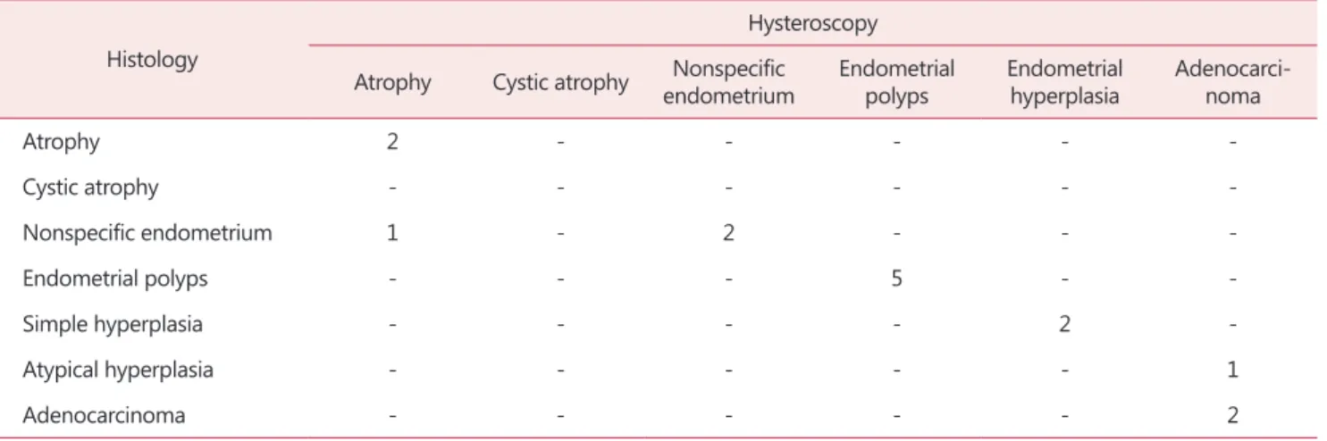
Hysteroscopic examination in the AUB group revealed 3 (20%) with atrophic endometrium, 2 (13.3%) with nonspecific endometrium, 5 (33.3%) with polyps, 2 (13.3%) with hyper- plasia, and 3 (20%) with areas suspicious for adenocarci-
Journal of Menopausal Medicine 2018;24:81-86 Hyuk Jung, et al. Postmenopausal Women Taking Tamoxifen
noma.
One of the 3 suspicious adenocarcinomas was found to be atypical hyperplasia on histologic examination (Table 2).
When the 2 groups were compared, the incidence of ma- lignancy was high in cases with bleeding. However, more
research is needed because of the small sample size.
Hysteroscopy had a sensitivity of 0.85, specificity of 0.83, positive predictive value of 0.79, and negative predictive value of 0.87. A significant difference was found between the presence or absence of endometrial pathology and dura- Table 3. Comparison of histologic findings and endometrial thickness with months of tamoxifen therapy
Histologic findings
No pathology Hyperplasia Polyp Adenocarcinoma P value
No. of patients 13 8 12 3
Endometrial thickness 9.8 ± 7.42 10.2 ± 5.6 21.3 ± 6.9 - NS
Tamoxifen therapy (months) 16.0 ± 7.4 28.0 ± 14.4 31.0 ± 16.4 - 0.05
NS: not significant
Table 1. Comparison of hysteroscopic and histologic diagnoses in 31 patients without abnormal uterine bleeding Histology
Hysteroscopy Atrophy Cystic atrophy Nonspecific
endometrium Endometrial
polyps Endometrial
hyperplasia Adenocarci- noma
Atrophy 5 - - - - -
Cystic atrophy - 2 - - - -
Nonspecific endometrium - 1 8 - 2 -
Endometrial polyps - - - 7 - -
Simple hyperplasia - - - - 4 -
Atypical hyperplasia - - - - - 1
Adenocarcinoma - - - - - 1
Nonspecific endometrium refers to all cases in which no pathologic lesions are seen
Table 2. Comparison of hysteroscopic and histologic diagnoses in 15 patients with abnormal uterine bleeding Histology
Hysteroscopy Atrophy Cystic atrophy Nonspecific
endometrium Endometrial
polyps Endometrial
hyperplasia Adenocarci- noma
Atrophy 2 - - - - -
Cystic atrophy - - - - - -
Nonspecific endometrium 1 - 2 - - -
Endometrial polyps - - - 5 - -
Simple hyperplasia - - - - 2 -
Atypical hyperplasia - - - - - 1
Adenocarcinoma - - - - - 2
Journal of Menopausal Medicine 2018;24:81-86
J MM
tion of tamoxifen therapy (in months; P < 0.05), with longer therapy associated with more pathologic findings on histol- ogy (Table 3).
Discussion
Tamoxifen is a nonsteroidal selective estrogen receptor modulator that is used primarily for adjuvant treatment of estrogen receptor-positive breast cancer in premenopausal women, and in some postmenopausal women.9 It is also used for chemoprevention in women at increased risk of breast cancer. Tamoxifen is associated with increased risks of uterine pathology, including endometrial polyps, endome- trial carcinoma, hyperplasia, uterine sarcoma, and uterine carcinosarcoma.
Ozşener et al.10 showed that tamoxifen use increases the risk of endometrial cancer and premalignant change. They also noted a significant association between endometrial thickness and duration of tamoxifen treatment (P = 0.025).
Hann et al.11 reported abnormal endometrial biopsies in 44% of women treated with tamoxifen for less than 5 years, whereas 58% of endometrial biopsies revealed abnormal re- sults when duration of tamoxifen treatment was > 5 years.
Cohen et al.12 showed that 28.6% of patients on tamoxifen had endometrial pathology. The incidence was significantly more in symptomatic patients. Seoud et al.13 concluded that the value of routine screening for endometrial pathology in patients on tamoxifen is controversial. They found that all patients who developed an abnormal endometrium had ab- normal vaginal bleeding.
Clinical trials confirm that long-term tamoxifen therapy for breast cancer for at least 5 years, is more effective than short-term treatment (< 2 years).3,14 During that time, tamoxifen acts on the endometrium as an estrogen receptor agonist. Its effects vary depending on dosage, duration of treatment, and patient age and menopausal status.15
The frequency of endometrial cancer was reported to dou- ble in trials of 1 or 2 years of tamoxifen and approximately quadrupled in trials of 5 years of therapy.14 Therefore, screening of postmenopausal asymptomatic patients taking tamoxifen is necessary to identify those who may develop significant endometrial pathology.15
In these patients, endometrial pathology may be diag- nosed earlier because they are likely to have symptoms such as AUB;16 however, it is not known whether the increase in endometrial carcinoma represents a true increase. TVS is considered accurate for screening of these patients for endo- metrial changes and is used to suggest additional diagnostic evaluations.15
TVS may fail, however, due to the echogenic, irregular, cystic effect produced by tamoxifen on endometrial stroma and on the myometrium, without necessarily causing epi- thelial disease.17 Therefore endometrial thickness cannot be used in these patients to define abnormalities.18 A high prevalence of polyps (40%) in patients with thickened en- dometrium was reported in another study and could cor- relate with duration of tamoxifen intake.19 It does not seem likely that endometrial polyps are premalignant lesions, but malignancies in polyps of women taking tamoxifen were re- cently reported.20
Women on tamoxifen therapy with AUB or a thick en- dometrium require evaluation for uterine pathology. The approach to evaluation differs between pre- and postmeno- pausal women.
The evaluation typically includes TVS and/or endometrial sampling. American College of Obstetricians and Gynecolo- gists (ACOG) guidelines advise that the initial test used to evaluate postmenopausal bleeding in average-risk women may be either endometrial sampling or TVS.21
Endometrial thickness ≤4 mm on TVS in postmeno- pausal women at average risk has been demonstrated to be an effective test to exclude endometrial cancer. The ACOG also advises that blind endometrial biopsy is most effective for detecting global (pathology occupies at least 50% of the surface area of the endometrial cavity), but may miss focal pathology.22
If focal pathology is suspected, a saline infusion sonogram or hysteroscopy should be performed. For postmenopausal women on tamoxifen, most experts advise endometrial bi- opsy rather than TVS alone; however, some find expectant management in the setting of a thin endometrial echo (≤4 mm) to be acceptable. On the other hand, as noted, endo- metrial biopsy also has limitations, since it may miss focal pathology. For these reasons, we perform both hysteroscopic endometrial biopsy and TVS.
Journal of Menopausal Medicine 2018;24:81-86 Hyuk Jung, et al. Postmenopausal Women Taking Tamoxifen
Conclusion
Our experience indicates that hysteroscopic biopsy can get accurate information on endometrial thickening by tamoxi- fen. We believe that all women undergoing tamoxifen ther- apy for breast cancer should have regular TVS assessment of the endometrium. TVS is helpful for screening patients receiving long-term therapy. For early diagnosis of endo- metrial abnormalities, hysteroscopy is required if patients become symptomatic or if TVS reveals thickened endome- trium.
Acknowledgement
This study was supported by a research fund from Chosun University, 2017.
Conflict of Interest
No potential conflict of interest relevant to this article was reported.
References
1. Suh-Burgmann EJ, Goodman A. Surveillance for endome- trial cancer in women receiving tamoxifen. Ann Intern Med 1999; 131: 127-35.
2. Markovitch O, Tepper R, Fishman A, Aviram R, Cohen I.
Long-term follow-up of postmenopausal breast cancer patients following discontinuation of tamoxifen therapy.
Maturitas 2008; 59: 387-93.
3. Cohen I, Azaria R, Bernheim J, Shapira J, Beyth Y. Risk factors of endometrial polyps resected from postmenopausal patients with breast carcinoma treated with tamoxifen.
Cancer 2001; 92: 1151-5.
4. Cohen I, Azaria R, Shapira J, Yigael D, Tepper R. Signifi- cance of secondary ultrasonographic endometrial thicken- ing in postmenopausal tamoxifen-treated women. Cancer 2002; 94: 3101-6.
5. Hwang KR, Jun HW, Sung JY, Hwang KT, Choi IS, Bae KB. The endometrial ultrasonographic changes in post- menopausal breast cancer patients taking aromatase in-
hibitors after stopping of tamoxifen treatment. J Korean Soc Menopause 2008; 14: 130-8.
6. Achiron R, Grisaru D, Golan-Porat N, Lipitz S. Tamoxifen and the uterus: an old drug tested by new modalities. Ul- trasound Obstet Gynecol 1996; 7: 374-8.
7. Sin CH, Kim SA, Ki WS, Jeoung HY, Song SY, Jung H.
The diagnostic role of hysteroscopy in postmenopausal bleeding. Korean J Obstet Gynecol 2007; 50: 1240-6.
8. Ceci O, Bettocchi S, Marello F, Di Venere R, Pellegrino AR, Laricchia L, et al. Hysteroscopic evaluation of the endome- trium in postmenopausal women taking tamoxifen. J Am Assoc Gynecol Laparosc 2000; 7: 185-9.
9. Early Breast Cancer Trialists’ Collaborative Group (EBCTCG).
Aromatase inhibitors versus tamoxifen in early breast can- cer: patient-level meta-analysis of the randomised trials.
Lancet 2015; 386: 1341-52.
10. Ozşener S, Ozaran A, Itil I, Dikmen Y. Endometrial pathol- ogy of 104 postmenopausal breast cancer patients treated with tamoxifen. Eur J Gynaecol Oncol 1998; 19: 580-3.
11. Hann LE, Giess CS, Bach AM, Tao Y, Baum HJ, Barakat RR. Endometrial thickness in tamoxifen-treated patients:
correlation with clinical and pathologic findings. AJR Am J Roentgenol 1997; 168: 657-61.
12. Cohen I, Perel E, Flex D, Tepper R, Altaras MM, Cordoba M, et al. Endometrial pathology in postmenopausal tamoxifen treatment: comparison between gynaecologically symptom- atic and asymptomatic breast cancer patients. J Clin Pathol 1999; 52: 278-82.
13. Seoud M, Shamseddine A, Khalil A, Salem Z, Saghir N, Bikhazi K, et al. Tamoxifen and endometrial pathologies: a prospective study. Gynecol Oncol 1999; 75: 15-9.
14. Lee DO, Choo CW, Lee JY, Moon KY, Chung YK. Com- parison of histologic results from hysteroscopic biopsy and blinded biopsy by currettage in women with tamoxifen therapy after breast cancer. J Korean Soc Menopause 2008;
14: 246-50.
15. Cheng WF, Lin HH, Torng PL, Huang SC. Comparison of endometrial changes among symptomatic tamoxifen- treated and nontreated premenopausal and postmenopausal breast cancer patients. Gynecol Oncol 1997; 66: 233-7.
16. Kedar RP, Bourne TH, Powles TJ, Collins WP, Ashley SE, Cosgrove DO, et al. Effects of tamoxifen on uterus and ovaries of postmenopausal women in a randomised breast cancer prevention trial. Lancet 1994; 343: 1318-21.
17. Dijkhuizen FP, Brölmann HA, Oddens BJ, Roumen RM, Coebergh JW, Heintz AP. Transvaginal ultrasonography and endometrial changes in postmenopausal breast cancer patients receiving tamoxifen. Maturitas 1996; 25: 45-50.
18. Neven P, Vergote I. Should tamoxifen users be screened for
Journal of Menopausal Medicine 2018;24:81-86
J MM
endometrial lesions? Lancet 1998; 351: 155-7.
19. Timmerman D, Deprest J, Bourne T, Van den Berghe I, Collins WP, Vergote I. A randomized trial on the use of ultrasonography or office hysteroscopy for endometrial as- sessment in postmenopausal patients with breast cancer who were treated with tamoxifen. Am J Obstet Gynecol 1998; 179: 62-70.
20. Ramondetta LM, Sherwood JB, Dunton CJ, Palazzo JP.
Endometrial cancer in polyps associated with tamoxifen
use. Am J Obstet Gynecol 1999; 180: 340-1.
21. American College of Obstetricians and Gynecologists. ACOG committee opinion No. 440: The role of transvaginal ultra- sonography in the evaluation of postmenopausal bleeding.
Obstet Gynecol 2009; 114: 409-11.
22. Committee on Practice Bulletins-Gynecology. Practice bul- letin no. 128: diagnosis of abnormal uterine bleeding in reproductive-aged women. Obstet Gynecol 2012; 120: 197- 206.