http://dx.doi.org/10.5468/ogs.2015.58.2.124 pISSN 2287-8572 · eISSN 2287-8580
Introduction
Epithelial ovarian cancer (EOC) is the common female cancer worldwide and one of the most lethal malignancies [1]. The incidence is reported to be relatively high in developed coun- tries [2] and the incidence of ovarian cancer has been increas- ing in Korea according to the Korea Central Cancer Registry's nationwide cancer incidence monitor since 1999 [3]. Several reasons for poor prognosis related to EOC are suspected but the advanced disease at initial visit is presumed as major factor for poor survival in EOC. Stage is known for one of the power- ful prognostic factor for survival and 5-year overall survival (OS) rates of stage I and II EOC is approximately 90% and 60%, respectively, and these rates decline to about 30% in more ad-
vanced stages (III and IV) [4].
Surgical exploration and pathological staging is standard pro-
Survival analysis of revised 2013 FIGO staging classification of epithelial ovarian cancer and comparison with previous FIGO staging classification
E Sun Paik, Yoo-Young Lee, Eun-Jung Lee, Chel Hun Choi, Tae-Joong Kim, Jeong-Won Lee, Duk-Soo Bae, Byoung-Gie Kim
Department of Obstetrics and Gynecology, Samsung Medical Center, Sungkyunkwan University School of Medicine, Seoul, Korea
Objective
To analyze the prognostic role of revised version of International Federation of Gynecology and Obstetrics (FIGO) stage (2013) in epithelial ovarian cancer and compare with previous version staging classification
Methods
We retrospectively enrolled patients with epithelial ovarian cancer treated at Samsung Medical Center from 2002 to 2012.
We reclassified the patients based on the revised FIGO staging classification.
Results
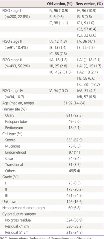
Eight hundred seventy-eight patients were enrolled (stage I, 22.8%; stage II, 10.4%; stage III, 56.2%; stage IV, 10.7%).
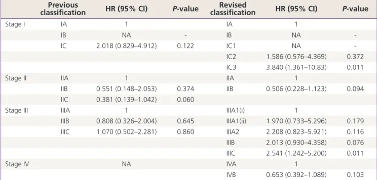
Previous stage IC (98, 11.1%) was subdivided into IC1 (9, 1.0%), IC2 (57, 6.4%), and IC3 (32, 4.1%). In addition, previous stage IV (94, 1.7%) was categorized into IVA (37, 4.2%) and IVB (57, 6.5%) in new staging classification. Stage IIC (66, 7.5%) has been eliminated and integrated into IIA (36, 4.1%) and IIB (55, 6.2%) in revised classification. Revised FIGO stage IC3 had significant prognostic impact on PFS (hazard ratio [HR], 3.840; 95% confidence interval [CI], 1.361 to 10.83; P=0.011) and revised FIGO stage IIIC appears to be an independent, significant poor prognostic factor for PFS (HR, 2.541; 95% CI, 1.242 to 5.200; P=0.011) but not in the case of previous version of FIGO stage IIIC (HR, 1.070; 95%
CI, 0.502 to 2.281; P=0.860). However, any sub-stages of both previous and revised version in stage II and IV, there was no significant prognostic role.
Conclusion
Revised FIGO stage has more progressed utility for informing prognosis than previous version, especially in stage I and III. For stage II and IV, further validation should be needed in large population based study in the future.
Keywords: Neoplasm staging; New classification; Ovarian neoplasms
Received: 2014.5.29. Revised: 2014.7.27. Accepted: 2014.10.9.
Corresponding author: Byoung-Gie Kim
Department of Obstetrics and Gynecology, Samsung Medical Center, Sungkyunkwan University School of Medicine, 81 Irwon- ro, Gangnam-gu, Seoul 135-710, Korea
Tel: +82-2-3410-3513 Fax: +82-2-3410-0630 E-mail: [email protected]
Articles published in Obstet Gynecol Sci are open-access, distributed under the terms of the Creative Commons Attribution Non-Commercial License (http://creativecommons.
org/licenses/by-nc/3.0/) which permits unrestricted non-commercial use, distribution, and reproduction in any medium, provided the original work is properly cited.
Copyright © 2015 Korean Society of Obstetrics and Gynecology
cedure for classification of EOC and International Federation of Gynecology and Obstetrics (FIGO) classification is widely used.
However, there have been debates on the issues that existing FIGO staging of EOC classifies heterogeneous groups of popu- lation as same stage [5,6]. For example, FIGO stage IIIC patients with only isolated lymph node metastasis have been shown better prognosis compared to the patients with stage IIIC with peritoneal carcinomatosis or abdominal metastatic lesion more than 2 cm [6-12]. Unfortunately FIGO staging classification of EOC has not been revised since 1988 under these critical issues but recently, FIGO announced revised FIGO staging classifica- tion [13] reflecting recent evidences. This study is designed to investigate the clinical relevance of new FIGO staging system and to compare revised classification with previous version of FIGO staging system in EOC.
Materials and methods
1. Patients
With the institutional review board approval (2014-05-083), we retrospectively searched the patients who were diagnosed with EOC at Samsung Medical Center, Seoul, Korea from 2002 to 2012. Using the electrical medical records, data of patients such as age, stage, cell type, tumor grade, adjuvant chemo- therapeutic regimen, type of surgery, surgical outcomes, etc.
were gathered. The patients who had synchronous cancers were excluded. All cases were staged based on the operation records and final pathological reports according to last and revised versions of FIGO staging classification, respectively.
Stage IC1 was reclassified for patients with iatrogenic rup- ture occurred during operation. Stage IC1 was confirmed by operation record in which incident of iatrogenic rupture was recorded separately by operator. The cases of iatrogenic rup- ture without previously proven positive result of malignancy for washing cytology were classified as stage IC1. The cases with positive result of malignancy for washing cytology were subdivided as stage IC3 irrelevant to status of iatrogenic rup- ture. Regarding stage IV, clinically suspected lung lesions on imaging studies such as computed tomography, magnetic resonance imaging, and positron emission tomography or cyto-pathological confirmation of distant organs was required for diagnosis. Primary tubal or peritoneal cancers were also included in this study. Surgical outcomes were categorized as no gross residual, optimal residual (<1 cm), and suboptimal (≥1 cm). Complete surgical staging includes washing cytology,
hysterectomy, bilateral salpingo-oophporectomy, pelvic and/
or para-aortic lymphadenectomy, omentectomy, and multiple biopsies on suspicious lesions. For suspected stage I EOC, fertil- ity saving or comprehensive surgical staging was permitted via laparotomy or laparoscopy based on the attending physicians' preference. Adjuvant chemotherapy consisted of platinum based chemotherapy in all cases. Adjuvant chemotherapy can be omitted in the case of stage IA or IB with grade 1. Platinum resistance was defined as less than 6months of platinum free interval. Patients were followed up every 3 months for the first 2 years, then 6 months for up to 5 years and annually there- after. Patients were monitored based on clinical, radiological, biochemical and imaging techniques.
2. Statistical analysis
The Kaplan-Meier method was used to estimate progression free survivals (PFSs) and OSs and comparison of survival curves between groups were carried out with log-rank test. We de- fined PFS as the time from the initial treatment to relapse or the last follow-up visit; OS was the time from the initial treat- ment to death or the last follow-up visit. Multivariate analyses of prognostic factors were carried out using Cox regression models. Factors included in multivariate analysis in stage I were age, cell type (serous vs. non-serous), grade, surgical staging methods (complete staging vs. comprehensive staging), and platinum sensitivity. For each stage II, III, and IV, multivariate analysis was performed by adjusting age, cell type (serous vs.
non-serous), grade, and surgical outcomes (no gross residual, residual <1 cm, and residual ≥1 cm), and platinum sensitivity.
Statistical analyses were performed by IBM SPSS ver. 21.0 (IBM Corp., Armonk, NY, USA). A P-value of ≤0.05 was considered statistically significant and all P-values were two-sided.
Results
Overall, we were able to enroll 878 patients with EOC. The
basic characteristics of patients are presented in Table 1. The
median age of the population was 51.9 years old with the
range of 14 and 84. Most of the patients (66.9%, 587/878)
were diagnosed as advanced disease (stage III and IV) and
portion of stage II was the least (10.4% ,91/878). 92.3% of
patients (811/878) had primary site from ovary, 5.6% (49/878)
from fallopian tube, and 2.1% (18/878) from peritoneum. Se-
rous adenocarcinoma (62.9%, 553/878) and grade III (54.8%,
481/878) was the most common type for histology and tumor
grade, respectively. For treatment, 60 patients (6.8%) had neo- adjuvant chemotherapy and 52 patients (5.9%) had surgical treatment alone without chemotherapy. Optimal surgical cyto- reduction was achieved in 75.1% (660/878) in all patients and 63.4% (372/587) among stage III and IV. The median follow up time was 42.1 month with the range of 0.2 to 136.6.
Compared to previous FIGO staging system, revised one is di- vided into more sub-stages. Previous stage IC (98, 11.1%) sub- divided into IC1 (9, 1.0%), IC2 (57, 6.4%), and IC3 (32, 4.1%).
In addition, previous stage IV (94, 1.7%) was categorized into IVA (37, 4.2%) and IVB (57, 6.5%) in new staging classifica- tion. Stage IIC (66, 7.5%) has been eliminated and integrated into IIA (36, 4.1%) and IIB (55, 6.2%) in revised classification.
Redistribution of stage III has been observed in new staging classification. Number of patients with stage IIIC in previous staging system (452, 51.8%) decreased in new staging system (384, 43,7%). Relatively, combined number of patients with stage IIIA1(i) (18, 2.1%), IIIA1(ii) (15, 1.7%), IIIA2 (18, 2.1%), IIIB (58, 6.6%) was increased in new staging system (Table 1).
1. Stage I and II
The median age of the patients with stage I was 45.5 years old (range, 14 to 81). Among the EOC patients with stage I, clear cell (20.0%, 40/200), endometrioid (23.0%, 46/200) and mu- cinous (28.5%, 57/200) were relatively common histologic type compared with whole population and serous type consisted of 20.5% (41/200) in stage I. Number of low grade tumor was also higher in stage I (grade I, 27.5%, 55/200; grade II, 24.0%, 48/200) and about one third of the patients (37.5%, 75/200) had comprehensive surgical management including fertility saving surgery. There was no recurrence or death in FIGO stage IB of previous version and FIGO stage IB and IC1 of revised version. In univariate analysis, either previous or revised FIGO stages did not show statistical significance in terms of PFS and OS (Fig. 1). When we performed multivariate analysis to adjust age, cell type (serous vs. non-serous), grade, and surgical stag- ing methods (complete staging vs. comprehensive staging), only revised FIGO stage IC3 had significant prognostic impact on PFS (hazard ratio [HR], 3.840; 95% confidence interval [CI], 1.361 to 10.83; P=0.011), but not on OS (Table 1).
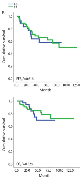
The median age of the patient with stage II was 49.0 years old (range, 31 to 77). Serous histologic type was the most common (47.3%, 43/91) followed by endometrioid (19.8%, 18/91), mucinous (11.0%, 10/91), and clear cell (7.7%, 7/91).
63.7% of patients (58/91) showed grade III and only 6.6%
(6/91) had grade I. Most of the patients received complete
surgical staging (78.0%, 71/91) and suboptimal cytoreduction was shown in 3.3% (3/91). None of the survival analyses ap- proved prognostic role of either FIGO stage classifications in terms of PFS and OS in stage II (Fig. 2, Tables 2 and 3).
Table 1. Patients characteristics (n=878)
Old version, (%) New version, (%) FIGO stage I IA, 96 (10.9) IA, 96 (10.9)
(n=200, 22.8%) IB, 6 (0.6) IB, 6 (0.6) IC, 98 (11.1) IC1, 9 (1.0)
IC2, 57 (6.4) IC3, 32 (3.6) FIGO stage II IIA, 12 (1.3) IIA, 36 (4.1)
(n=91, 10.4%) IIB, 13 (1.4) IIB, 55 (6.2) IIC, 66 (7.5)
FIGO stage III IIIA, 16 (1.8) IIIA1(i), 18 (2.1) (n=493, 56.2%) IIIB, 25 (2.8) IIIA1(ii), 15 (1.7)
IIIC, 452 (51.8) IIIA2, 18 (2.1) IIIB, 58 (6.6) IIIC, 384 (43.7) FIGO stage IV IV, 94 (10.7) IVA, 37 (4.2)
(n=94, 10.7) IVB, 57 (6.5)
Age (median, range) 51.92 (14–84)
Primary site (%)
Ovary 811 (92.3)
Fallopian tube 49 (5.6)
Peritoneum 18 (2.1)
Cell type (%)
Serous 553 (62.9)
Mucinous 75 (8.5)
Endometrioid 97 (11)
Clear 74 (8.4)
Transitional 31 (3.5)
Others 48(5.4)
Grade (%)
I 73 (8.3)
II 178 (20.2)
III 481 (54.8)
Unknown 146 (16.6)
Neoadjuvant chemotherapy 60 (6.8) Cytoreductive surgery
No gross residual 324 (36.9)
Residual <1 cm 336 (38.2)
Residual ≥1 cm 218 (24.8)
FIGO, International Federation of Gynecology and Obstetrics.
2. Stage III and IV
The median age of patients with FIGO stage III was 53 years old with the range of 22 to 84. Serous histologic type (80.9%, 399/493) and grade III (78.3%, 386/493) were most com- mon. 5.7% (28/493) had neoadjuvant chemotherapy and suboptimal cytoreduction was shown in 35.3% (174/493). As shown in Fig. 3, revised FIGO stage classification is associated with significant stratification among stage III patients based on
PFS (P=0.005) and OS (P=0.025). These findings were not ob- served in previous version of FIGO stage. After adjusting age, cell type (serous vs. non-serous), grade, and surgical outcomes (no gross residual, residual <1 cm, and residual ≥1 cm), revised FIGO stage IIIC appears to be an independent, significant poor prognostic factor for PFS (HR, 2.541; 95% CI, 1.242 to 5.200;
P=0.011) (Table 2) but not in previous version of FIGO stage
IIIC (HR, 1.070; 95% CI, 0.502 to 2.281; P=0.860) (Table 2). In
Fig. 1. Comparison of previous and revised staging classification of stage I ovarian cancer with progression free survival (PFS) and overall survival (OS).(A) Old version and (B) revised version.
Cumulative survival Cumulative survival
Cumulative survival Cumulative survival
Month Month
Month Month
IAIB
IC1 IC2
IC3
A B
IAIB IC
terms of OS, the significant prognostic impact of revised FIGO stage IIIC, observed in PFS, turned out to be not significant, just showing trend of poor survival (HR, 3.390; 95% CI, 0.830 to 13.85; P=0.089).
The median age of patients with stage IV was 56.0 years old with the range of 23 to 81. Majority of stage IV patients had grade III (91.5%, 86/94) and serous histologic type (74.5%, 70/94) ovarian cancer. About one fourth (24.5%, 23/94) had
neoadjuvant chemotherapy and optimal cytoreduction was achieved in 56.4% (53/94). When stage IV patients were divided into IVA and IVB according to revised FIGO stage, we could not find any differences in terms of proportion of neoadjuvant chemotherapy and optimal cytoreduction between two groups.
As shown in Fig. 4, we could not find any statistical significant survival differences in PFS and OS and these non-significant find- ings still remained in multivariate analysis as well (Tables 2, 3).
Fig. 2. Comparison of previous and revised staging classification of stage II ovarian cancer with progression free survival (PFS) and overall survival (OS).
(A) Old version and (B) revised version.
Cumulative survival
Month
IIAIIB
Cumulative survivalCumulative survival
Cumulative survival
Month Month Month
A B
IIAIIB IIC
Table 2. Progression free survival in multivariate analysis Previous
classification HR (95% CI) P-value Revised
classification HR (95% CI) P-value
Stage I IA 1 IA 1
IB NA - IB NA -
IC 2.018 (0.829–4.912) 0.122 IC1 NA -
IC2 1.586 (0.576–4.369) 0.372
IC3 3.840 (1.361–10.83) 0.011
Stage II IIA 1 IIA 1
IIB 0.551 (0.148–2.053) 0.374 IIB 0.506 (0.228–1.123) 0.094
IIC 0.381 (0.139–1.042) 0.060
Stage III IIIA 1 IIIA1(i) 1
IIIB 0.808 (0.326–2.004) 0.645 IIIA1(ii) 1.970 (0.733–5.296) 0.179
IIIC 1.070 (0.502–2.281) 0.860 IIIA2 2.208 (0.823–5.921) 0.116
IIIB 2.013 (0.930–4.358) 0.076
IIIC 2.541 (1.242–5.200) 0.011
Stage IV NA IVA 1
IVB 0.653 (0.392–1.089) 0.103
Multivariate analysis was performed in stage I by adjusting age, cell type (serous vs. non-serous), grade, and surgical staging methods (complete staging vs. comprehensive staging). Multivariate analysis was performed in stage II, III, and IV, respectively, by adjusting age, cell type (serous vs. non-serous), grade, and surgical outcomes (no gross residual, residual <1 cm, and residual ≥1 cm).
HR, hazard ratio; CI, confidence interval; NA, not applicable.
Table 3. Overall survival in multivariate analysis Previous
classification HR (95% CI) P-value Revised
classification HR (95% CI) P-value
Stage I IA 1 IA 1
IB NA - IB NA -
IC 0.930 (0.241–3.585) 0.916 IC1 NA -
IC2 2.543 (0.484–13.36) 0.270
IC3 0.287 (0.036–2.257) 0.235
Stage II IIA 1 IIA 1
IIB 0.972 (0.151–6.245) 0.976 IIB 1.607 (0.309–8.370) 0.573
IIC 0.634 (0.152–2.643) 0.531
Stage III IIIA 1 IIIA1(i) 1
IIIB 0.295 (0.102–0.850) 0.024 IIIA1(ii) 2.162 (0.357–13.10) 0.402
IIIC 0.420 (0.184–0.960) 0.040 IIIA2 8.480 (1.748–41.12) 0.008
IIIB 2.529 (0.588–10.87) 0.212
IIIC 3.390 (0.830–13.85) 0.089
Stage IV NA IVA 1
IVB 1.139 (0.826–1.569) 0.428
Multivariate analysis was performed in stage I by adjusting age, cell type (serous vs. non-serous), grade, and surgical staging methods (complete staging vs. comprehensive staging). Multivariate analysis was performed in stage II, III, and IV, respectively, by adjusting age, cell type (serous vs. non-serous), grade, surgical outcomes (no gross residual, residual <1 cm, and residual ≥1 cm), and chemosensitivity.
HR, hazard ratio; CI, confidence interval; NA, not applicable.
Discussion
In this study, we could find that revised 2013 FIGO staging classification in EOC is acceptable and has an independent prognostic role especially in IC3 and IIIC, which were not shown in IC and IIIC of previous FIGO stage. However, the prognostic significance remains uncertain in stage II and IV.
EOC is staged surgically and surgical staging should be
confirmed based on pathological findings. The major role of staging system is not only to provide universal terminol- ogy to be able to use in different centers worldwide, but also to give information about the prognosis of the patients and outcome prediction after specific treatment. Since the last version of ovarian cancer FIGO staging classification in 1988, there have been concerns that FIGO staging cannot delineate the heterogeneity of EOC patients especially in IC and IIIC. For
Fig. 3. Comparison of previous and revised staging classification of stage III ovarian cancer with progression free survival (PFS) and overall survival (OS).(A) Old version and (B) revised version.
Cumulative survivalCumulative survival Cumulative survivalCumulative survival
Month Month
Month Month
IIIAIIIB IIIC
A B
IIIA(i) IIIB(ii)
IIIC IIIB
IIIC
example, whether intra-operative iatrogenic rupture of ovary in stage I EOC might have an effect on the survival outcome or not is long standing controversy [14-18]. Recent meta- analysis reported that iatrogenic rupture might not decrease recurrence compared to early-stage EOC without rupture in which complete surgical staging followed by platinum-based chemotherapy [19]. This suggests that stage IC with iatrogenic rupture might have better prognosis than the other stage IC.
In our study, there is no case of recurrence in stage IC1, which showed even better prognosis than stage IA EOC, and these findings also support that iatrogenic ruptured IC EOC should have been allocated to different category. In the revised FIGO stage classification, stage IC3 became significant independent prognostic factor, as previous FIGO stage IC with iatrogenic rupture was re-categorized to IC1.
It has been also suggested that Stage IIIC in previous FIGO
Fig. 4. Comparison of previous and revised staging classification of stage IV ovarian cancer with progression free survival (PFS) and overall survival (OS).(A) Old version and (B) revised version.
Cumulative survivalCumulative survival Cumulative survivalCumulative survival
Month Month
Month Month
IV IVA
IVB