390
CASE REPORTKorean Circ J 2008;38:390-392
Print ISSN 1738-5520 / On-line ISSN 1738-5555 Copyright ⓒ 2008 The Korean Society of Cardiology
Anomalous Origin of a Right Coronary Artery With Extrinsic Compression Between the Great Vessels: The Intravascular Ultrasound Images
Jae-Youn Moon, MD, Hae Chang Jeong, MD, Jae Yeong Cho, MD, Doo Sun Sim, MD, Hyung Wook Park, MD, Young Joon Hong, MD, Ju Han Kim, MD, Young Keun Ahn, MD and Myung Ho Jeong, MD
Cardiovascular Research Institute of Chonnam National University, The Heart Center of Chonnam National University Hospital, Gwangju, Korea
ABSTRACT
The anomalous origin of the right coronary artery is a rare condition, but it has clinical importance because there have been reports of nonfatal or fatal myocardial infarction and sudden death associated with exercise for patients with this anatomy. We describe here a patient for whom 64 channel multi-detector row computed tomography was useful to identify this anomaly, and intravascular ultrasound was used to evaluate the myocardial ischemia by vi- sualizing the coronary lumen.
(Korean Circ J 2008;38:390-392)KEY WORDS:
Anomalies; Computed tomography; Intravascular ultrasonography.
Introduction
The anomalous origin of the right coronary artery right coronary artery (RCA) is a rare condition,
1)but it has clinical importance because there have been reports of nonfatal or fatal myocardial infarction and sudden death associated with exercise for patients with this anatomy.
1-3)Because of the high fatality rate associated with this anomaly, most of the published reports that studied large groups of patients have used postmortem diagnoses. The conventional coronary angiogram has lim- itations with regard to clear visualization of the slitlike ostium of a coronary artery with an anomalous origin.
Therefore, intravascular ultrasound (IVUS) for obtain- ing the cross-sectional luminal image and multidetector row computed tomography (MDCT), which allows three- dimensional visualization of the coronary artery with high spatial resolution, may be promising imaging mo- dalities for diagnosing and evaluating this anomaly.
4)5)We describe here a patient for whom MDCT was use- ful to identify this anomaly and IVUS was used for eval-
uating the myocardial ischemia by visualizing the cor- onary lumen.
Case
A 22-year-old man presented for evaluation of his effort angina, and he had experienced this for several months. He had no risk factors for coronary artery dis- ease and the resting electrocardiogram (ECG) was nor- mal. He had no history of syncope. Because of the typical angina symptoms, we checked the 64-channel MDCT images for coronary artery disease even though he was young. The 64-channel MDCT scan (Fig. 1) revealed an anomalous origin of the RCA from the left coronary cusp (LCC) with subsequent extrinsic compression bet- ween the great vessels (the aorta and main pulmonary artery). After the CT scan, conventional coronary angio- graphy and an IVUS study were performed. According to visual estimation, there was no critical stenotic lesion on the left anterior oblique (LAO) 45°projection view (Fig. 2A); however, a critical stenosis was noted on the right anterior oblique (RAO) 45°projection image (Fig.
2B). The IVUS study showed no atherosclerotic plaque burden on the entire RCA, but a spindle shaped arte- rial lumen (a slit-like lumen) caused by extrinsic com- pression was noted from the proximal portion of the RCA to the ostium (Fig. 3A).
Finally, a bypass graft using the right internal mam- mary artery was performed to prevent myocardial ische-
Received: January 9, 2008
Revision Received: February 25, 2008 Accepted: March 2, 2008
Correspondence: Myung Ho Jeong, MD,Cardiovascular Research Institute of Chonnam National University, The Heart Center of Chonnam National Uni- versity Hospital, 8 Hak-dong, Dong-gu, Gwangju 501-757, Korea Tel: 82-62-220-7578, Fax: 82-62-223-3105
E-mail: [email protected]
Jae-Youn Moon, et al.·
391
mia or sudden cardiac death. The patient has remained asymptomatic after surgery.
Discussion
The causes of myocardial ischemia for this anomaly remain unclear, but the acute angle of the take-off and kinking of the RCA as it arises from the left coronary cusps and also compression of the RCA when it courses within the aortic wall or between the aorta and the pulmonary artery have been thought to be the possible mechanisms.
6)7)The myocardial ischemia in this case was presumed to be caused by compression of the anom- alous right coronary artery as it coursed between the
pulmonary artery and the aorta. Because of the extrinsic compression between the two large vessels, the lumen was squeezed to a spindle shape (a slit-like lumen). This very eccentric lumen narrowing showed relative normal images on the LAO projection views, but a critical ste- nosis could be seen on the counter RAO projection views in the transverse plane. Therefore, this anomaly can be missed during the conventional angiogram due to this eccentric luminal narrowing.
Several clinical reports on RCA anomalies have been previously reported in Korea,
8-10)yet this particular RCA anomaly has not yet been reported on. Invasive coronary angiography was not suitable for our young patient who had no risk factors for a therosclerosis. With the advent
A B
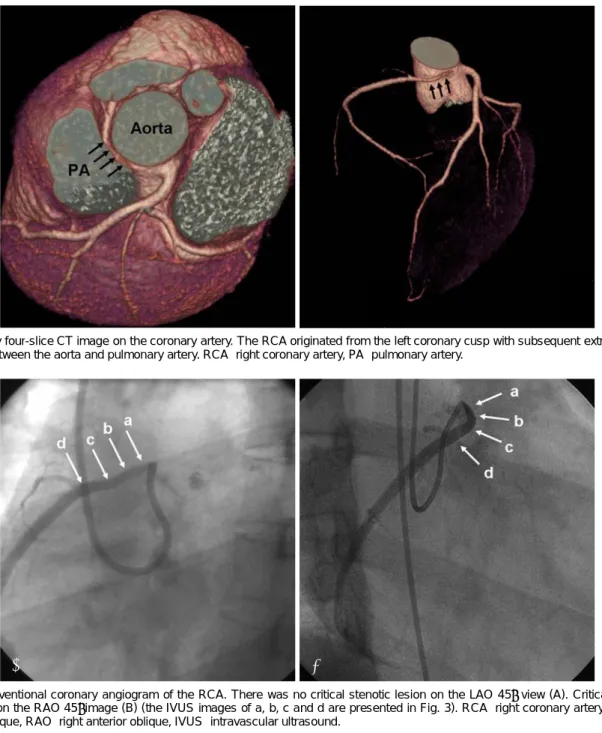
Fig. 2. Conventional coronary angiogram of the RCA. There was no critical stenotic lesion on the LAO 45° view (A). Critical stenosis was noted on the RAO 45°image (B) (the IVUS images of a, b, c and d are presented in Fig. 3). RCA: right coronary artery, LAO: left anterior oblique, RAO: right anterior oblique, IVUS: intravascular ultrasound.
Fig. 1. Sixty four-slice CT image on the coronary artery. The RCA originated from the left coronary cusp with subsequent extrinsic com- pression between the aorta and pulmonary artery. RCA: right coronary artery, PA: pulmonary artery.
392
·Extrinsic Compression of Anomalous RCAof MDCT, noninvasive imaging of coronary artery anat- omy has recently become possible. The anomaly of the coronary artery in our case was also easily detected by this noninvasive technique. Future development of MDCT technology should provide higher spatial resolution and this would be more informative for evaluating the me- chanisms by which myocardial ischemia is provoked in patients with an anomalous origin of the RCA.
REFERENCES
1) Yamanaka O, Hobbs RE. Coronary artery anomalies in 126,595 patients undergoing coronary arteriography. Cathet Cardiovasc Diagn 1990;21:28-40.
2) Roberts WC, Siegel RJ, Zipes DP. Origin of the right coronary artery from the left sinus of valsalva and its functional conse- quences: analysis of 10 necropsy patients. Am J Cardiol 1982;
49:863-8.
3) Kragel AH, Roberts WC. Anomalous origin of either the right or left main coronary artery from the aorta with subsequent cours- ing between aorta and pulmonary trunk: analysis of 32 necropsy cases. Am J Cardiol 1988;62:771-7.
4) Ichikawa M, Sato Y, Komatsu S, Hirayama A, Kodama K, Saito S.
Multislice computed tomographic findings of the anomalous ori- gins of the right coronary artery: evaluation of possible causes of myocardial ischemia. Int J Cardiovasc Imaging 2007;23:353-60.
5) Sato Y, Inoue F, Kunimasa T, et al. Diagnosis of anomalous origin of the right coronary artery using multislice computed tomogra- phy: evaluation of possible causes of myocardial ischemia. Heart Vessels 2005;20:298-300.
6) Benge W, Martins JB, Funk DC. Morbidity associated with ano- malous origin of the right coronary artery from the left sinus of valsalva. Am Heart J 1980;99:96-100.
7) Virmani R, Chun PK, Goldstein RE, Robinowitz M, McAllister HA. Acute takeoffs of the coronary arteries along the aortic wall and congenital coronary ostial valve-like ridges: association with sudden death. J Am Coll Cardiol 1984;3:766-71.
8) Kim MS, Han JK, Lee SE, et al. Cases of right ventricular myo- cardial infarction in patients with an absent or hypoplastic right coronary artery. Korean Circ J 2007;37:84-6.
9) Shin SH, Park CG, Kim YH, et al. Anomalous origin of the right coronary artery from the diagonal branch of the left anterior de- scending coronary artery. Korean Circ J 2004;34:615-7.
10) Hyun DW, Hur SH, Han SW. A case of right coronary artery originating from distal left circumflex (single coronary artery).
Korean Circ J 2003;33:1044-7.
A B
D C
Fig. 3. Intravascular ultrasound (IVUS) of the RCA. The IVUS study showed no atherosclerotic plaque burdens on the entire RCA. How- ever, the lumen was squeezed to a spindle shape (A) in the proximal portions because of the extrinsic compression between two large vessels (angiographic images of A, B, C and D are presented in Fig. 2). RCA: right coronary artery, CSA: cross sectional area.