• 검색 결과가 없습니다.

Cardiac Sarcoidosis Presenting as Complete Atrioventricular Block: Findings on PET/MRI

N/A
N/A
Protected

Academic year: 2022

Share "Cardiac Sarcoidosis Presenting as Complete Atrioventricular Block: Findings on PET/MRI"

Copied!
2
0
0

로드 중.... (전체 텍스트 보기)

전체 글

(1)Korean Circ J. 2018 Oct;48(10):947-948 https://doi.org/10.4070/kcj.2018.0045 pISSN 1738-5520·eISSN 1738-5555. Images in Cardiovascular Medicine. Cardiac Sarcoidosis Presenting as Complete Atrioventricular Block: Findings on PET/MRI Chan-Hee Lee , MD, PhD1, Jang-Won Son , MD, PhD1, and Eun-Jung Kong , MD, PhD2 Division of Cardiology, Department of Internal Medicine, Yeungnam University Medical Center, Daegu, Korea Department of Nuclear Medicine, Yeungnam University Medical Center, Daegu, Korea. 1. 2. Received: Feb 4, 2018 Revised: May 4, 2018 Accepted: May 30, 2018 Correspondence to Chan-Hee Lee, MD, PhD Division of Cardiology, Department of Internal Medicine, Yeungnam University Medical Center, Yeungnam University College of Medicine, 170, Hyeonchung-ro, Nam-gu, Daegu 42415, Korea. E-mail: [email protected] Copyright © 2018. The Korean Society of Cardiology This is an Open Access article distributed under the terms of the Creative Commons Attribution Non-Commercial License (https:// creativecommons.org/licenses/by-nc/4.0) which permits unrestricted noncommercial use, distribution, and reproduction in any medium, provided the original work is properly cited. ORCID iDs Chan-Hee Lee https://orcid.org/0000-0001-9338-0679 Jang-Won Son https://orcid.org/0000-0002-8109-5018 Eun-Jung Kong https://orcid.org/0000-0002-2300-4675 Conflict of Interest The authors have no financial conflicts of interest. Author Contributions Data curation: Son JW; Methodology: Kong EJ; Software: Kong EJ; Writing - original draft: Lee CH; Writing - review & editing: Lee CH.. https://e-kcj.org. A 72-year-old male was transferred from a health promotion center with complete atrioventricular block (CAVB, Figure 1A). He complained of dyspnea, and had a 1-year history of hypertension and 50 pack-year history of smoking. A whole body fluorodeoxyglucose (FDG) positron emission tomography/magnetic resonance imaging (PET/MRI), taken during a medical check-up, revealed multiple supraclavicular and mediastinal lymphadenopathies, and a hot FDG uptake in the septal area of the heart (Figure 1B, arrow). An FDG cardiac PET/MRI was performed for a further evaluation 3 weeks later. The PET/MRI revealed a late gadolinium enhancement (LGE) in the mid-wall of the basal septum (Figure 1C and 1D, arrowheads) with a corresponding FDG uptake (Figure 1E and 1F, arrowheads). Bronchoscopy was performed for an endobronchial ultrasound guided lymph node (LN) biopsy, and an endobronchial mass was incidentally found in the right upper apical segment. The endobronchial mass was confirmed as a squamous cell carcinoma. A permanent pacemaker implantation was performed with steroid therapy under planning for an operation. A right upper lobectomy was performed with a mediastinal LN dissection (2R, 4R, 10R, 3A, 7, 8, and 9) by video-assisted thoracoscopic surgery. The LN biopsy revealed chronic granulomatous inflammation without a tumor metastasis. He had a final diagnosis of sarcoidosis involving the heart and mediastinal LN, concurrent with lung cancer. In this case, the cardiac PET/MRI well-visualized the myocardial inflammation and fibrosis related to the CAVB. Comprehensive imaging with cardiac PET/MRI could be the gold standard for a clinical diagnosis1)2) in cardiac sarcoidosis.. REFERENCES 1. Dweck MR, Abgral R, Trivieri MG, et al. Hybrid magnetic resonance imaging and positron emission tomography with fluorodeoxyglucose to diagnose active cardiac sarcoidosis. JACC Cardiovasc Imaging 2018;11:94-107. PUBMED | CROSSREF. 2. Terasaki F, Yoshinaga K. New guidelines for diagnosis of cardiac sarcoidosis in Japan. Ann Nucl Cardiol 2017;3:42-5. CROSSREF. 947.

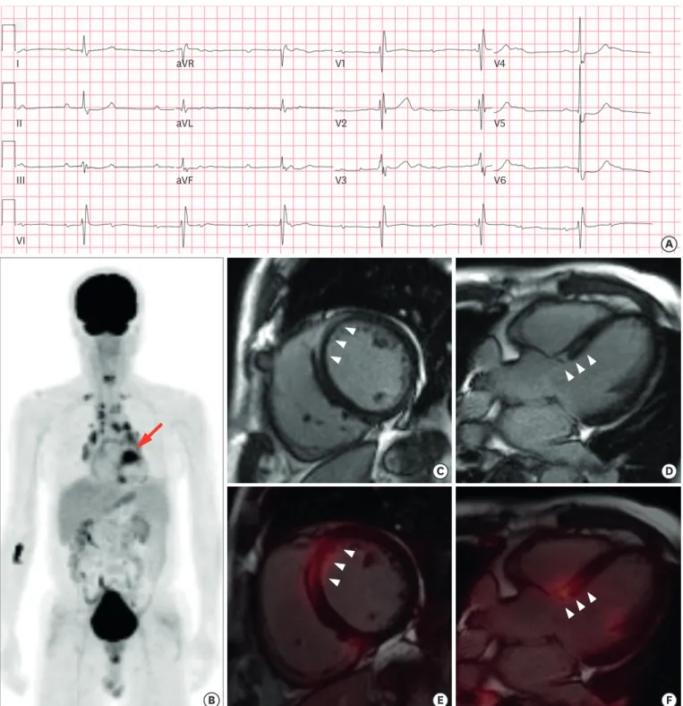
(2) PET/MRI in Cardiac Sarcoidosis. I. aVR. V1. V4. II. aVL. V2. V5. III. aVF. V3. V6. VI. A. B. C. D. E. F. Figure 1. (A) Electrocardiogram; PET/MRI. (B) A whole-body image of the FDG uptake. T1-weighted image; (C) short-axis and (D) long-axis, and cardiac PET/MRI image; (E) short-axis and (F) long-axis. FDG = fluorodeoxyglucose; MRI = magnetic resonance imaging; PET = positron emission tomography.. https://e-kcj.org. https://doi.org/10.4070/kcj.2018.0045. 948.

(3)

수치

Figure 1. (A) Electrocardiogram; PET/MRI. (B) A whole-body image of the FDG uptake. T1-weighted image; (C) short-axis and (D) long-axis, and cardiac PET/MRI  image; (E) short-axis and (F) long-axis

참조

관련 문서

A study on galvanizing of Start-ups atmosphere based on Smart specialization and the entrepreneurial university - Technion institute of Technology, Hebrew University-.. Han, Jung

– Mechanisms of overpressure generation – Estimating pore pressure at depth.. Zoback MD, 2007, Reservoir Geomechanics,

In order to get the feature of pedestrian, a block-by-block histogram is created using the direction of the gradient based on HOG (Histogram of

Seung Soo Jang Sung Gyun Shin Min Jae Lee Sang Soo Han Chan Ho Choi Sungkyum Kim Woo Sung Cho and Song Hyun Kim POSTECH Yeong Rok Kang Wol Soon Jo Soo Kyung Jeong and

Purpose: We investigated the impact of treatment on electroencephalogram (EEG) findings, and determined efficacy of antiepileptic drugs according to EEG findings

Dong Won Lee, Young Dug Bae, Suk Kwon Kim, Hee Yun Shin, Bong Guen Hong, Hyun Kyu Jung, Yang Il Jung, Jeong Yong Park, Byung Kwon Choi, and Yong Hwan Jeong(KAERI). P07B04

P04D06 Microstructure Analysis on Beryllium Reflector Blocks of Research Reactors Suk Hoon Kang, Jinsung Jang, Yong-Hwan Jeong, Chang-Hee Han, Yang-Il Jung, and Tae Kyu

녹내장의 진행정도는 시야검사 상 평균 mean deviation (MD) 값이 –11.96 dB 으로 중기 녹내장 소견 을 보였고, 녹내장의 종류는 일차성개방우각녹내장 (primary