한국컴퓨터정보학회 동계학술대회 논문집 제25권 제1호 (2017. 1)
235
엑스선촬영 각도를 측정할 수 있는 장치 개발과 흉부 X선 영상촬영에서의 적용
김태훈O, 허동운*, 류종현*, 정창원*, 전홍영*, 김규겸*, 홍지민*, 장미연*, 김대원**, 윤권하***
O*원광대학교, 영상과학 연구 센터, **원광대학교 의과대학, 신경외과
***원광대학교 의과대학, 영상의학과
e-mail: [email protected],[email protected], {jhryu, mediblue, zip80,kgkim}@wku.ac.kr, [email protected], {wkdaldus5, kimdw, khy1646}@wku.ac.kr
Development of portable digital radiography system with device for sensing X-ray source-detector angle and its
application in chest imaging
Tae-Hoon KimO, Dong-Woon Heo*, Jong-Hyun Ryu*, Chang-Won Jeong*, Hong Young Jun*, Kyu Gyeom Kim*, Jee Min Hong*, Mi Yeon Jang*, Dae Won Kim**, Kwon-Ha Yoon***
O*Imaging Science Research Center, Wonkwang University
**Dept. of Neurosurgery, Wonkwang University School of Medicine
***Dept. of Radiology, Wonkwang University School of Medicine
● 요 약 ●
This study was to develop a portable digital radiography (PDR) system with a function measuring the X-ray source-with-detector angle (SDA) and to evaluate the imaging performance for the diagnosis of chest imaging. The SDA device consisted of an Arduino, an accelerometer and gyro sensor, and a Bluetooth module. According to different angle degrees, five anatomical landmarks on chest images were assessed using a 5-point scale. Mean signal-to-noise ratio and contrast-to-noise ratio were 182.47 and 141.43. Spatial resolution (10% MTF) and entrance surface dose were 3.17 lp/mm (157μm) and 0.266mGy. The angle values of SDA device were not significant difference as compared to those of the digital angle meter. In chest imaging, SNR and CNR values were not significantly different according to different angle degrees (repeated-measures ANOVA, p>0.05). The visibility scores of the border of heart, 5th rib and scapula showed significant differences according to different angles (rmANOVA, p<0.05), whereas the scores of the clavicle and 1st rib were not significant. It is noticeable that the increase in SDA degree was consistent with the increase of visibility score. Our PDR with SDA device would be useful to be applicable to clinical radiography setting according to the standard radiography guideline at various fields.
키워드: portable digital radiography (PDR) system, X-ray source-detector angle (SDA), radiography setting.
한국컴퓨터정보학회 동계학술대회 논문집 제25권 제1호 (2017. 1)
236
I. Introduction
In clinical radiography, X-ray source-to- detector distance (SDD) and X-ray source-with- detector angle (SDA) are important factors for acquiring appropriate image quality. According to standard clinical radiography guidelines [1,2], it is recommended for imaging positions as follows: SDD of 180 cm, and SDA of 0° (vertical incidence) for chest PA (posterior to anterior) imaging; 100 cm SDD and 15-20° SDA (head side direction incidence) for cervical spine AP (anterior to posterior) imaging;
and 100 cm SDD and 5-7° SDA (head side direction incidence) for lateral knee imaging. Recently, most of commercial portable digital radiography (PDR) systems are well-equipped a tape measure for SDD, whereas are relatively less equipped a specific SDA device [3]. In clinical situations, it has often been encountered the unexpected conditions or extreme medical situations such as imaging experiments for immobilized bedridden patients, post-stroke hemiplegic patients and severely injured patients [4]. From these viewpoints, radiographic imaging system automatically determining SDD and SDA can be helpful for clinical radiographic settings. However, there were few studies focusing on the SDA measurement in clinical radiography.
Therefore, the purpose of this study was to develop a PDR system including a device for obtaining the SDA and to assess the imaging performance for the diagnosis of chest imaging.
II. Materials and Methods
1. Development and performance test of X-ray SDA device
To determine the flip angle degree between an X-ray source and a detector, we developed an X-ray SDA device that consisted of a main board (Arduino Uno Rev.3), a 6 degrees of freedom (DOF) inertial measurement unit (IMU) shield (embedded in the ADXL345 accelerometer and the ITG-3200 gyro), and a Bluetooth module (HC-05 master, HC-06 slave) [5].
Fig. 1. System block of the device for obtaining the X-ray source-with-detector angle (SDA).
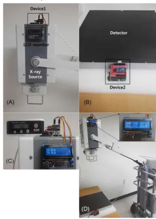
This device was attached to the X-ray source (device 1) and detector (device 2), respectively. The Arduino main board was the core processing component coded by the Sketch software tool. The 6 DOF IMU shield included an accelerometer and gyro sensor, and transmitted the angle data to the Arduino through I2C communication after sensing the angle data from the X-ray source and detector. The Bluetooth module was connected to the Arduino core processor.
Fig. 2. The device for obtaining the SDA, which was attached on an X-ray source (A) and a detector (B). Photography showed the initial setting for obtaining the angle degree using
the SDA device and digital angle meter (C). The measured angle degree using PDR with SDA device displayed on a
LCD monitor (D).
The SDA value was measured as following steps. The Arduinos of devices 1 and 2 received angle data through I2C communication from the 6 DOF IMU Shield. Then, the Arduinos calculated the angle values for device 1 (XS and YS) and device 2 (XD and YD) by applying an algorithm to the angle data. The Bluetooth module (HC-06) on device 2 received the angle values (XD and YD) via serial communication from the Arduino, and transmitted the angle values (XD and YD) to the Bluetooth module (HC-05) of device 1 in real-time. The Bluetooth module (HC-05) of device 1 transmitted the angle values (XD and YD) to the Arduino via serial communication. Finally, the Arduino of device 1 calculated the SDA difference (XSDA and YSDA)
한국컴퓨터정보학회 동계학술대회 논문집 제25권 제1호 (2017. 1)
237 as follows: XSDA = XS - XD, YSDA = YS – YD.
To test the performance of SDA device, we used a digital angle meter (DL-155V, STS, Tokyo, Japan) with accuracies of 0.1%/degree (0-10°/80-90° degree), and 0.2%/degree (10-80°) as a standard reference device.
2. Measurements of image quality and radiation dose
The image quality of the PDR system was evaluated as signal-to-noise ratio (SNR), contrast-to-noise ratio (CNR), and spatial resolution using a bar phantom (X-ray test pattern type 18; FUNK, Berlin, Germany). The SNR and CNR were calculated as the ratio of the lead bar (0.05-mm thick) value to noise and the ratio of the lead bar-air contrast to noise, respectively.
Mean SNR and CNR values were obtained from six image sets using the developed PDR system. The modulation transfer function (MTF) has been used to evaluate spatial resolution of imaging systems. This study was used a bar phantom to generate the MTF curve and was measured image resolution at 10% on MTF curve.
The radiation dose was calculated using the method described by the International Commission on Radiological Protection.
The ESD measurement was performed using a dosimeter (Piranha, RTI Electronics, Molndal, Sweden). This study was measured the ESD under the conditions of 80 kVp tube voltage, 4 mAs current, 100 ms and 1 meter SDD.
3. Chest radiography according to different angle degrees
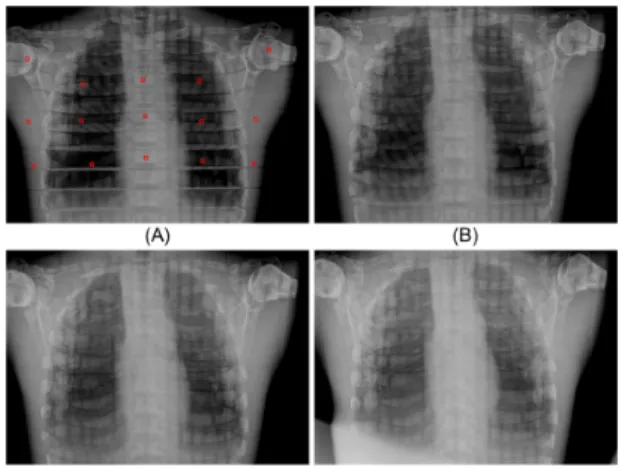
Chest radiographs were performed six times under the conditions of different angles using the developed PDR system with SDA device. For the analysis of chest images, SNR and CNR according to different angle degrees were measured from 15 different points (Fig. 3A).
Two expert radiologists (more than 10 years of experience) blindly evaluated each chest image, and reached a consensus regarding the anatomic landmarks [19]. The 5 anatomic landmarks were the border of the heart, clavicle, 1st rib, 5th rib and scapula.
Each anatomic landmark on chest image data was analyzed according to the radiological diagnosis on a 5-point scale: 1, definitely seen; 2, probably seen; 3, equivocal; 4, probably not seen; and 5, definitely not seen.
Fig. 3. Representative chest AP images obtained from the developed PDR according to different angles: (A) 0°, (B) 10°, (C) 20° and (D) 30° angle degree. The squares of red line (30×30 pixels) on chest image indicated 15 points
for SNR and CNR measurements.
4. Statistical analysis
All statistical analyses were performed using the Statistical Package for the Social Sciences (SPSS ver. 20, Chicago, IL, USA) software. The angle difference between both angle meters (SDA device and digital angle meter) was analyzed with the independent two sample t-test. According to the different angle degrees, the variation in image qualities and visibility scores were analyzed with the repeated- measures analysis of variance (rmANOVA) and Tukey's post hoc tests.
Ⅲ. Results
1. Performances of PDR
The size and weight of the PDR system were 723(Wh)×650(L)×1376 (H) mm and approximately 60 kg, respectively. Mean SNR and CNR were 182.47±6.75 and 141.43±6.08. The spatial resolution at 10% MTF was 3.17 lp/mm (157 μm) and the ESD was 0.266 mGy.
2. Performance of SDA device
The SDA device was attached to the X-ray source and detector, respectively, and its size was 55 (W)×80 (L)×35 (H) mm. The angle values obtained from the SDA device were displayed real-time on the LCD monitor (1 times/sec). There was no significant angle difference between digital angle meter and our SDA device (p > 0.05).
한국컴퓨터정보학회 동계학술대회 논문집 제25권 제1호 (2017. 1)
238
Degree (˚) 0˚ 10˚ 20˚ 30˚ p-value*
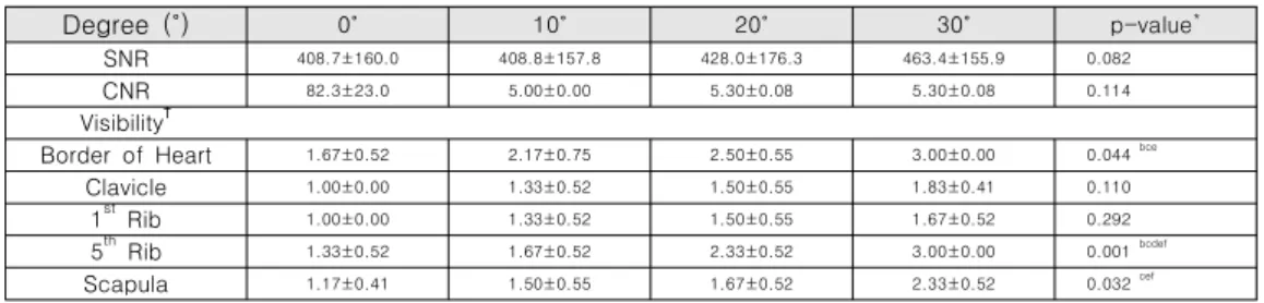
SNR 408.7±160.0 408.8±157.8 428.0±176.3 463.4±155.9 0.082
CNR 82.3±23.0 5.00±0.00 5.30±0.08 5.30±0.08 0.114
Visibility†
Border of Heart 1.67±0.52 2.17±0.75 2.50±0.55 3.00±0.00 0.044 bce
Clavicle 1.00±0.00 1.33±0.52 1.50±0.55 1.83±0.41 0.110
1st Rib 1.00±0.00 1.33±0.52 1.50±0.55 1.67±0.52 0.292
5th Rib 1.33±0.52 1.67±0.52 2.33±0.52 3.00±0.00 0.001 bcdef
Scapula 1.17±0.41 1.50±0.55 1.67±0.52 2.33±0.52 0.032 cef
Data are presented as mean ± SD after six times measurements.
* The significant difference between different angle degrees was analyzed with repeated-measures ANOVA with Tukey’s post hoc test: a, 0 vs 10; b, 0 vs 20; c, 0 vs 30; d, 10 vs 20; e, 10 vs 30; and f, 20 vs 30.
Table 2. SNR, CNR and visibility values on chest AP images according to different angles Degree
(˚) Digital angle meter Developed SDA device
0 0.00±0.00 0.00±0.00
5 5.00±0.00 5.30±0.08
10 10.00±0.00 10.70±0.08
15 15.00±0.00 15.50±0.08
20 20.00±0.00 20.87±0.04
25 25.00±0.00 25.80±0.08
30 30.00±0.00 30.70±0.08
35 35.00±0.00 35.77±0.04
40 40.00±0.00 40.77±0.12
45 45.00±0.00 45.77±0.04
50 50.00±0.00 50.80±0.08
55 55.00±0.00 55.77±0.04
60 60.00±0.00 60.73±0.04
p-value* 0.865
* The difference between angle values of digital angle meter and developed SDA device was analyzed with the independent two sample t-test.
Table 1. Angle values and angle difference of digital angle meter and developed X-ray source-with- detector angle (SDA) device
3. Chest radiographic study according to different angles for clinical application
Imaging quality and visibility scores were summarized in Table 2. SNR and CNR values were not significantly different from different angle degrees (rmANOVA, p>0.05). The visibility score of the border of the heart, 5th rib and scapula were significantly different according to different angles (rmANOVA, p<0.05), whereas the clavicle and 1st rib were not significant.
Objective image qualities were not significantly different in this study, thus it could be considered to be applicable appropriated angle degrees in clinical experiments. However, it is noticeable that the increase in SDA degree was consistent with the increase of visibility score.
Ⅳ. Conclusions
Our PDR system with SDA device provided accurate SDA and SDD values according to the clinical radiography standard guidelines. This system would be useful for the applications in clinical radiography.
Acknowledgment
This study was supported by the grants of the Korean Health Technology R&D Project (HI12C0110, Ministry of Health &
Welfare), and the National Research Foundation of Korea (NRF) (2016M3A9E9941547 and 2016M3A9A7918499)
References
[1] Poletti JL, McLean D, "The effect of source to image-receptor distance on effective dose for some common X-ray projections," Br J Radiol, Vol. 78, No.
933, pp. 810-815, 2005.
[2] Bontrager KL, Lampignano J, "Textbook of radiographic positioning and related anatomy," 8th ed. St. Louis, MO:
Elsevier/Mosby, 2014.
[3] Lin PJ, Schueler BA, Balter S, et al, "Accuracy and calibration of integrated radiation output indicators in diagnostic radiology: A report of the AAPM Imaging Physics Committee Task Group 190," Med Phys, Vol.
42, No. 12, pp. 6815-6829, 2015.
[4] Alkatout I, "Communicative and ethical aspects of physician-patient relationship in extreme situations,"
Wien Med Wochenschr, Vol. 165(23-24): 491-498, 2015.
[5] D'Ausilio A, "Arduino: a low-cost multipurpose lab equipment," Behav Res Methods, Vol. 44, No. 2, pp.
305-313, 2012.