Anti-inflammatory Activity of Antimicrobial Peptide Papiliocin 3 Derived from the Swallowtail Butterfly, Papilio xuthus
Yong Pyo Shin†, Joon Ha Lee†, In-Woo Kim, Minchul Seo, Mi-Ae Kim, Hwa Jeong Lee, Minhee Baek, Seong Hyun Kim and Jae Sam Hwang*
Department of Agricultural Biology, National Institute of Agricultural Sciences, Rural Development Administration, Wanju 55365, Korea Received August 10, 2020 /Revised September 9, 2020 /Accepted September 15, 2020
The development of novel peptide antibiotics with potent antimicrobial activity and anti-inflammatory activity is urgently needed. In a previous work, we performed an in-silico analysis of the Papilio xuthus transcriptome to identify putative antimicrobial peptides and identified several candidates. In this study, we investigated the antibacterial and anti-inflammatory activities of papiliocin 3, which was se- lected bioinformatically based on its physicochemical properties against bacteria and mouse macro- phage Raw264.7 cells. Papiliocin 3 showed antibacterial activities against E. coli and S. aureus without inducing hemolysis and decreased the nitric oxide production of the lipopolysaccharide-induced Raw264.7 cells. Moreover, ELISA and Western blot analysis revealed that papiliocin 3 reduced the ex- pression levels of pro-inflammatory enzymes, such as inducible nitric oxide synthase (iNOS), cyclo- oxygenase-2 (COX-2), and prostaglandin E2 (PGE2). In addition, we examined whether papiliocin 3 could inhibit the expression of pro-inflammatory cytokines (interleukin-6 and interleukin-1β) in LPS- induced Raw264.7 cells. We found that papiliocin 3 markedly reduced the expression level of cyto- kines through the regulation of mitogen-activated protein kinases (MAPK) and nuclear factor kappa B (NF-κB) signaling. We also confirmed that papiliocin 3 binds to bacterial cell membranes via a spe- cific interaction with lipopolysaccharides. Collectively, these findings suggest that papiliocin 3 could be a promising molecule for development as a novel peptide antibiotic.
Key words : Anti-inflammatory activity, antimicrobial activity, antimicrobial peptide, Papilio xuthus, RNA sequencing
†Authors contributed equally.
*Corresponding author
*Tel : +82-63-238-2974, Fax : +82-63-238-3833
*E-mail : [email protected]
This is an Open-Access article distributed under the terms of the Creative Commons Attribution Non-Commercial License (http://creativecommons.org/licenses/by-nc/3.0) which permits unrestricted non-commercial use, distribution, and reproduction in any medium, provided the original work is properly cited.
서 론
광범위한 항생제의 사용으로 인해 내성이 증가함에 따라 지금까지 미생물에 노출되지 않은 새로운 계열의 항생제 개발 이 필요하게 됐다[6, 14]. 항균 펩타이드(antimicrobial peptides, AMPs)는 광범위한 항균(antibacterial)과 항진균(antifungal) 및 항바이러스(antiviral) 활성을 가지고 있어 기존 항생제를 대체할 수 있는 새로운 후보물질로 주목 받고 있다. 현재까지 식물과 곤충에서부터 척추동물에 이르기까지 다양한 종에서 1,500 종류 이상의 AMPs이 발견됐으며[13], 이 분자들은 대부 분의 생명체에서 선천성 면역의 중요한 성분으로 인식되고 있다[1, 5, 13, 15]. 이들 AMPs의 작용기전(action mechanism) 은 박테리아 세포막의 탈분극(depolarization)의 유도나 투과 (permeabilization)하여 활성을 나타내며, 몇몇 AMPs는 온전
한 막을 통과하여 세포 내 물질과 상호작용을 한다[4, 7, 16, 46].
AMPs는 액체 상태에 존재할 때 나타내는 2차 구조를 기반 으로 여러 유형으로 나눠진다[18, 21]. β-sheet 형태의 구조를 갖는 펩타이드는 분자 내 구조를 안정화 시키고 세포막 통과 에 도움을 줄 수 있는 이황화 결합(disulfide bond)을 가지고 있으며, α-나선 형태의 구조를 갖는 일부 펩타이드는 시스테인 잔기를 가지고 있어 분자 내 이황화 브릿지(disulfide bridge) 형성한다[11, 45]. 대부분의 AMPs는 다수의 소수성 잔기들이 존재하며 소수성 잔기들 사이에 상호작용을 통해 중합체 형태 를 만들 수 있으며 이를 통해 박테리아 막에 대한 친화성을 높인다[27, 35, 39]. AMPs가 최적의 항균 활성을 나타내는데 있어 분자 내 전하의 밀도, 소수성 및 폴리머 사슬의 길이 사이 의 균형이 중요한 것으로 사료된다[32, 33]. 양전하로 하전된 아미노산의 수를 증가시키거나 펩타이드 사슬에서 이들의 위 치를 변화시키는 것은 AMPs의 2차 구조에 영향을 미쳐 그들 의 항균활성에 추가로 영향을 줄 수 있다. 따라서, 펩타이드의 전하, 소수성 및 길이의 조합은 AMPs의 활성에 있어 매우 중요하다[8, 43].
곤충들은 다양한 항균 펩타이드와 단백질을 생산한다. 곤충 의 항균활성에 대한 연구는 박테리아에 의해 면역화된 가중나 무고치나방(Samia Cynthia) [2]과 세크로피아나방(Hyalophora cecropia) [9]의 번데기에서 처음 관찰되었으며, 그 후 면역화를
유도한 노랑초파리(Drosophila melanogaster)의 성충에서 추가 로 확인되었다[40]. 곤충에서 발견된 최초의 AMPs는 1980년 세크로피아나방의 번데기로부터 정제된 세크로핀으로 이 후 다양한 곤충으로부터 150 종류 이상의 AMPs가 발견되었다 [20]. 대부분의 곤충 AMPs는 분자량이 작고 양이온성 및 염기 성 펩타이드로 박테리아와 진균에 대해 활성을 나타내며, 몇 몇 AMPs는 선충이나 바이러스들에 대해서도 활성을 갖는다.
척추동물의 AMPs 중 포유동물에서 발견된 디페신(hBD-2, hBD-3, hBD-4)과 카델리시딘(LL-37, PR-39)은 면역자극 및 조 절에 관한 기능과 작용기전(mechanism)에 대해 연구가 잘 되 어 있다[18, 36]. 이들 AMPs는 면역세포로부터 케모카인(che- mokines)과 사이토카인(cytokines)의 생산을 조절하고, 패혈 증 발생을 제한하며, 상처치유 및 혈관형성에 관여하는 것으 로 in vivo와 in vitro 연구를 통해 확인되었다[3, 29]. 최근 연구 들에서 트리아토민 노린재(triatomin bug)와 깔따구(midge) 의 AMPs와 같은 일부 곤충의 AMPs도 면역반응에 관여하는 것으로 확인되었다[36, 42]. 그람음성균과 같은 병원성 미생물 의 감염 시 다량의 LPS 방출은 과도하게 선천성 면역반응을 자극할 수 있으며 패혈성 쇼크와 같은 심각한 문제들을 야기 할 수 있다[28]. 곤충의 세크로핀과 세크로핀-유도체는 LPS로 결합할 수 있는 능력을 가지고 있으며 in vitro와 in vivo 연구를 통해 항염증 활성을 구명하였다. 나비목의 세크로피아나방과 호랑나비(Papilio xuthus) 그리고 산누에나방(Antheraea pernyi) 으로부터 확인된 AMPs는 nitric oxide의 생성을 억제하고 LPS 로부터 유도된 면역세포에서 전염증성 사이토카인 유전자의 발현을 억제하는 것으로 알려졌다[24, 31, 44]. 또한, 감염동물 모델을 이용한 in vivo 실험에서 복강 내로 주입한 세크로피아 나방의 AMPs에 의해 혈액 내 내독소(endotoxin)의 감소와 생 존율이 증가함을 확인 할 수 있었다[12, 23]. 이러한 연구결과들 은 곤충의 AMPs가 항균 및 항염증 기능을 통해 생의학적 치료 제로 적용할 수 있는 잠재성을 증명하였다. 이러한 관점에서 본 연구는 호랑나비 유충의 전사체 분석을 통해 선별한 항균 펩타이드 파필리오신 3의 항균활성 및 항염증 기능을 입증하 여 파필리오신 3의 의학적 활용 가능성을 확인해 보았다.
재료 및 방법
실험곤충
호랑나비(Papilio xuthus)는 국립농업과학원의 곤충사육동 에서 계대사육을 통해 유지되어오고 있다. 호랑나비 유충의 먹이원은 기주식물(탱자 또는 산초나무)를 사용하였으며 23
℃, 65%의 상대습도 조건으로 16L : 8D의 광주기로 사육되었다.
펩타이드
파필리오신 3은 애니젠(주)에서 고상법을 이용하여 합성하 였고, 95% 이상 순도의 펩타이드만을 사용하였다. 합성한 펩
타이드는 산성화시킨 증류수(0.01% 아세트산용액)에 녹여서 사용하였으며 사용 전 까지 -20℃에 보관하였다.
항균활성
파필리오신 3의 항균활성은 radial diffusion assay [41] 방 법을 통해 테스트하였다. 펩타이드 시료는 0.01% 아세트산용 액으로 준비하였으며 펩타이드 시료의 농도는 일정량으로 se- rial dilution 하였다. Radial diffusion assay에 사용한 Escher- ichia coli, Staphylococcus aureus는 3%(w/v) tryptic soy broth (TSB)에서 37℃, 180 rpm의 조건으로 18시간 배양한 후 대수 증식기(log phase)에 들어가도록 동일한 조건에서 2시간 30분 간 2차 배양하였다. Citrate phosphate buffer (9 mM sodium phosphate, 1 mM sodium citrate, pH7.4)와 1%(w/v) type Ⅰ (low EEO) agarose, 0.03% TSB로 구성된 멸균시킨 underlay gel에 배양된 미생물(4×10⁶ colony forming units)을 각각 넣고 혼합해준 뒤 사각플레이트에 붓고 underlay gel이 굳으면 지 름 3 mm의 펀치를 이용하여 구멍을 내어 준비하였다. Serial dilution을 통해 준비한 농도별 펩타이드들은 5 µl씩 구멍에 넣었다. 시료들이 확산되도록 37℃에서 3시간 동안 배양한 후, 멸균된 overlay agar (6% TSB, 1% agarose) 10 ml을 붓고 37℃
에서 하룻밤 동안 추가 배양하였다. 다음날 미생물이 성장하 지 못한 명확한 영역(clear zone)의 부위들을 확인하고 지름을 0.1 mm까지 측정하여 그 값을 units로 표현하였다(0.1 mm=1 units).
용혈활성
다양한 농도(6.25, 12.5, 25, 50, 100 μg/ml)의 펩타이드 시료 20 μl로 인산완충식염수(phosphate buffered saline, PBS)에 있 는 2.5% (v/v) ICR 마우스 적혈구 용액을 180 μl 넣어 주었다.
벌 독으로부터 분리된 α-나선 구조의 항균 펩타이드인 멜리틴 (melittin)은 적혈구를 파괴하는 양성 대조군으로 사용하였다.
펩타이드와 적혈구의 혼합물은 37℃에서 30분간 반응시킨 후 600 μl의 PBS를 각각의 튜브에 추가로 넣어주었다. 원심분리 기를 이용하여 10,000×g에서 3분간 원심분리 후 상등액을 취 하여 분광 광도계로 540 nm에서 흡광도를 측정하였다. 마우스 적혈구는 농촌진흥청 국립농업과학원 동물윤리위원회의 승 인을 받아 실험이 진행되었다(승인번호: NAS-202014).
세포배양
Raw264.7 세포주는 Dulbecco’s Modified Eagle Medium (DMEM)에 10% fetal bovine serum (FBS)과 1% penicillin G- streptomycin을 첨가하여 37℃, 5% CO2 조건하에서 계대배양 하고 유지하였다.
세포생존율 측정
파필리오신 3의 세포독성을 확인하기 위하여 Raw264.7 세
Table 1. Sequences of primers used for qRT-PCR
cDNAs Primer sequences Accession number
iNOS Forward, 5’-CAGCACAGGAAATGTTTCAGC-3’
Reverse, 5’-TAGCCAGCGTACCGGATGA-3’
NM_010927
Cox-2 Forward, 5’-CAGACAACATAAACTGCGCCTT-3’
Reverse, 5’-GATACACCTCTCCACCAATGACC-3’
NM_011198
Il-6 Forward, 5’-GAGGATACCACTCCCAACAGACC-3’
Reverse, 5’-AAGTGCATCATCGTTGTTCATACA-3’
NM_031168
Il-1β Forward, 5’-CCTTCCAGGATGAGGACATGA-3’
Reverse, 5’-TGAGTCACAGAGGATGGGCTC-3’
NM_008361
Tnf-α Forward, 5’-ATGAGAAGTTCCCAAATGGC-3’
Reverse, 5’-CTCCACTTGGTGGTGGTTTGCTA-3’
NM_013693
Gapdh Forward, 5’-AAGGTCATCCCAGAGCTGAA-3’
Reverse, 5’-CTGCTTCACCACCTTCTTGA-3’
NM_008084 포를 96-well cell culture plate에 2×10⁴ cells/well로 분주하고
18시간 동안 배양하였다. 그 후 파필리오신 3을 25, 50, 100, 200 μg/ml의 농도로 처리하여 24시간 동안 추가 배양한 후 MTS (3-(4,5-dimethylthiazol-2-yl)-5-(3-carboxymethoxyphenyl)- 2-(4-sulfophenyl)-2H-tetrazolium) reagent를 사용하여 세포 생존율을 측정하였다. 흡광도는 multi detector (Beckman, DTX 8800, CA, USA)를 이용하여 490 nm 파장에서 측정하였다.
Nitric oxide (NO) assay
Raw264.7 세포로부터 lipopolysaccharide (LPS)에 의해 생 성된 nitric oxide (NO)의 양은 Griess reagent를 이용하여 측 정하였다. Raw264.7 세포는 8×10⁴ cells/well로 96-well cell culture plate에 분주하여 5% CO2 incubator에서 18시간 동안 배양하였다. NO 생성 유도를 위해 세포에 LPS를 처리하기 전 파필리오신 3을 25, 50, 100, 200 μg/ml의 농도로 1시간 동안 전 처리 한 뒤 100 ng/ml의 LPS를 처리하여 24시간 배양하였 다. 배양액의 상등액 100 μl를 취하여 Griess 시약과 반응 시킨 후 multi detector (Beckman, DTX8800, CA, USA)로 540 nm 에서 흡광도를 측정하여 NO 생성량을 측정하였다. 생성된 NO의 퍼센트는 LPS를 처리한 샘플을 최대 NO 생성량 기준 으로 하여 계산하였다.
Quantitative real-time PCR (qRT-PCR)
Raw264.7 세포는 1×10⁶ cells/well로 6-well plate에 분주하 여 5% CO2 incubator에서 18시간 동안 배양한 후 파필리오신 3을 25, 50, 100, 200 μg/ml의 농도로 1시간 전처리 한 후에 100 ng/ml의 LPS를 처리하여 17시간 배양하였다. 배양한 Raw264.7 세포는 phosphate buffered saline (PBS)로 2회 세척 하고 TRIzol reagent (Invitrogen, Carlsbad, CA) 1 ml을 첨가 하여 total RNA를 추출하였다. 동량의 RNA (1 μg)로부터 High Capacity cDNA Reverse Transcription Kit (Applied
Biosystems, Foster city, CA)를 이용하여 cDNA를 합성하였 고, 염증관련 유전자의 발현은 Table 1에서 제시한 각각의 pri- mer와 함께 AMPIGENE® qPCR Green Mix Lo-ROX (Enzo Life Sciences, USA)를 이용하여 ABI 7500 Real Time PCR System (PE Applied Biosystems, Foster City, CA, USA)으로 확인하였다.
Enzyme-linked immunosorbent assay (ELISA) Raw264.7 세포는 1×10⁶ cells/well로 6-well plate에 분주하 여 5% CO2 incubator에서 18시간 동안 배양한 후 파필리오신 3을 25, 50, 100, 200 μg/ml의 농도로 1시간 전처리 한 후에 100 ng/ml의 LPS를 처리하여 24시간 배양하였다. 그 후 배양 액으로부터 유리된 TNF-α, IL-6, IL-1β (ThermoFisher, Wal- tham, MA)와 PGE2 (RD systems, Minneapolis, USA) ELISA kit를 이용하여 측정하였다.
Limulus Amebocyte Lysate (LAL) assay
파필리오신 3과 LPS의 결합 여부를 확인하기 위해 Pierce Chromogenic Endotoxin Quant Kit (ThermoFisher, Wal- tham, MA)를 이용하여 제공된 시험법에 따라 분석하였다. 고 정된 농도의 LPS (0.5 pg/ml)와 PBS (control) 그리고 파필리 오신 3을 25, 50, 100, 200 µg/ml의 농도로 37℃에서 30분간 반응시킨 후 LAL 시료를 첨가하여 10분간 동일 조건에서 추 가로 배양하였다. 그 후 발색기질을 첨가하여 LPS로부터 활성 화된 효소에 의한 발색정도를 multi detector (Beckman, DTX8800, CA, USA)로 405 nm에서 흡광도를 측정하였다.
Western blot analysis
Raw264.7 세포는 1×10⁶ cells/well로 6-well plate에 분주하 여 5% CO2 incubator에서 18시간 동안 배양한 후 파필리오신 3을 25, 50, 100, 200 μg/ml의 농도로 1시간 전처리 한 후에
A B
Fig. 1. Antibacterial and hemolytic activities of papiliocin 3 against bacteria and mouse erythrocytes determined through radial diffusion assay and hemolysis assay. (A) Papiliocin 3 concentration (x-axis) was plotted against the diameter of the bacteria growth inhibition zone (y-axis) after incubation for 16 hr, and is expressed in units (1 mm=10 units). Data are presented as mean ± S.D. with three separate experiments. (B) Melittin was used as a control. Percentage hemolysis was calculated with the following equation: % Hemolysis=(A540 of sample–A540 of peptide-free control)/(A540 of 100% control–A540 of pep- tide-free control)×100.
100 ng/ml의 LPS를 처리하여 24시간 동안 배양하였다. 그 후 배양액을 제거 한 뒤 M-PER™ Mammalian Protein Extrac- tion Reagent (ThermoFisher, Waltham, MA)과 protease &
phosphatase inhibitor cocktail (ThermoFisher, Waltham, MA) 를 이용하여 세포를 lysis 시킨 후 원심분리(12,000 rpm, 15 min)하여 상등액을 수거하였다. BCA protein assay kit (Pierce, Rockford, IL, USA)로 정량한 동일양의 단백질을 sodium do- decyl sulfate-polyacrylamide gel electrophoresis (SDS-PAGE) 로 분리한 후, nitrocellulose membrane으로 transfer하였다.
Membrane을 5% skim milk로 1시간 동안 반응시켜 비특이적 단백질에 대한 반응을 차단하고 β-actin, iNOS, COX-2, anti- phospho and total-p44/p42 MAPK, anti-phospho and total- p38 MAPK, anti-phospho and total JNK, and IκB (Cell sig- naling, MA, USA) 항체를 각각 4℃에서 하룻밤 동안 반응시킨 후 horseradish peroxidase (HRP)가 결합되어 있는 2차 항체 로 1시간 반응시켰다. 각 반응 사이에 0.05% TBST로 10분씩 3회 수세 하였다. 그 후 항체에 대한 대응 단백질을 Western Lightning Plus (PerkinElmer, Boston, MA, USA)를 사용하여 FluorChem (Alpha Innotech Corporation, San Leandro, CA, USA) 이미지 분석장비로 확인하였다.
통계처리
모든 실험은 3회 반복하여 측정하였으며, 그 결과는 평균과 표준편차(mean ± SD)로 나타냈다. 실험군 간의 유의성은 Dun- can post-hoc ANOVA analysis를 통해 검정하였고, p<0.05일 때 군 간의 차이가 유의적인 것으로 판단하였다.
결과 및 고찰
파필리오신 3의 항균활성 및 용혈활성
호랑나비 유충의 전사체분석을 통해 선별한 항균 펩타이드 인 파필리오신 3은 radial diffusion assay 기법을 이용하여 미생물에 대한 항균활성을 확인하였다(Fig. 1A). 그 결과 대표 적 그람음성균인 대장균(E. coli)과 그람양성균인 황색포도상 구균(S. aureus)에 대해 파필리오신 3이 농도의존적으로 광범 위한 활성을 나타내었다. 세포독성을 확인하기 위한 용혈활성 테스트에서는 대조군으로 사용된 멜리틴(melittin)은 12.5 μg/
ml에서 약 90%의 용혈활성을 나타낸 반면 파필리오신 3은 최고농도인 100 μg/ml에서도 용혈활성이 없는 것으로 확인되 었다(Fig. 1B). 파필리오신 3은 14개의 아미노산으로 이루어져 있으며 중성 pH에서 총전하(net charge) +3의 값을 가지며 C-말단이 아미드화(amidation) 되어 있다. 양전하를 갖는 이러 한 항균 펩타이드들은 미생물의 세포막 표면에 존재하는 음전 하의 성분들과 상호작용을 통해 세포막에 이온채널(ion chan- nel)을 형성하거나 막을 가로지르는 구멍을 뚫어 항균활성을 나타내는 것으로 알려져 있다[16].
파필리오신 3의 세포독성 및 Nitric Oxide 생성 억제 파필리오신은 그람음성균(Escherichia coli JM 109)으로 면역 화를 유도한 호랑나비 유충으로부터 처음 유전자 및 존재를 확인했으며, 세크로피아나방으로부터 분리된 항균 펩타이드 cecropin A와 구조적으로 78.4%가 유사한 것으로 밝혀졌다 [25]. 현재까지 호랑나비로부터 파필리오신 1, 2의 존재가 확인
A B
Fig. 2. Cell viability and inhibition of NO production in Raw264.7 cells after papiliocin 3 treatment. (A) Cell viability was measured through the MTS assay after 24 hr incubation with the indicated concentrations of the peptides. (B) Raw264.7 cells were pretreated with papiliocin 3 for 1 hr prior to incubation with LPS for 24 hr. The nitrite in the culture media was measured with the Griess reagent. Data are presented as mean ± S.D. with three separate experiments. Statistical analyses were performed as described. #p<0.05 in comparison with control group. ***p<0.001 compared to the LPS-treated group. CTR, control; LPS, lipopolysaccharide.
됐으며 다양한 미생물들에 대해 광범위한 항균활성을 가지고 있는 것으로 밝혀졌다[25, 26]. 하지만, 기존에 밝혀진 펩타이 드의 아미노산 서열이 길어 새로운 치료제로서 개발하는데 한계가 있다. 이러한 문제를 해결하기 위해 최근 연구들에서 는 파필리오신의 아미노산 서열을 기초로 한 유도체를 고안하 고 그들의 항균 및 항염증 효능을 입증하는 연구들이 진행되 었다[23, 24].
호랑나비 유충으로부터 새로운 항균 펩타이드를 선별하기 위한 노력의 일환으로 전사체분석을 통해 선별된 파필리오신 3의 항염증 효능을 검정하기 위해 유효농도 설정을 위한 세포 독성 시험을 진행하였다. 세포독성 여부는 MTS assay로 확인 하였으며, Raw264.7 세포에 25, 50, 100, 200 μg/ml의 농도로 파필리오신 3을 처리하고 24시간 뒤에 MTS 시약을 처리하였 다. 그 결과 파필리오신 3은 200 μg/ml의 농도까지 세포생존 율이 90% 이상 유지하였으며(Fig. 2A), 이후 진행될 항염증 실험들은 200 μg/ml을 최대농도로 설정하여 실험을 진행하였 다. 파필리오신 3이 LPS로 활성화된 Raw264.7 세포에서 nitric oxide (NO)의 생성 정도에 미치는 영향을 확인하였다. 그 결과 최저농도인 25 μg/ml에서는 NO의 생성을 억제하는 효과를 보이지 못하였지만 50 μg/ml부터는 농도의존적으로 NO의 생성이 억제되는 것을 확인할 수 있었다(Fig. 2B).
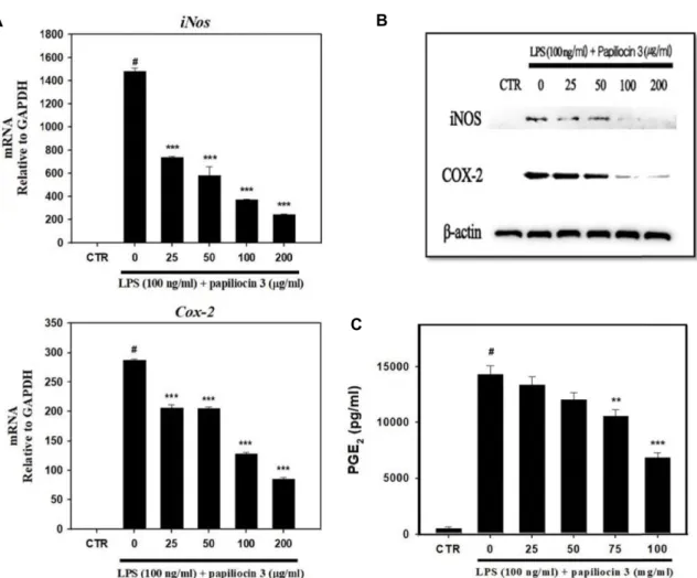
파필리오신 3이 iNOS와 COX-2 그리고 PGE2 단백질 발 현에 미치는 영향
파필리오신 3의 NO 생성억제 효과뿐만 아니라 염증반응의 신호전달과정에서 중요한 매개인자의 iNOS와 COX-2 그리고 PGE2 발현 양상에 미치는 영향을 확인하였다. 세포내 iNOS 단백질은 염증반응 동안에 NF-κB 활성으로 발현이 유도 생성 되며, COX-2는 arachidonic acid를 PGE2로 전환시키는 효소
로 염증과 같은 외부 스트레스가 발생할 경우 발현이 증가하 는 것으로 보고되어 있다[38]. PGE2는 대식세포에서 TNF-α, IL-1β 등의 염증성 사이토카인의 생성 억제와, IL-10과 같은 항염증성 사이토카인의 생성을 촉진하는 면역반응의 조절자 로 잘 알려져 있으며 염증반응 동안에 COX-2에 의해 생성되 게 된다[17]. LPS로부터 염증이 유도된 Raw264.7 세포에서 파 필리오신 3에 의한 iNOS와 COX-2 발현 양상을 확인하기 위한 qRT-PCR과 western blot 분석에서 25, 50, 100, 200 μg/ml의 농도로 파필리오신 3을 처리한 결과 iNOS와 COX-2 유전자 모두 발현이 감소하는 것을 확인할 수 있었으며, 단백질 수준 또한 현저히 감소되는 것을 확인할 수 있었다(Fig. 3A, Fig.
3B). 앞서 언급한 바와 같이 PGE2는 COX-2의 발현이 증가할수 록 농도가 증가하므로 ELISA 분석을 통해 LPS로부터 유도된 염증반응에서 파필리오신 3에 의한 PGE2의 생성억제능을 확 인하였다. LPS로 염증이 유도된 Raw264.7 세포에 파필리오신 3을 앞선 농도와 동일하게 처리한 결과 COX-2의 효과와 유사 하게 PGE2의 생성억제효과를 관찰할 수 있었다.
파필리오신 3이 염증성 사이토카인 발현에 미치는 영향 염증반응은 생체를 방어하기 위한 필수적인 면역반응이지 만, 조절되지 않고 과도하게 염증관련 물질들이 발현되면 급 성 패혈성 쇼크를 유발하게 되어 심할 경우 사망에까지 이르 게 하는 원인이 되기도 한다[10]. 그러므로 적절한 수준의 염증 반응은 매우 중요한데 파필리오신 3이 전염증성 사이토카인 (proinflammatory cytokine)인 TNF-α, IL-6 그리고 IL-1β의 발 현을 저해할 수 있는지 qRT-PCR과 ELISA 실험을 통해 유전 자 및 단백질 수준에서 각각 확인하였다. 먼저 유전자 수준에 서 분석한 결과 IL-6의 경우 25 μg/ml, IL-1β의 경우 50 μg/ml 의 농도부터 농도의존적으로 전염증성 사이토카인의 발현이
A B
C
Fig. 3. Inhibitory effects of papiliocin 3 on iNOS and COX-2 expression and PGE2 production in LPS-induced Raw264.7 cells. (A) Raw264.7 cells were pretreated with papiliocin 3 for 1 hr prior to incubation with LPS. Total RNA was isolated 18 hr after LPS treatment. The levels of iNOS and COX-2 mRNA were determined by qRT-PCR. The data were normalized to Gapdh mRNA levels. (B) Raw264.7 cells were pretreated with papiliocin 3 for 1 hr prior to incubation with LPS. Protein was isolated 24 hr after LPS treatment. The protein expression levels of iNOS and COX-2 were determined through Western blot analysis.
The data were normalized to β-actin protein levels. (C) Raw264.7 cells were pretreated with papiliocin 3 for 1 hr prior to iuncubation with LPS for 24 hr. PGE2 levels in the culture media were measured through ELISA. Data are presented as mean ± S.D. with three separate experiments. #p<0.05 in comparison with control group. ***p<0.001, **p<0.01, in comparison with LPS group. CTR, control; LPS, lipopolysaccharide.
저해되는 효과를 나타내었다(Fig. 4A). 또한 ELISA를 통해 단 백질 수준에서도 저해되는지 확인하였으며, 그 결과 IL-6와 IL-1β 모두 유전자 수준과 유사하게 25 μg/ml의 농도부터 농 도의존적으로 저해효과를 나타내었다(Fig. 4B). 그러나 TNF-α 의 경우에는 유전자와 단백질 수준에서 모두 현저한 저해효과 를 확인할 수는 없었다.
파필리오신 3과 LPS의 상호작용
내독소(endotoxin)로 잘 알려진 LPS는 그람음성균의 대표 적인 세포벽 성분으로 항균 펩타이드가 세균의 내막으로 삽입 되는 것을 막는 장벽으로 작용한다. 사멸된 세균으로부터 다 형체(oligomer)로 방출된 LPS는 선천성 면역에 관여하는 세포 나 다양한 방어기전과 관련된 세포들에 의해 인식되며 전염증
성 사이토카인의 방출을 유도한다. 이러한 관점에서 파필리오 신 3이 LPS와 결합을 통해 내독소를 중화시킴으로써 염증반 응을 제어할 수 있는지 여부를 확인하였다. 파필리오신 3과 LPS의 결합 유무는 Limulus Amebocyte Lysate (LAL) assay 를 통해 조사하였다. 그 결과 파필리오신 3의 농도가 증가함에 따라 농도의존적으로 LPS를 중화시키는 능력이 증가하는 것 을 알 수 있었다(Fig. 5). 이러한 결과는 파필리오신 3이 LPS와 강하게 결합한다는 것으로 보여주는 증거로 과도한 염증반응 을 제어하기 위해 항균 펩타이드가 사용될 수 있음을 보여주 는 증거라 할 수 있다.
파필리오신 3이 Raw264.7 세포에서 MAPKs 인산화에 미 치는 영향
A
B
Fig. 4. Inhibitory effects of papiliocin 3 on the production of proinflammatory cytokines in LPS-induced Raw264.7 cells. (A) Raw264.7 cells were pretreated with papiliocin 3 for 1 hr prior to incubation with LPS. Total RNA was isolated at 18 hr after LPS treatment. The levels of IL-6 and IL-1β mRNA were determined through quantitative real-time-PCR. The data were normalized to Gapdh mRNA level. (B) Raw264.7 cells were pretreated with papiliocin 3 for 1 hr prior to incubation with LPS for 24 hr. IL-6 and IL-1β levels in the culture media were measured through ELISA. Data are presented as mean ± S.D. with three separate experiments. #p<0.05 in comparison with control group. ***p<0.001 in comparison with LPS group. CTR, control;
LPS, lipopolysaccharide.
Fig. 5. Neutralization effect of LPS by papiliocin 3. LPS from E.
coli 0111:B4 (0.5 pg/ml) was incubated with papiliocin 3 and PBS for 30 min at 37℃. The LPS-neutralization activity was evaluated using an endotoxin quantification kit. Data are presented as mean ± S.D. with three separate experi- ments. LPS neutralization activity was calculated with the following equation: % LPS neutralization=(Ablank–Apeptide)/
Ablank×100. ***p<0.001, **p<0.01, in comparison with PBS group.
A B
Fig. 6. Inhibitory effect of papiliocin 3 on MAPKs and NF-kB signaling pathways in Raw264.7 cells. (A) Raw264.7 cells (1×10⁶ cells/well in a 6-well plate) were incubated with the indicated concentration of papiliocin 3. Protein was then isolated at 30 min after LPS treatment, and phosphorylation of ERK, p38 and JNK was detected using Western blot analysis. The data were normalized to total protein, respectively. (B) Degradation of IκB was detected using Western blot analysis. The data were normalized to β-actin. CTR, control; LPS, lipopolysaccharide.
대식세포의 염증반응에서 LPS로부터 개시되는 TLR (toll like receptor)-4 신호전달경로는 mitogen-activated protein kinases (MAPKs)와 NF (nuclear factor) κB의 신호전달경로와 같은 다양한 신호전달경로를 활성화 시킨다[34]. MAP kin- ases는 인산화 되지 않은 상태로 세포질 내에 존재하다가 자극 으로부터 활성화 되면 인산화가 이뤄져 염증반응에 관여하게 된다[30]. NF-κB 신호전달과정은 IκB kinase (IKK) complex에 의해 매개되며, 세포질에 NF-κB/IκB complex로 존재하다 염 증반응이 개시되면 NF-κB로부터 IκB가 분리되어 세포질로 떨 어져 나오며 단백질분해효소들에 의해 분해가 이뤄지게 된다 [22]. 그러므로 파필리오신 3이 Raw264.7 세포에서 LPS로부터 유도된 MAPKs의 인산화와 NFκB 신호전달경로에서 IκB 분 해에 어떠한 영향을 주는지 Western blot analysis를 통해 확인 하였다. Raw264.7 세포로 파필리오신 3을 1시간 동안 전처리 한 후 LPS를 30분 동안 처리하여 염증을 유발하였다. 그 결과 MAPKs의 ERK, p38, JNK의 인산화는 LPS에 의해 유도 발현 되지만 파필리오신 3의 농도가 증가함에 따라 인산화 정도가 농도의존적으로 감소하는 결과를 확인할 수 있었다(Fig. 6A).
또한, IκB 단백질의 분해에 있어서 파필리오신 3을 처리하지 않은 LPS군은 IκB의 분해가 최대인 것에 비해 파필리오신 3를 처리한 군에서는 농도가 증가할수록 IκB의 분해가 감소하는 것을 알 수 있었다(Fig. 6B). 본 연구에서 수행된 결과들을 종합 했을 때 이러한 결과들은 전사체분석을 통해 선별된 항균 펩 타이드인 파필리오신 3이 염증성 질환에 도움을 줄 수 있는
물질로 개발될 수 있을 것이라 사료된다.
감사의 글
본 성과물은 농촌진흥청 차세대바이오그린21사업(세부과 제번호: PJ01325601)의 지원에 의해 이루어진 것임.
The Conflict of Interest Statement
The authors declare that they have no conflict of interest with the contents of this article.
References
1. Blondelle, S. E., Lohner, K. and Aguilar, M. 1999. Lipid-in- duced conformation and lipid-binding properties of cyto- lytic and antimicrobial peptides: determination and biologi- cal specificity. Biochim. Biophys. Acta. 15, 89-108.
2. Boman, H. G., Nilsson-Faye, I., Paul, K. and Rasmuson, Jr.
T. 1974. Insect immunity. I. Characteristics of an inducible cell-free antibacterial reaction in hemolymph of Samia cyn- thia pupae. Infect. Immun. 10, 136-145.
3. Bowdish, D. M. E., Davidson, D. J., Scott, M. G. and Han- cock, R. E. 2005. Immunomodulatory activities of small host defense peptides. Antimicrob. Agents Chemother. 49, 1727- 1732.
4. Brogden, K. A. 2005. Antimicrobial peptides: pore formers or metabolic inhibitors in bacteria? Nat. Rev. Microbiol. 3, 238-250.
5. Bulet, P., Stöcklin, R. and Menin, L. 2004. Anti-microbial peptides: from invertebrates to vertebrates. Immunol. Rev.
198, 169-184.
6. Cruz, J., Ortiz, C., Guzmán, F., Fernández-Lafuente, R. and Torres, R. 2014. Antimicrobial peptides: promising com- pounds against pathogenic microorganisms. Curr. Med. Chem.
21, 2299-2321.
7. Dathe, M. and Wieprecht, T. 1999. Structural features of heli- cal antimicrobial peptides: their potential to modulate activ- ity on model membranes and biological cells. Biochim. Bio- phys. Acta. 15, 71-87.
8. Derache, C., Meudal, H., Aucagne, V., Mark, K. J., Cadène, M., Delmas, A. F., Lalmanach, A. C. and Céline, L. 2012.
Initial insights into structure-activity relationships of avian defensins. J. Biol. Chem. 287, 7746-7755.
9. Faye, I., Pye, A., Rasmuson, T., Boman, H. G. and Boman, I. A. 1975. Insect immunity. 11. Simultaneous induction of antibacterial activity and selection synthesis of some hemo- lymph proteins in diapausing pupae of Hyalophora cecropia and Samia cynthia. Infect. Immun. 12, 1426-1438.
10. Ferrero-Miliani, L., Nielsen, O. H., Andersen, P. S. and Girardin, S. E. 2007. Chronic inflammation: importance of NOD2 and NALP3 in interleukin-1beta generation. Clin. Exp.
Immunol. 147, 227-235.
11. Freudenthal, O., Quilès, F. and Francius G. 2017. Discrepan- cies between cyclic and linear antimicrobial peptide actions on the spectrochemical and nanomechanical fingerprints of a young biofilm. ACS. Omega 2, 5861-5872.
12. Giacometti, A., Cirioni, O., Ghiselli, R., Viticchi, C., Mocche- giani, F., Riva, A., Saba, V. and Scalise, G. 2001. Effect of mono-dose intraperitoneal cecropins in experimental septic shock. Crit. Care Med. 29, 1666-1669.
13. Guaní-Guerra, E., Santos-Mendoza, T., Lugo-Reyes, S. O.
and Terán, L. M. 2010. Antimicrobial peptides: general over- view and clinical implications in human health and disease.
Clin. Immunol. 135, 1-11.
14. Hancock, R. E., Nijnik, A. and Philpott, D. J. 2012. Modulating immunity as a therapy for bacterial infections. Nat. Rev.
Microbiol. 10, 243-254.
15. Hancock, R. E. and Lehrer, R. 1998. Cationic peptides: a new source of antibiotics. Trends Biotechnol. 16, 82-88.
16. Hancock, R. E. and Sahl, H. G. 2006. Antimicrobial and host- defense peptides as new anti-infective therapeutic strategies.
Nat. Biotechnol. 24, 1551-1557.
17. Harris S. G., Padilla, J., Koumas, L., Ray, D. and Phipps, R.
P. 2002. Prostaglandins as modulators of immunity. Trends Immunol. 23, 144-150.
18. Hollmann, A., Martinez, M., Maturana, P., Semorile, L. C.
and Maffia, P. C. 2018. Antimicrobial peptides: Interaction with model and biological membranes and synergism with chemical antibiotics. Front. Chem. 6, 204.
19. Heilborn, J. D., Nilsson, M. F., Kratz, G., Weber, G., Sørensen, O., Borregaard, N. and Ståhle-Bäckdahl, M. 2003. The cath-
elicidin anti-microbial peptide LL-37 is involved in re-epi- thelialization of human skin wounds and is lacking in chronic ulcer epithelium. J. Invest. Dermatol. 120, 379-389.
20. Hultmark, D., Steiner, H., Rasmuson, T. and Boman, H. G.
1980. Insect immunity. Purification and properties of three inducible bactericidal proteins from hemolymph of immu- nized pupae of Hyalophora cecropia. Eur. J. Biochem. 106, 7-16.
21. Jozefiak, A. and Engberg, R. M. 2017. Insect proteins as a potential source of antimicrobial peptides in livestock pro- duction. A review. J. Anim. Feed Sci. 26, 87-99.
22. Kim, D. H. and Chung, J. Y. 2002. Akt: versatile mediator of cell survival and beyond. J. Biochem. Mol. Biol. 35, 106-115.
23. Kim, J. E., Jacob, B., Jang, M. H., Kwak, C. H., Lee, Y. J., Son, K., Lee, S. J., Jung, I. D., Jeong, M. S., Kwon, S. H.
and Kim, Y. M. 2019. Development of a novel short 12-meric papiliocin-derived peptide that is effective against Gram- negative sepsis. Sci. Rep. 9, 3817.
24. Kim, J. K., Lee, E. J., Shin, S. Y., Jeong, K. W., Lee, J. Y., Bae, S. Y., Kim, S. H., Lee, J. Y., Kim, S. R., Lee, D. G., Hwang, J. S. and Kim, Y. M. 2011. Structure and function of papiliocin with antimicrobial and anti-inflammatory ac- tivities isolated from the swallowtail butterfly, Papilio xuthus.
J. Biol. Chem. 286, 41296-41311.
25. Kim, S. R., Hong, M. Y., Park, S. W., Choi, K. H., Yun, E.
Y., Goo, T. W., Kang, S. W., Suh, H. J., Kim, I. S. and Hwang, J. S. 2010. Characterization and cDNA cloning of a cecro- pin-like antimicrobial peptide, papiliocin, from the swallow- tail butterfly, Papilio xuthus. Mol. Cells 29, 419-423.
26. Kim, S. Y., Park, S. W., Goo, T. W., Kang, S. W., Yun, E.
Y. and Hwang, J. S. 2013. Recombinant antibacterial peptide papillocin 2 and its mass production method using the gene of papillocin 2 from Papilio xuthus. Korea patent 10-2013- 0007783.
27. Kindrachuk, J. and Napper, S. 2010. Structure-activity rela- tionships of multifunctional host defence peptides. Mini Rev.
Med. Chem. 10, 596-614.
28. Kell, D. B. and Pretorius, E. 2015. On the translocation of bacteria and their lipopolysaccharides between blood and peripheral locations in chronic, inflammatory diseases: the central roles of LPS and LPS-induced cell death. Integr. Biol.
(Camb). 7, 1339-1377.
29. Lai, Y. and Gallo, R. L. 2009. AMPed up immunity: how antimicrobial peptides have multiple roles in immune de- fense. Trends Immunol. 30, 131-141.
30. Lasa, M., Mahtani, K. R., Finch, A., Brewer, G., Saklatvala, J. and Clark, A. R. 2000. Regulation of cyclooxygenase 2 mRNA stability by the mitogen-activated protein kinase p38 signaling cascade. Mol. Cell. Biol. 20, 4265-4274.
31. Lee, E. J., Shin, A. R. and Kim, Y. M. 2015. Anti-in- flammatory activities of cecropin A and its mechanism of action. Arch. Insect Biochem. Physiol. 88, 31-44.
32. Leptihn, S., Har, J. Y., Wohland, T. and Ding, J. L. 2010.
Correlation of charge, hydrophobicity, and structure with antimicrobial activity of S1 and MIRIAM peptides. Biochem- istry 2, 9161-9170.
33. Locock, K. E. S., Michl, T. D., Valentin, J. D. P., Vasilev, K.,
초록:호랑나비 유래 항균 펩타이드 파필리오신 3의 항염증 활성
신용표†․이준하†․김인우․서민철․김미애․이화정․백민희․김성현․황재삼*
(농촌진흥청 국립농업과학원 농업생물부 곤충산업과)
본 연구에서는 호랑나비 유충의 유전체 분석을 통해 선별된 파필리오신 3의 항균 및 항염증 활성을 확인하였 다. 선행연구에서 RNA 시퀀싱 분석을 통해 호랑나비의 전사체를 분석하였으며, 결과를 바탕으로 인실리코(in sili- co) 분석을 진행하여 전사체 유래 항균 펩타이드를 스크리닝하고 선발하였다. 수행된 항균 활성 및 용혈 활성 테스 트에서 파필리오신 3은 그람음성균인 E. coli와 그람양성균인 S. aureus에 대해 강력한 항균활성을 나타낸 반면 마우스 적혈구에 대한 용혈 활성은 전혀 없었다. 다음으로 마우스 대식세포주 Raw264.7 세포를 이용하여 파필리 오신 3의 항염증 활성을 확인하였다. 그 결과 파필리오신 3은 LPS로부터 유도된 Raw264.7 세포들의 산화질소 생 성을 감소시키는 결과를 보여주었다. 뿐만 아니라 실시간 역전사 중합효소 연쇄반응(qRT-PCR) 방법과 효소결합 면역흡착측정법(ELISA)을 통해 파필리오신 3이 Raw264.7 세포에서 전염증성 사이토카인(IL-6, IL-1β)의 발현을 감소시킨다는 것을 확인할 수 있었다. 또한, 염증반응의 신호전달인자들(MAPKs, NF-κB)의 인산화를 억제하는 것을 확인하였는데, 이는 파필리오신 3이 LPS와의 상호작용을 통해 결합하여 효과를 나타낸다는 것을 확인할 수 있었다. 이러한 결과들은 호랑나비 유전체 분석을 통해 확인된 파필리오신 3이 새로운 항균 및 항염증 치료제로서 개발하는데 가능성 있는 물질로 사료된다.
Hayball, J. D., Qu, Y., Traven, A., Griesser, H. J., Meagher, L. and Haeussler, M. 2013. Guanylated polymethacrylates:
a class of potent antimicrobial polymers with low hemolytic activity. Biomacromolecules 14, 4021-4031.
34. Lu, Y. C., Yeh, W. C. and Ohashi, P. S. 2008. LPS/TLR4 signal transduction pathway. Cytokine 42, 145-151.
35. McDonald, M., Mannion, M., Pike, D., Lewis, K., Flynn, A., Brannan, A. M., Browne, M. J., Jackman, D., Madera, L., Coombs, M. R. P., Hoskin, D. W., Rise, M. L. and Booth, V. 2015. Structure-function relationships in histidine-rich an- timicrobial peptides from Atlantic cod. Biochim. Biophys. Acta.
1848, 1451-1461.
36. Nayduch, D., Lee, M. B. and Saski, C. A. 2014. Gene discov- ery and differential expression analysis of humoral immune response elements in female Culicoides sonorensis (Diptera:
Ceratopogonidae). Parasit. Vectors 7, 388.
37. Niyonsaba, F., Ushio, H., Nakano, N., Ng, W., Sayama, K., Hashimoto, K., Nagaoka, I., Okumura, K. and Ogawa, H.
2007. Antimicrobial peptides human beta-defensins stim- ulate epidermal keratinocyte migration, proliferation and production of proinflammatory cytokines and chemokines.
J. Invest. Dermatol. 127, 594-604.
38. NǾrregaard, R., Kwon, T. H. and Frokiǽr, J. 2015. Physiology and pathophysiology of cyclooxygenase-2 and prostaglandin E2 in the kidney. Res. Clin. Pract. 34, 194-200.
39. Patocka, J., Nepovimova, E., Klimova, B., Wu, Q. and Kuca, K. 2019. Antimicrobial peptides: amphibian host defense peptides. Curr. Med. Chem. 26, 5924-5946.
40. Robertson, M. and Postlethwait, J. H. 1986. The humoral antibacterial response of drosophila adults. Dev. Comp.
Immunol. 10, 167-179.
41. Steinberg, D. A. and Lehrer, R. I. 1997. Designer assays for antimicrobial peptides. disputing the "one-size-fits-all" theory.
Methods Mol. Biol. 78, 169-186.
42. Vieira, C. S., Mattos, D. P., Waniek, P. J., Santangelo, J. M., Figueiredo, M. B., Gumiel, M., da Mota, F. F., Castro, D.
P., Garcia, E. S. and Patrícia, A. 2015. Rhodnius prolixus interaction with Trypanosoma rangeli: modulation of the im- mune system and microbiota population. Parasit. Vectors 8, 135.
43. Walkenhorst, W. F., Klein, J. W., Vo, P. and Wimley, W.
C. 2013. pH Dependence of microbe sterilization by cationic antimicrobial peptides. Antimicrob. Agents Chemother. 57, 3312-3320.
44. Wang, J., Ma, K., Ruan, M., Wang, Y., Li, Y., Fu, Y. V., Song, Y., Sun, H. B. and Wang, J. 2018. A novel cecropin B-derived peptide with antibacterial and potential anti-inflammatory properties. PeerJ 6, e5369.
45. Yaakobi, K., Liebes-Peer, Y., Kushmaro, A. and Rapaport, H. 2013. Designed amphiphilic β-sheet peptides as tem- plates for paraoxon adsorption and detection. Langmuir 29, 6840-6848.
46. Yeaman, M. R. and Yount, N. Y. 2003. Mechanisms of anti- microbial peptide action and resistance. Pharmacol. Rev. 55, 27-55.