• 검색 결과가 없습니다.

Physeal Remodeling after Internal Fixation of Slipped Capital Femoral Epiphysis

N/A
N/A
Protected

Academic year: 2021

Share "Physeal Remodeling after Internal Fixation of Slipped Capital Femoral Epiphysis"

Copied!
8
0
0

로드 중.... (전체 텍스트 보기)

전체 글

(1)

Vol.20 No.1. 28-35. June, 2003.

1)

책임저자:김세동, 대구시 남구 대명동 317-1, 영남대학교병원 정형외과 TEL.(053) 620-3642 FAX.(053) 628-4020 E-mail: [email protected]

Running title:대퇴골두 골단분리증 환자의 골단판 재형성

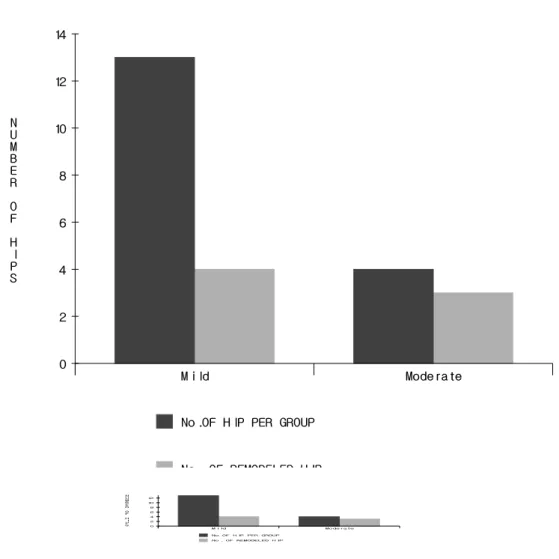
대퇴골두골단분리증 환자 중 금속내고정술을 시행받은 환자에서의 골단판의 재형성

영남대학교 의과대학 정형외과학교실

김세동․박병원

Physeal Remodeling after Internal Fixation of Slipped Capital Femoral Epiphysis Se Dong Kim, Byung Won Park

Department of Orthopaedic Surgery,

College of Medicine, Yeungnam University, Daegu, Korea

-Abstract-

Purpose: To evaluate physeal remodeling after internal fixation of slipped capital femoral epiphysis, We performed a retrospective review of the medical records and radiographs of 14 children (17 cases) who had had slipped capital femoral epiphysis.

Materials and Methods: We reviewed 14 patients who had slipped capital femoral epiphysis.

They were divided two groups. Group I were in situ pinning with single screw and group II were corrective osteotomy with multiple pinning. We identified physeal remodeling camparing with their preoperative, postoperative and last follow-up radiographs with measuring physeal- shaft angle.

Results: Early closure of the physis was observed in 6 cases among 14 patients (17 hips).

Compared with their postoperative radiographs, the last follow up radiographic study revealed that physeal remodeling was observed in 4 patients (4 hips) at group I (mean, 7.8°), in 2 patients (3 hips) at group II (mean, 10.7°).

Conclusion: In this study, physeal remodeling was observed in 4 patients (4 hips) in group I

and 2 patients (3 hips) in group II. The incidence of physeal remodeling was related with

degree of epiphyseal slippage, and age was not related with physeal remodeling potential.

(2)

서 론

대퇴골두 골단분리증은 급성장하는 청소년 기에 생길 수 있는 고관절질환의 하나로써 전 상방으로 향하고 있는 대퇴경부에 대해 대퇴골 두가 급성 혹은 만성적으로 후 내방으로 전위 되는 질환이나 그 원인은 아직까지 확실히 밝 혀진 바가 없다. 외상 혹은 성장판에 가해지는 전단력 등의 기계적 요인, 사춘기의 체중증가 및 성장판 경사도의 증가, 내분비장애 등 여러 가지 원인에 의한 성장판의 약화, 유전, 영양적 원인 등 다인자가 관여하는 것으로 생각하고 있다(1). 치료의 목적은 골단이 더 이상 분리되 는 것을 방지하고 골단판의 조기폐쇄를 유도하 여, 대퇴골두 무혈성괴사, 연골용해증과 골관절

염 같은 심각한 합병증을 방지하여 정상적인 고관절의 기능을 회복하는 것이다(2).

대퇴골두 골단분리증의 치료방법은 분리의 정도에 따라 다양하며 아직도 논란의 여지가 있다. 대개 초기의 분리정도에 따라 치료방법 이 결정되며 그 결과 또한 다양하다. 이것은 골단판 재형성의 영향을 고려하지 않은 것이 다. 이에 저자들은 1989년부터 2001년까지 영 남대학교 의과대학 부속병원 정형외과에 입원 후 대퇴골두 골단분리증으로 진단받고 수술적 치료를 받은 14명(17 고관절)에 대하여 입원기 록, 검사소견 및 방사선사진을 후향적으로 분 석하여 금속내고정술 시행 후 이 환자들의 골 단판의 변화와 골단판 재형성의 정도를 알아보 고자 한다.

Key Words: Slipped capital femoral epiphysis, Physeal remodeling

Table 1. Change of physeal-shaft angle after internal fixation of slipped capital femoral epiphysis

No. Sex Age Side PSA*

Degree (HSA

) Treatment postop. last F/U

1 F 12 Lt. 28° 28° mild (25°) in situ pinning

2 M 12 Lt. 54° 44° mild (7°) in situ pinning

3 F 10 Lt. 65° 60° mild (14°) in situ pinning

4 M 13 Lt. 60° 50° mild (7°) in situ pinning

5 M 12 Lt. 53° 50° mild (12°) in situ pinning

6 M 12 Lt. 48° 48° mild (15°) in situ pinning

7 M 12 Lt. 63° 59° mild (9°) in situ pinning

8 M 12 Lt. 60° 58° mild (10°) in situ pinning

9 M 6 both 44°/ 70° 45°/ 74° mild (18°/ 21°)  in situ pinning

10 M 12 both 53°/ 55° 50°/ 55° mild (13°/ 11°) in situ pinning

11 F 8 Lt. 45° 39° moderate (33°) in situ pinning

12 M 14 both 65°/ 50° 48°/ 41° moderate (51°/ 34°) multiple pinning

13 M 13 Rt. 46° 40° moderate (38°) multiple pinning

14 F 17 Lt. 42° 44° moderate (39°) multiple pinning

*: Physeal-Shaft Angle, †: Head-Shaft Angle

(3)

연구 대상 및 방법

1989년 6월부터 2001년 2월까지 영남대학교 의과대학 부속병원 정형외과에 대퇴골두 골단 분리증으로 인한 고관절동통 및 보행장애를 주 소로 내원한 총 14명(17고관절)의 환자를 대상 으로 하였다. 총 14명 중 남자가 10명, 여자가 4명이였으며 남자 중 3례가 양측성이었다. 발 병 후 진단 당시의 연령은 6세에서 17세까지였 으며 평균연령은 11.8세였다.

O’Brien과 Fahey의 분류(3)에 따라 임상증 상의 발현시기와 외상의 병력을 기준으로 하였 을 때 급성기(acute)가 5례, 만성기(chronic)가 8례, 분리전기(preslip)가 1례였다. 방사선학적

으로 Bernard의 분류(4)에 따라 고관절 전후면 사진에서 대퇴골두의 경부에 대한 상대적 전이 를 나타내는 골두-골간각(Head-Shaft Angle) 을 측정하여 대퇴골두의 경부에 대한 상대적 전위의 정도를 구분하였는데 경도(mild)의 분 리가 10례, 중등도(moderate)의 분리가 4례였 다. 1개의 나사못를 이용하여 정위치 고정술(in situ pinning)을 시행받은 환자(1군)는 11례(13 고관절)이였고 평균 추시기간은 17.2개월 이였 다. 대퇴경부 절골술 및 다발성 핀 삽입술을 함께 시행받은 환자(2군)는 3례(4 고관절)이였 고 평균 추시기간은 27.3개월 이였다.

골단-간부 각도는 술 전, 술 후 및 최종 추 시 때의 골반 전후면 방사선사진에서 대퇴골의

Fig. 1. physeal-shaft angle defined as an angle between long axis of femoral shaft

and a line connecting each end of capital epiphyseal plate.

(4)

간부 중앙을 지나는 선과 골단판의 상하 경계 점을 지나는 선을 그어 측정하였다(Fig. 1.) 5°

미만의 변화는 계측자의 실수로 생각하여 5°

이상의 변화만을 골단판 재형성의 증거로 평가 하였다.

결 과

골단-간부각의 변화는 0°에서 17°로 평균

4.8°였다. 1개의 나사못을 이용한 정위치고정술 을 시행한 환자군(1군)의 골단-간부각의 변화 는 평균 3.7°였고 이들 중 4명(4 고관절, 31%) 에서 골단판 재형성(평균 7.8°)을, 대퇴경부 절 골술 및 다발성 핀 삽입술을 함께 시행한 환자 군(2군)의 골단-간부각의 변화는 평균 8.5°였 으며 이들 중 2명(3 고관절, 75%)에서 골단판 재형성(평균 10.7°)을 확인할 수 있었다. 전체 환자 중 골단판 재형성이 확인된 7례 중 경도

Fig. 2. Incidence of remodeling in mild and moderate group :

4 hips (31%) were remodeled in mild group (head-shaft angle < 30°) and,

3 hips (75%) were remodeled in moderate group (30°< head-shaft angle < 60°).

(5)

의 분리가 3례(1군 3례), 중등도의 분리가 4례 (1군 1례, 2군 3례)로 골단판 재형성의 빈도는 분리의 정도가 심할수록 약간 증가되는 소견을 보였다(Fig. 2). 6례에서 정상측과 비교하여 조 기에 골단판 폐쇄가 발견되었다.

고 찰

대퇴골두 골단분리증은 국소적 혹은 전신적 인 원인에 의해서 골단판을 통한 대퇴골두가 후내방으로 전위되는 질환으로 국내에서도 보 고가 증가되는 질환이다.

대부분의 환자들이 10∼16세 사이이며, 남자 가 12∼15세, 여자가 10∼13세로 남자의 발병 나이가 약간 더 높다, 성별로는 남자가 2∼2.5 배 더 많으며 좌측이 우측보다 약 2배 더 많다(1).

대퇴골두 골단분리증의 원인으로는 여러 가 지 가설이 있으나 아직도 확실한 원인을 밝혀 내지 못하고 있다. 급성 및 만성 전단력에 의 한 외상, 비만, 단백질 대사장애 등이 원인으로 꼽히지만 가장 많이 거론되는 것이 내분비장애

이다(1).

치료방법의 선택에는 아직도 논란의 여지가 많다. 경도의 분리일 경우 정위치 고정술(in situ pinning)을 통해 비교적 만족스러운 결과를 얻 었으나 분리의 정도가 심한 경우에는 치료방법 의 선택에 어려움이 있다. 대개 초기 분리 정 도에 따라 치료 방향이 결정되는 데 이는 재형 성(remodeling)의 영향을 완전히 배제하는 것 이 된다. 대퇴골두 골단분리증에서 재형성과정 은 1926년 Key(5)에 의해 처음 언급되었으며 그 이후 많은 연구가 진행되었다. Lacroix 등(6) 은 노출된 대퇴골 골간부에서 파골성 흡수 (osteoclastic resorption)에 의해 재형성이 일어 난다고 하였고, Bellemans 등(7)은 국소적인 골 흡수와 부가성장(apposition)이 일어나 광범 위한 재형성 과정이 진행된다고 생각하였다.

Clarke 등(8)은 지속적인 골단판 성장(physeal growth)이 재형성 과정에 중요한 역할을 한 다고 보았다.

O’Brien 등(3)은 대퇴골 경부의 앞쪽 부위에 서 재형성이 일어나 볼록하던 윤곽이 오목하

Fig. 3. 13 year-old male with unilateral slipped capital femoral epiphysis patient.

Left: Physeal-shaft angle was 60°after in situ pinning fixation.

Right: Physeal-shaft angle was 50°after 19 months.

(6)

게 변한다고 하였으며 개방 상태의 삼방연골 (triradiate cartilage)이 재형성과 연관이 있다 고 보았고 고관절의 기능 회복에 대퇴 경부의 재형성이 중요한 역할을 한다고 보고하였다.

실제로 저자들의 연구에서도 재형성이 확인된 6명 모두 Heyman과 Herdon(9)의 분류상 우수 (Good)이상의 임상적 결과를 보였다.

Jonathan 등(10)은 환자의 나이, 성, 체중, 증상 발현 기간 등은 재형성에 큰 영향이 없으 나 분리의 정도가 심할수록 재형성의 가능성은 낮아지며 삼방연골이 개방되어 있을 경우 재형 성의 가능성은 매우 증가한다고 하여 O’Brien 등(3)의 주장을 뒷받침하였다. 그러나, Worng- Chung과 Shong(11)은 환자의 나이, 삼방연골 의 상태, 대퇴골두의 나사못 위치와 숫자는 재 형성 결과(remodeling potential)에 별 영향을 주지 않는 것으로 보고하였고 분리의 정도는 경 도(mild)나 중등도(moderate)에 비해 중도(severe) 의 분리군에서 재형성의 빈도가 더 높다고 보

고하였다. 저자들의 연구에서는 경도의 분리에 비해 중등도 분리군에서 재형성의 빈도가 약간 더 높은 것으로 나타났다. 대퇴경부 절골술 및 다발성 핀 삽입술을 함께 시행한 환자군에서 재형성의 빈도가 약간 더 높은 것으로 나타났 고 환자의 나이는 큰 연관성이 없는 것으로 보 였다.

이렇게 재형성이 일어나는 과정에 대해서도 아직 논쟁의 여지가 있지만 대퇴골두 골단분 리증으로 금속내고정술을 시행받은 환자의 상 당수에서 재형성이 일어난 것을 확인할 수 있 었다. 따라서 초기 치료 과정에서 재형성을 고려하지 않고 분리의 정도만을 기준으로 치 료 방향을 결정하기보다는 중도의 분리라도 초기에는 정위치 고정술을 시행하고 일정 기 간 재형성 과정을 지켜본 뒤 2차적인 치료방 법을 선택하는 것도 나쁘지는 않으리라 생각 한다.

Fig. 4. 14 year-old male patient with bilateral slipped capital femoral epiphysis.

Left: Right side was treated by extracapsular neck base osteotomy with cannulated screw, and left side treated by femoral neck wedge osteotomy with cannulated screw. Postoperative physeal-shaft angle was 65°and 50°, respectively.

Right: Physis-shaft angle decreased to 48°and 41°after 2 year.

(7)

요 약

대퇴골두 골단분리증으로 진단받고 수술적 치료를 받았던 14례(17 고관절)을 대상으로 이 들을 1개의 나사못을 이용한 정위치고정술을 시행한 환자군(1군)과 대퇴경부 절골술 및 다 발성 핀 삽입술을 함께 시행한 환자군(2군)으 로 나누어 골단-간부각을 측정하여 수치를 술 전, 술 후 및 마지막 추시를 비교하여 금속 내 고정술 시행 후 골단판 재형성을 확인하여 다 음과 같은 결과를 얻었다.

총 14명(17 고관절) 중 6명(7 고관절)에서 골단판 재형성을 확인 할 수 있었다. 정위치고 정술을 시행한 환자 11명(13례) 중, 4명(4 고관 절, 평균 7.75°)에서, 대퇴경부 절골술 및 다발 성 핀 삽입술을 함께 시행한 환자 3명(4례) 중 2명(3 고관절, 평균 10.7°)에서 골단판 재형성 을 확인할 수 있었다. 골단판 재형성이 확인된 7례 중 경도의 분리가 3례(1군 3례), 중등도의 분리가 4례(1군 1례, 2군 3례)로 골단판 재형성 의 빈도는 분리의 정도가 심할수록 증가되는 소견을 보였고 발병 당시 나이와는 연관성이 없었다. 골단판 재형성이 확인된 7례 중 6례에 서 골단판 조기 폐쇄를 확인하였다.

참 고 문 헌

1. 석세일, 정문상, 조재림, 김기수, 김남현, 이 석현, 우영균 등: 정형외과학, 제5판, 최신 의학사, 서울, 1999, pp. 492.

2. Canale ST, Daugherty K, Jones L et al.:

Campbell’s Operative Orthopaedics, 10th ed., Mosby, St. Louis, 2002, pp. 1483.

3. O’Brien ET, Fahey JJ: Remodeling of the

femoral neck after in situ pinning for slipped capital femoral epiphysis. Am J Bone Joint Surg 59-A: 62-68, 1977.

4. Jacobs B: Diagnosis and natural history of slipped capital femoral epiphysis. In Instructional Course Lecture, The American Academy of Orthopaedic Surgeons. Vol 21, Mosby, St. Louis, 1972, pp. 167-173.

5. Key JA: Epiphyseal coxa vara or displa- cement of the capital epiphysis of the femur in adolescence. J Bone Joint Surg 8: 53-117, 1926.

6. Lacroix P, Verbrugge J: Slipping of the upper femoral epiphysis : a pathologic study. Am J Bone Joint Surg 33-A:

371-381, 1951.

7. Bellemans J, Fabry G, Molenaers G, Lammens J, Moens P: Slipped capital femoral epiphysis : A long term follow up, with special emphasis on the capacities for remodeling. J Pediatr Orthop 5: 151- 157, 1996.

8. Clarke NMP, Harrison MHM: Slipped upper femoral epiphysis : a potential for spontaneous recovery. Br J Bone Joint Surg 68-B: 541-544, 1986.

9. Heyman CH, Herdon CH: Epiphyseodesis for early slipping of the upper femoral epiphysis. Am J Bone Joint Surg 36: 539 -554, 1954.

10. Jonathan RJ, Dennis CP, Terence MH,

Brouce KF: Remodeling after pinning

for slipped capital femoral epiphysis. Br

J Bone Joint Surg 72-B: 568-573, 1990.

(8)

11. Womg-Chung J, Strong ML: Physeal remodeling after fixation of slippred

capital femoral epiphysis. J Pediatr

Orthop 11: 2-5, 1991.

수치

Table  1.  Change  of  physeal-shaft  angle  after  internal  fixation  of  slipped  capital  femoral          epiphysis
Fig. 1. physeal-shaft  angle  defined  as  an  angle  between  long  axis  of  femoral  shaft  and  a  line  connecting  each  end  of  capital  epiphyseal  plate.
Fig.  2.  Incidence  of  remodeling  in  mild  and  moderate  group  :
Fig. 3. 13  year-old  male  with  unilateral  slipped  capital  femoral  epiphysis  patient.
+2

참조

관련 문서

1 John Owen, Justification by Faith Alone, in The Works of John Owen, ed. John Bolt, trans. Scott Clark, &#34;Do This and Live: Christ's Active Obedience as the

In this way, the origin of capital gain tax which is to fulfill the security of internal revenue and property holdback functions has a lot of natural

measured in clockwise from the north to the vertical plane containing the given line - Plunge: An acute angle measured in a vertical. plane between the given line and the

The basic goals of the capital system is to maintain the capital of corporations strictly in order to protect creditors of the corporation. Such capital

The switching of gecko toes between attachment and detach- ment, utilization of the nanotape-like functions of the spatula pads, the orienting actions of the seta shaft,

The clinical outcome and complication for treating proximal femoral shaft fracture were compared and analyzed through the group treated with closed

à An element of material near the center of the shaft has a low stress and a small moment arm and thus contributes less to the twisting moment than an

Therefore, in the linear elasticity theory, the shear stress also varies linearly from the axis of a circular shaft.... FIGURE 5.3 Distribution