Ⅰ. 서 론
악교정 수술은 심한 치열안면 변형(dentofacial deformi- ties)을 갖는 환자에서 흔히 고려되고, 악안면 골격의 3차원 적 부조화 정도 즉, 전후방적, 수직적, 횡적인 문제에 따라 편악 혹은 양악 수술을 시행하게 되며, 점점 후자의 빈도가 증가되고 있다. 통상적으로 악교정 수술은 환자의 임상검사 및 방사선 검사 자료를 통해 수립된 치료 계획에 따라 수술 전 교정치료가 선행되고, 그 이후 악교정 수술이 이루어지 며, 최종적으로 마무리 교정 치료가 이루어진다. 이 때 만족 할 만한 악교정 수술 결과를 얻고 또한 이를 예측하기 위해
정확히 결정하게 된다. 이와 같은 수술 계획이 수술장에서 신속 정확하게 반영되기 위해서는 계획에 따라 미리 제작된 splint가 필요하며, 상하악을 동반 수술하는 양악 수술에서 는 적어도 상악골을 재위치 시키기 위한 intermediate splint 와 이 후 하악골을 재위치시키기 위한 final splint가 필요함은 두 말할 필요가 없다. Intermediate splint를 제 작하기 위해서는 일반적으로 교합기에 탑재된 상악 석고 모 형을 삼차원적으로 이동시키는 모형 수술(model surgery) 이 필요한데, 모형 수술에 있어 무엇보다도 중요한 것은 paper surgery 등을 통해 결정된 상악골의 이동 방향과 양 을 정확히 반영하는 것이다. 그리고 모형 수술을 위해 안궁 이남기*∙최동순*∙차봉근*∙박영욱**∙김지혁**
*강릉대학교 치과대학 교정학교실, **강릉대학교 치과대학 구강악안면외과학교실
Model Repositioning Instrument를 이용한 악교정 수술의 치험례
ORTHOGNATHIC SURGERY USING MODEL REPOSITIONING INSTRUMENT:
A CASE REPORT
Nam-Ki Lee*, Dong-Soon Choi*, Bong-Kuen Cha*, Young-Wook Park**, Ji-Hyuck Kim**
*Department of Orthodontics, College of Dentistry, Kangnung National University
**Department of Oral and Maxillofacial Surgery, College of Dentistry, Kangnung National University
Moderate to severe dentofacial deformities usually require combined orthodontic treatment and orthog- nathic surgery to obtain the most stable result with optimal function and facial esthetics. Accordingly, the orthodontist and oral maxillofacial surgeon must be able to exactly diagnose existing deformities, establish an appropriate treatment plan, and execute the recommended treatment. Especially, to obtain optimal result of the maxillary surgery, model surgery is essential. But, the preoperatively planned position of the maxillary dental arch often cannot be sufficiently achieved during actual surgery, and deviations in the sagittal and vertical dimensions are common. To achieve three dimensional repositioning of the maxilla exactly, several methods have been introduced so far. Recently Model Repositioning Instrument (MRI, SAM, Inc., Mu¨nchen, Germany), one of these methods, has been introduced and applied clinically, which is reported as accurate, effective and prompt method for three dimensional repositioning of the maxilla.
This article describes an introduction and a clinical application of this MRI.
Key words : Orthognathic surgery, Model surgery, Model Repositioning Instrument(MRI) Abstract
없기에 이 장비를 사용하여 양호한 결과를 보인 증례와 함 께 장비의 사용 방법과 임상적 중요성을 알아보고자 한다.
Ⅱ. Model Repositioning Instrument를 이용한 모형 수술 과정
1. 구성요소
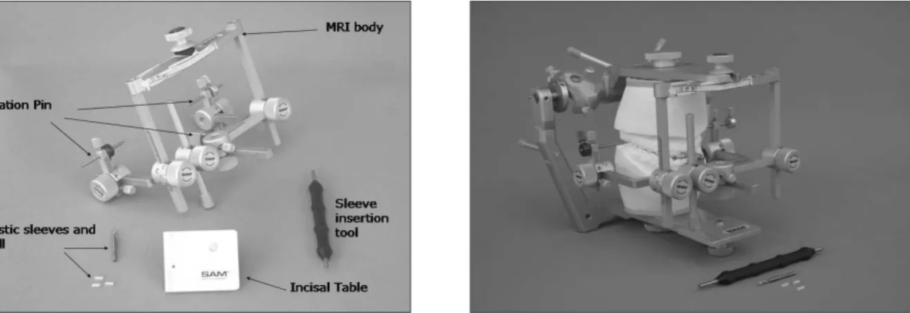
Model Repositioning Instrument(이하 MRI)는 본체 (MRI body)와 테이블(incisal table)로 구성되어 있고, 상 악 모형을 고정하기 위한 MRI 플라스틱 관 삽입 키트(MRI Plastic sleeve insert kit)와 같은 부속품이 있다(Fig. 3).
또한 본체에는 모형을 고정하는 핀(Fixation Pin)이 전방 부와 후방부에 총 3개가 있고 3차원적으로 조절할 수 있는 스크류와 눈금이 있다.
Fig. 1.Conventional method of model surgery. The hori- zontal and vertical reference line and osteotomy line is drawn on the base of the maxillary cast. And then the base which will be removed for repositioning the maxil- lary cast is trimmed.
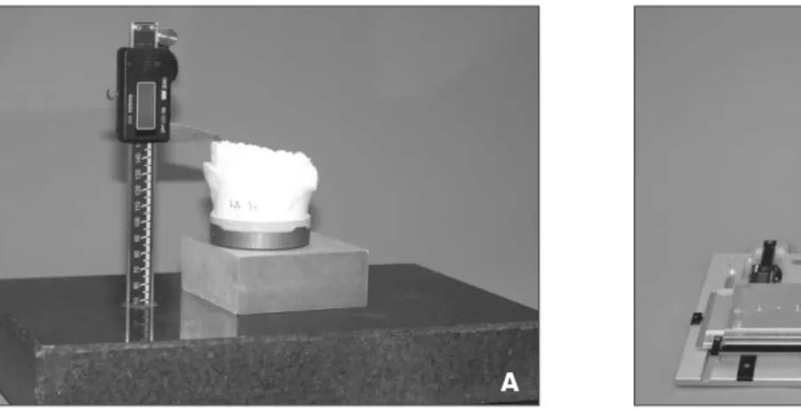
Fig. 2.Model platform and model block.
A. Erickson Model Surgery Platform (Great Lakes Orthodontic Ltd, Tonawanda, NY).
B. Reference Measuring Instrument�(SAM, Inc., Mu¨nchen, Germany).
A B
Fig. 3.Components and accessories of Model Repositioning Instrument�(SAM, Inc., Mu¨nchen, Germany).
Fig. 4.Procedure of model surgery using MRI.
A. Facebow transfer on Axis-Orbitale plane.
B. Mounting of the upper and lower cast on the SAM 3 semi-adjustable articulator.
C. Drilling for the MRI plastic sleeve at the level of brackets on the upper right and left first molars and right central incisor.
D. Insertion of the MRI plastic sleeve.
E. Placement of the MRI body and incisal table. The base of the upper cast was removed arbitrarily for repositioning.
F. Before reposition of the upper cast.
G. After reposition of the upper cast. Upper right first molar was moved to 3.5mm superiorly and 1.0mm posteriorly.
A B C
D E F
G H I
에 플라스틱 관(MRI sleeve)을 끼운다(Fig. 4C, 4D).
3) 상악 모형은 다시 교합기에 탑재하고, 교합기의 상부에 MRI 본체를, 하부에는 테이블을 장착한 후, 좌우 제1대 구치와 전치부에 끼운 플라스틱 관과 고정용 핀이 정확 히 일치하도록 조정한다. Incisal Pin을 조정하여 수직 고경을 확실히 유지시키고, 마지막으로 상악 모형이 자 유롭게 움직일 수 있도록 모형의 기저부는 적당히 제거 한 후, 고정용 핀의 위치에 맞게 잘 위치시키고 모형을 단단히 고정한다(Fig. 4E).
4) 이제 전방부와 좌우 후방부에 있는 각 조절 나사들을 이 용하여 상악 모형을 3차원적으로 재위치시킬 수 있으며, 이동 방향과 그 양은 두부계측방사선사진 상에서 계획한
교하고 간편하게 모형을 재위치 시킬 수 있다(Fig. 4 F-H).
5) 상악 모형의 이동이 완료되면 모형의 기저부는 석고를 이용하여 교합기 상부에 다시 재고정 한 후 자가중합형 레진을 이용하여 intermediate splint를 제작하며(Fig.
4I), 하악은 통법대로 상하악 최종 교합상태에서 final splint를 제작한다.
6) 이렇게 제작된 splint를 이용하여 실제 상악골을 포함한 양악 수술 후, 두부계측방사선사진을 촬영하여 수술 전 과 비교해 보았을 때, 매우 정확하게 수술이 이루어진 것 을 확인할 수 있었다(Fig. 5B).
Fig. 5.A. Pretreatment cephalometric radiograph and surgical treatment plan.
B. Superimposition of the pretreatment and posttreatment cephalometric radiographs.
A B
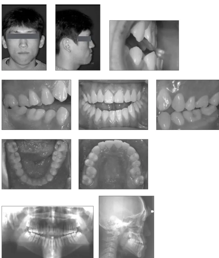
Fig. 6.A 17 years 2 months patient present with mandibular prognathism and vertical excess.
Fig. 9. Superimposition of pretreatment and posttreatment cephalometric radiographs.
Fig. 10.Potential errors occurring by measuring at the base of the cast during conventional model surgery. The incisal edge of an upper incisor moves posterior away from the planned position8).
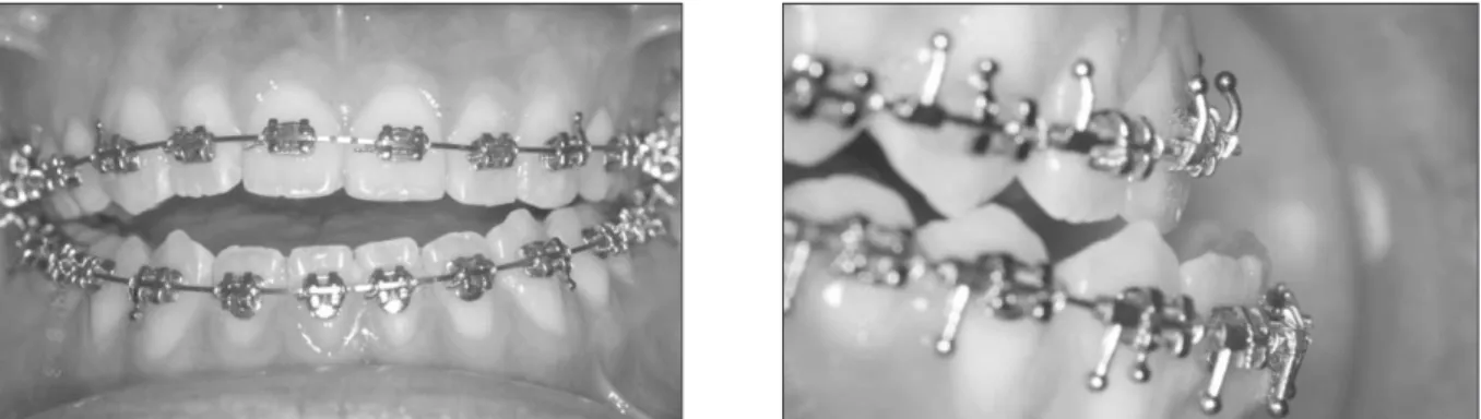
Fig. 8.Extraoral and intraoral photos on end of treatment.
Ⅲ. 증 례
17세 2개월의 남환으로 하악의 돌출과 개방교합을 주소 로 내원하였으며, 특별한 과거 병력은 없었다. 하악골의 전 돌과 과도한 하안면 고경으로 인한 장안모 양상을 보였으 며, 구내소견에서는 전치부 개방교합 및 반대교합, III급 구 치 관계를 보였고, 상악은 약간의 총생(crowding), 하악에 는 약간의 공극(spacing)이 있었으며, 미약한 치열 중심선 의 불일치를 보이고 있었다. 파노라마 방사선사진에서는 제 3대구치 매복과 뭉툭한 상악 중절치 치근단을 관찰할 수 있 었다. 임상검사와 측모 두부계측방사선사진 분석을 토대로 하악골의 과성장에 의한 골격성 III급 부정교합으로 진단되 었으며, 악관절 부위의 특이할만한 증상은 없었다(Fig. 6, Table 1).
상악궁에 미약한 총생이 있었고, 심미적으로도 비순각을 개선할 필요가 있어 상악 소구치 발치가 고려되었으나, 환 자가 치료 기간의 단축을 원하였고, 중절치 치근단도 짧은 상태라 제3대구치를 제외하고 비발치에 의한 악교정수술 치료를 하기로 결정하였다. 한편 하악골의 후방이동과 함께 안모의 수직적 조절과 pterygomasseteric sling에 따른 수 술 후의 불안정성을 예방할 목적으로 상악골 후방부를 상방 이동시키기로 하였으며, 이와 같이 시행될 경우 상악골과 함께 상악 전치는 시계방향으로 회전되어 비순각은 어느 정 도 개선되는 효과가 있을 것이라 예상되었다.
에지와이즈 장치와 연속호선을 이용하여 12개월간 통법
부위에서 5 mm 상방 이동시킴으로써 시계방향으로 회전시 켰으며, 하악골은 양측시상분할골절단술(bilateral sagittal split ramus osteotomy)과 함께 원심 골편을 12 mm 후방 이동시켰다. 상악골을 재위치시키기 위한 intermediate splint는 앞에서 설명한 방법대로 MRI를 이용하여 제작하 였고, 하악의 원심 골편을 고정하기 위한 final splint는 통 법대로 simple 교합기를 이용하여 제작하였다.
수술 후 교정 치료는 약 6개월이 소요되었으며, 치료 결과 하악의 전돌 양상은 사라졌고, 하안면고경도 약간 감소되어 수직 비율상 보다 조화로운 안모가 되었다. 구치는 I급 교합 으로 긴밀하고, 전방부의 반대교합과 개방교합도 양호한 수 평, 수직 피개교합으로 개선되었으며, 정중선도 잘 일치되 었다(Fig. 8). 치료 전∙후 두부계측방사선사진을 중첩해 본 결과 ANB는 -3.2�에서 0.9�로 개선되고, 안면각(facial contour angle; G-Sn-sPog) 역시 -5.5�에서 8�로 개선되 어 오목한 양상이 사라졌고, 하안면고경(ANS-Me)은 2.5 mm 감소되었으며, 또한 상악골의 시계방향 회전으로 비순 각은 90.1�에서 94.5�로 개선되었다(Fig. 9, Table 1).
Ⅳ. 총괄 및 고찰
흔히 이용되는 모형 수술 방법 중 교합기에 탑재된 모형의 기저부에 임의의 수평선과 수직선의 눈금을 그리거나 계측 자(ruler)를 이용하여 모형의 이동 양을 계측하는 방법은 여러 가지 문제점이 발생 가능하다. 첫째, 악골의 이동 방향 Table 1.Cephalometric Data of Pretreatment and Posttreatment. Cephalometric Norms of the Soft Tissues from Kim et al.9)
Norm Pretreatment Posttreatment
SNA(°) 82.0 81.9 84.5
SNB(°) 80.0 85.1 83.6
ANB(°) 2.0 -3.2 0.9
A to N⊥FH(mm) -1.1 2.5 4.6
Mandibular Length(mm) 120.0 142.5 136.9
Midfacial Length(mm) 92.6 92.2 94.2
Mandibular Plane Angle(°) 23.0 21.4 24.7
Lower Ant. Fac. height(mm) 65.2 86.7 84.2
Mx.1 - APo(mm) 6.2 3.2 8.6
Mn.1 - APo(mm) 3.0 8.8 5.2
G-Sn-sPog (°) 12.2 -5.5 8
G-Sn/Sn-sMe 0.98 0.94 1.03
Nasolabial angle(°) 93.9 90.1 94.5
Esth. Plane U.L.(mm) -0.74 -6.1 -2.4
Esth. Plane L.L.(mm) 0.93 -0.7 -0.4
sPog: soft tissue pogonion, sMe: soft tissue menton.
이와 같은 단점을 보완하기 위해 모형 플랫폼(model platform)과 모형 블록(model block)을 사용하는 방법이 소개되었으며, 이는 기준 평면이 수평, 수직적으로 모형 수 술에 정확히 반영되도록 함으로써 3차원 공간에서의 정확 한 모형 수술이 가능하다는 장점이 있다3-5). 하지만 계측과 모형을 재위치시키기 위해서는 교합기에서 모형을 분리해 야만 하고, 이동량 만큼의 석고를 수작업으로 정교하게 제 거하고 계측해야 하는 번거로움과 이로 인한 오차는 개선되 지 못한다.
이에 반해, MRI를 이용하면 석고 모형의 기저부 측면에 기준선과 골절단선을 그릴 필요가 없으며, 계측자를 이용하 여 계측할 필요도 없다. 또한 재위치시키기 위하여 석고 기 저부를 정확히 제거해야하는 번거로움도 없다6-8). 다만 앞에 서도 설명했듯이 paper surgery를 통해 Axis-Orbitale 선 을 기준으로 좌우 제1대구치와 중절치의 3차원적인 위치가 결정되면 MRI 본체에 있는 세부위의 조절 나사를 통해 모 형을 재위치시키면 된다. Schwestka-Polly R 등7)은 20명 의 환자에서 이 장비를 이용하여 splint를 제작하고 dou- ble-splint 방법으로 하악 과두의 위치를 고정하는 방법으 로 상악골 수술을 한 결과 수평, 수직적으로 0.5 mm 이내 의 매우 정교한 수술이 이루어졌음을 보고한 바 있다. 이처 럼 정확한 상악골 수술을 위해서는 정확한 splint의 제작이 필요하며, 동시에 외과의의 정확한 수술도 필수적이다. 추 후 국내에서도 이 장비를 이용하여 모형 수술 후 악교정수 술을 시행한 경우 수술의 정확성에 대한 연구가 필요하리라 사료된다.
한편 MRI를 이용한 상악골의 전후방 및 수직, 수평적 이 동은 가능하지만 상악골 분절골절단술(segment osteoto-
Orbitale 선)을 유지함으로써 3차원적인 수술의 양과 방향 을 정확하게 반영함과 동시에 신속한 모형 수술을 가능하게 하는 기구로 사료된다.
참고문헌
1. Bell WH, Creekmore TD, Alexander RG : Surgical correc- tion of the long face syndrome. Am J Orthod 71 : 40, 1977.
2. Epker BN, Fish LC : Surgical-orthodontic correction of open-bite deformity. Am J Orthod 71 : 278, 1977.
3. Erickson KM, Bell WH, Goldsmith DH : Analytical model surgery. In Bell WH: Modern practice in orthognathic and reconstructive surgery. Philadelphia, WB Saunders co.
154, 1992.
4. Ellis E III : Accuracy of model surgery: evaluation of an old technique and introduction of a new one. J Oral Maxillofac Surg 48 : 1161, 1990.
5. Lee SH, Lee SH, Ju HH et al : The accuracy of measure- ments during model surgery for orthognathic planning. J Kor Oral Maxillofac Surg 27 : 37, 2001.
6. Schwestka-Polly R, Kubein-Meesenburg D, Luhr HG : Application of the model-positioning appliance for three- dimensional positioning of the maxilla in cast surgery. Int J Adult Orthodon Orthognath Surg 8 : 25, 1993.
7. Schwestka-Polly R, Kubein-Meesenburg D, Luhr HG : Techniques for achieving three-dimensional positioning of the maxilla applied in conjunction with the Go¨ttingen con- cept. Int J Adult Orthod Orthognath Surg 13 : 248, 1998.
8. Schwestka-Polly R, Kubein-Meesenburg D, Luhr HG : From the 2-dimensional analysis in the lateral radiograph to the 3-dimensional repositioning in maxillary surgery.
Int Poster J Dent Oral Med 2 : Poster 41, 2000.
9. Kim KH, Choy KC, Kim GK et al : Cephalometric norms of the soft tissue of Korean for orthognathic surgery. J Kor Oral Maxillofac Surg 27 : 231, 2001.
저자 연락처
우편번호 210-702
강원도 강릉시 지변동 123번지 강릉대학교 치과대학 교정학교실 차 봉 근
원고 접수일 2006년 4월 5일
Reprint Requests Bong-Kuen Cha
Dept. of Orthodontics, Kangnung National University Dental Hospital 123, Chibyun-dong, Kangnung-city, Kangwon-do, 210-702, South Korea Tel: +82-33-640-3192 Fax: +82-33-640-3057
E-mail: [email protected] Paper received 5 April 2006