섬유화 진행 간경변 조직 모델에서
Thymosin β
4와C-myc
mRNA 융합 발현 연구김진수1*, 박운규2
1대전보건대학교 임상병리과 교수, 2대전대학교 임상병리과 박사과정
A Study on the Expression of
Thymosin-β
4 andc-Myc
mRNA in the Model of liver cirrhosis with fibrosisJean-Soo Kim1*, Un-Kyu Park2
1Professor, Department of Clinical Laboratory Science, Daejeon Health Institute of Technology
2Doctoral course, Department of Clinical Laboratory Science, Daejeon university
요 약 이 연구의 목적은 간 섬유화로 인한 간경변 모델에서 c-Myc과, Thymosin-β4유전자의 발현을 알아 보고자 실시하였으며 연구 방법은 2군으로 나누어 섬유화로 인한 간경변 모델 실험군과 증류수를 이용한 대조군으로 분류 한 후 실험을 하였다. 본 연구를 통한 결과는 간경변 모델 실험군은 대조군에 비하여 c-Myc 과 Thymosin-β4의 발현 이 증가 되는 것을 뚜렷하게 알 수 있었고, 전자현미경적 검경 외에 여러 특수 염색을 통하여 간조직의 변화를 관찰 할 수 있었다. 결론적으로 기존 간기능 검사 관련 임상검사에서 질환 예방과 질환 판정 시 혈청학적 변화와 조직학적인 검사와 더불어 c-Myc 과 Thymosin-β4의 분자 기법을 통한 유전자의 발현 상태를 확인함으로써 간 질환 판정에 기여 할 수 있을 것으로 사료 되며. 향후 이 연구를 기반으로 실험군의 종류와 관련 유전자를 추가 하여 심도 있는 연구를 진행할 예정이다.
주제어 : 헤마톡실린 에오진 염색, 마손삼색, 세망섬유, c-Myc, Thymosin-β4, 융합
Abstract The propose of this study has been conducted to examine expression of c-Myc and Thymosin- β4 in liver cirrhosis model from liver fibrosis and For the method of study, the experiment was conducted in 2 groups; liver cirrhosis model experiment group due to liver fibrosis and control group with distilled water. This study outcome showed that liver cirrhosis model experiment group had significantly higher expression of c-Myc and Thymosin-β4. with changes to hepatic tissue of special staining and electron microscopy. In conclusion, in clinical tests regarding liver function, molecular evaluation of c-Myc and Thymosin-β4 and their expression along with serological change and histological assessment can be utilized as a reference for diagnosing liver disease for prevention and diagnosis of the disease, Based on this research in the future, we will carry out an in-depth study by adding the types of experimental groups and related genes.
Key Words : Hematoxylin, Eosin, Masson's Trichrome, Reticulum, c-Myc, Thymosin-β4, Convergence
*This paper was supported by Daejeon Health Institute of Technology in 2018
*Corresponding Author : Jean–Soo Kim([email protected]) Received March 28, 2019
Accepted June 20, 2019
Revised April 25, 2019 Published June 28, 2019
1. 서론
최근 현대인들의 각종 스트레스와 과도한 업무로 인한 피로 누적과 지나친 음주로 인하여 간 질환에 많이 노출 되어 있으며 그 정도가 심한 즉, 간조직의 섬유화[1,2]로 인한 재생 결절로 간 표면이 불규칙하고 굳어지는 간경 변 환자도 상당히 많이 존재 한다[3]. 간경변은 그대로 방 치시 간경화를 통해 간암으로 진행하게 된다. 현재 이런 질병 관련 의학 진단 검사는 적은 비용으로 정확한 결과 를 얻을 수 있는 방향으로 발전하고 있으며 특히 요즘은 여러 검사 방법 중 분자기법이 각광 받고 있는 것이 현 흐름이다. 예를 들면 각종 암 진단에 있어 혈액 종양 지 표 검사나 관련 혈청학적 검사를 고전적으로 많이 시행 하였지만 최근에는 HER2, ALK, BRAF , BRCA, P53등 분자기법을 통한 유전자 검사를 통하여 유방암이나 난소 암, 폐암, 위암 등을 검사하고 있다. 그러나 간 질환 관련 검사는 대부분이 AST, AFP, ALP등 혈청학적 검사나 고 비용의 CT 검사, 초음파 검사를 통하여 진단이 대부분 이루어지고 있다. 따라서 간질환이 시작되었을 때 변화 되는 관련 유전자를 검사함으로써 상대적으로 간장기의 상태를 더 정확히 알 수 있다면 진단하는데 많은 도움이 있을 것으로 예측이 된다. 이 연구에 보고자 하는 Thymosin-β4 와 c-Myc 유전자의 간 질환이 있을 때 발현 관련성을 보고자 하는 것이다. Thymosin-β4 는 성 장인자[4-7]이며 4.9 kDa[8], G액틴의 분리과정[9,10], 조직 손상 시 또는 암생성시 혈관의 생성 과정에 관여 하 며[11,12] 배아세포 [13-15], 모낭 [16], 신생 혈관 초기 [17] 종양 또는 종양전이 [18.19], 특히 전이에서도 흑색 종 [20]과 섬유육종[21] 관련 전이 에서는 발현 양이 현 저히 증가 된다고 알려져 있다. c-Myc (c- Myelocyto matosis oncogene)은 염색체 8번 장완 24번에 있으며 세포증식, Telomerase 발현, 그리고 leukemia, 폐암 등에서 발현이 높다고 알려져 있다[22]. 기존 연구논문 김승조[23], 옥미선[24]에서 c-Myc이나 Thymosin-β4
는 위조직이나 자궁경부부위에서 병변부위 즉 암 발생이 있는 경우 각각 발현이 증가 되는 것으로 나타났다. 위 결과물을 토대로 간조직에서도 간질환에 노출 되었을 때 이런 유전자의 과발현 유형이 나타나지 않을까 예측이 되어졌고 연구를 하게 되었다.
따라서 본 연구는 간경변시 전자현미경적변화와 각종 특수 염색을 통한 광학 현미경적 조직학적인 변화를 알 아보고 간 경변시 정상 조직에 비해 간세포내의 Thymosin-β4, c-Myc, 1의 mRNA 발현의 차이를 관찰
함으로써 임상 진단 관련 참고 자료로 활용하고자 실시 하였다.
2. 재료 및 방법
2.1 간경변 모델
20~25g, 10주령 BALB/c를 공급받아 1주일간 최적 환경에 적응 적응시킨 후 실험모델 이주영 방법[25]을 채 택하여 CCl4 (Aldrich, USA) 및 Olive oil (Extra Virgin, ITali)의 혼합물 (2 volume : 1 volume, 1ml/kg)을 2회/주, 8주 동안 투여하여 간경변 실험 모델 을 만들었다. 실험은 대조군(5), 실험군(5) 나누어 진행 하였으며 구강 위관 영양법으로 투여한 후. 8주 경과 후 Ether로 마취하여 희생시켰다.
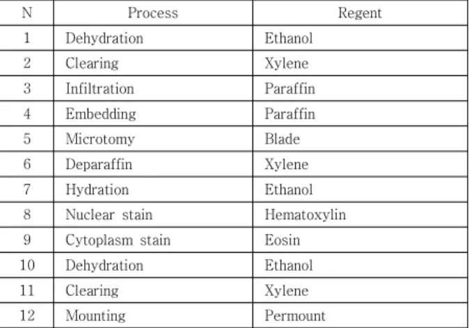
2.2 Hematoxylin & Eosin 염색
10% 포르말린을 이용하여 실험모델에서 적출한 간 조 직을 24시간 고정 하고 조직표본을 제작과정을 통하여 4~6μm 박절 하였다. 핵 염색은 Harris- Hematoxylin (영동제약, 한국), 세포질은 eosin Y (YD Diagnostics, korea)을 이용하여 염색을 하였다. Table 1
Table 1. Method of Hematoxylin & Eosin stain
N Process Regent
1 Dehydration Ethanol
2 Clearing Xylene
3 Infiltration Paraffin
4 Embedding Paraffin
5 Microtomy Blade
6 Deparaffin Xylene
7 Hydration Ethanol
8 Nuclear stain Hematoxylin
9 Cytoplasm stain Eosin
10 Dehydration Ethanol
11 Clearing Xylene
12 Mounting Permount
2.3 전자현미경적 조직 관찰
간 조직을 세절 한 후 1차 고정액 Glutaraldehyde, 2차 고정액 사산화 오스뮴 이용하여 고정 하였으며 탈수, 치환, 중합과정 등을 거쳐 전자현미경용 표본을 제작하고 투과 전자 현미경을 통하여 미세 구조를 관찰 하였다.
Table 2
Table 2. Transmission Electron Microscope : Method of TEM stain
N Process Content
1 Removal of specimen & slicing size 1x1x1 mm3 2 Prefixation & washing glutalaldehyde
3 Posfixation & washing osmium tetroxide (OsO4)
4 Dehydration Ethanol
5 Substitution propylene oxide
6 Infiltration Epon 812
7 Embedding polymerization oven Epon 812 8 Trimming
9 Semi-thin sectioning toluidine blue stain 10 Ultrathin sectioning 60~90 nm 11 Electron staining uranyl acetate 12 Observation
13 Photography 14 Interpretation
2.4 마손 삼색(Masson's Trichrome) 염색
박절하여 제작한 절편에 매염제로 Bouin 용액을 이 용 하였으며 Weigert iron hematoxylin에 10분, Biebrich scarlet-acid fuchsin 15분, aniline blue에 섬유질(아교섬유)를 염색 하였다. Table 3
Table 3. Method of Masson's Trichrom stain
N Process Regent
1 Dehydration Ethanol
2 Clearing Xylene
3 Infiltration Paraffin
4 Embedding Paraffin
5 Microtomy Blade
6 Deparaffin Xylene
7 Hydration Ethanol
8 Nuclear stain Hematoxylin
9 Cytoplasm stain Biebrich scarlet - acid fuchsin 10 Collagenous fiber stain Aniline blue
11 Dehydration Ethanol
12 Clearing Xylene
13 Mounting Permount
2.5 세망섬유(Gomori Reticulum) 염색
박절한 절편을 산화, 표백, 감작, 은 침투, 환원, 조색, 정착 순서대로 관련 시약을 사용하여 세망섬유를 염색 하였다. Table 4
Table 4. Method of Gomori Reticulum stain
N Process Regent
1 Dehydration Ethanol
2 Clearing Xylene
3 Infiltration Paraffin
4 Embedding Paraffin
5 microtomy Blade
6 Deparaffin Xylene
7 hydration Ethanol
8 Oxidation 1% potassiumpermanganate
9 Decolor 1% oxalic acid
10 Sensitization 2% ferric ammonium sulfate 11 Exposure to the silver solution Ammoniacal silver
12 Reduction 10 % formalin
13 Toning 0.2 % gold chloride
14 Removal of unreacted silver 5 % sodiumthiosulfate
15 Dehydration Ethanol
16 Clearing Xylene
17 Mounting Permount
2.6 유전자 검사
적출한 간 조직을 –70℃에 냉동 보관 후 Real time PCR를 실시하였으며 케니스 방법[26]을 이용하여 유전 자 결과를 분석하였다. RNA추출 과정은 조직 파쇄 후 RNA Prep Kit를 사용하였으며 RNA 20 μl을 이용 42°C 60 분 역전사 시킨 후 Accupower Greenstar qPCR preMix Kit로 cDNA 만들었으며 Primers를 Table 5와 같이 이용 하여 95°C에서 20초, 56°C에서 45초 과정으로 40번 증폭을 실시하였다.
3. 결과
3.1 전자현미경적 조직관찰
대조군에서는 핵 주위에 과립 내형질 세망, 능선이 뚜 렷한 미토콘드리아등 소기관들이 정상적으로 분포하고 있으나 실험군에서는 핵주위에 각종 소기관들의 분포가 적었으며 미토콘드리아의 수도 적은 것을 알 수 있었다.
특히 용해소체의 증가와 남아 있는 미토콘드리아 내막의 능선도 손상으로 인하여 뚜렷하지 못한 것을 알 수 있었 다. Fig. 1
3.2 Hematoxylin & Eosin 염색
대조군에 비하여 실험군에서는 간세포내의 핵 농축과 반응성 핵, 그리고 세포질내 액포 변성등 이상 세포들이 많이 분포하는 것을 알 수 있었다. Fig. 2
Ⓐ
Ⓑ
Fig. 1. Shapes of liver tissue in the Transmission Electron Microscope.
Ⓐ : Shapes of the control liver tissue in the Transmission Electron Microscope. This picture shows the structure of the mitochondria and the structure of endoplasmic reticulum
Ⓑ : Shapes of the liver tissue in the group administered with CCl4. in the Transmission Electron Mcroscope. This picture is not shows the structure of the mitochondria and the structure of endoplasmic reticulum
Ⓐ
Ⓑ
Fig. 2. H & E stain of liver tissue.
Ⓐ : A is a H E stain of control liver tissue. Sinusoids are visible well.
Ⓑ : B is a H & E stain of liver tissue administered with CCl4.. It is shown Pycnosis, size change of Nuclear and Vacuolar degeneration.
3.3 마손 삼색(Masson's Trichrome) 염색 M.T염색을 통하여 대조군과 실험군을 비교하였을 때 아교섬유 증식에 따른 섬유화로 실험군에서 간 소엽주위 와 중심정맥 부위에 교원섬유가 다량 분포하고 있는 것 을 알 수 있었다. Fig. 3
Ⓐ
Ⓑ
Fig .3. Masson's Trichrom stain of liver tissue.
Ⓐ : A is a MT stain of control liver tissue, Ⓑ :B is a Masson's Trichrom stain of liver tissue administered with CCl4. It is shown Collagen fiber and Bridge of fiber.
3.4 세망섬유(Gomori Reticulum) 염색 BALB/c mice의 세망섬유 염색은 대조군과 실험군에 서 모두 세망섬유 변화를 관찰할 수 없었다. 그러나 마손 삼색 염색과 같이 간세둥이, 중심정맥 주위에 교원섬유가 실험군에서 다량 분포 하였다. Fig. 5
3.5 유전자 검사
Real time PCR을 이용하여 케니스[26] 분석 방법을 통한 대조군과 실험군의 c-Myc및 Thymosin-β4 발현 을 비교 하였다. 보통 발현 연구 시 Ct값이 18~32로 나 타나는데 c-Myc과 Thymosin-β4는 Ct값이 대조군 각 각 28.49, 25.45이며 실험군은 24.98, 19.22 이었다.
Table 6 housekeeping gene인 β-actin 발현정도를 1 로 기준하였을때 CCl4를 투여한 실험군의 발현 양은 c-Myc 12.9, Thymosin-β4 85.3으로 실험군에서 뚜렸 하게 유전자의 발현이 증가됨을 알 수 있었다. Table 6
Ⓐ
Ⓑ
Fig. 4. Reticular fiber stain of liver tissue.
Ⓐ : A is a Masson's Trichrom of control liver tissue, Ⓑ :B is a Reticular fiber stain of liver tissue administered with CCl4. It is shown Collagen fiber of brown
Table 5. Primers used for the detection of gene expression
Target gene sequence (5´→3´) bp
Thymosin-β4
R AGA TGC AAA GAG GTT GG 111 F TCT TTT CCC TCT CTG CT
c-Myc
R GTG CTG CAT GAG GAG AC 84 F CAC ATC AAT TTC TTC CT
Table 6. Relative expression of β-actin, c-Myc and Thymosin-β4 gene expression
β-actin c-Myc ΔCt ΔΔCt 2-ΔΔCt
Control 18.76 28.49 9.73 0.00 1
CCl4 18.95 24.98 6.03 -3.70 12.9
β-actin Thymosin-β4 ΔCt ΔΔCt 2-ΔΔCt
Control 18.76 25.45 6.68 0.00 1
CCl4 18.95 19.22 0.27 -6.42 85.3
Table 7. Cq values of gene expression in groups administered with CCl4
Cq values = Ct
β-actin Thymosin-β4 c-Myc
Control 18.76 25.45 28.49
CCl4 18.95 19.22 24.98
4. 고찰 및 결론
본 연구는 암 전이나 혈관 재생에 관여 하는 Thymosin- β4 유전자와 c-Myc,종양 관련 유전자를 이용하여 간 경 변 모델을 통한 섬유화 된 간 조직 내에 조직학적 변화와 c-Myc,및 Thymosin-β4 발현 정도를 분석하고자 연구 하였다.
결과에 대하여 살펴보면 간조직 표면은 대조군에서는 외형적인 표면이 매끄러운 반면 섬유조직의 재생 결절로 실험군에서는 표면이 울퉁 불퉁 간경변 증상이 나타났다.
전자현미경적 조직관찰은 대조군에 비해 실험군에서는 핵주위에 각종 소기관들의 분포가 적었으며 미토콘드리 아의 수도 적은 것을 알 수 있었다. 특히 용해소체의 증 가와 남아 있는 미토콘드리아 내막의 능선도 손상으로 인하여 뚜렷하지 못한 것을 알 수 있었다. Hematoxylin
& Eosin 염색은 실험군에서 간세포내의 핵 농축과 반응 성 핵, 그리고 세포질내 액포 변성등 이상 세포들이 많이 분포하는 것을 알수 있었다. M.T염색은 대조군과 실험군 을 비교하였을 때 아교섬유 증식에 따른 섬유화로 실험 군에서 간 소엽주위와 중심정맥 부위에 교원섬유가 다량 분포하고 있는 것을 알 수 있었다. 세망섬유(Gomori Reticulum) 염색은 세망섬유 변화를 관찰할 수 없었으 나 간세둥이, 중심정맥 주위에 교원섬유가 실험군에서 다량 분포하고 있는 것을 알 수 있었다. c-Myc및 Thymosin-β4
발현은 housekeeping gene인 β-actin 유전자 발현을 1로 기준하였을 때 CCl4를 투여한 실험군의 발현 양은 c-Myc 12.9, Thymosin-β4 85.3으로 실험군에서 뚜렷 하게 유전자의 발현이 증가 됨을 알 수 있었다.
위와 같은 결과는 홍순호[27]연구와 사염화탄소를 통 한 간조직의 손상 확인시 특수염색과 전자현미경적 조직 변화로 연구한 유영빈[28]연구에서도 비슷한 결과를 얻 을 수 있었다. 즉 정상적인 간에서 어떤 이유에 의한 손 상으로 간경변 간경화 간암등 간 질환이 발생 하였을 때 일부 혈청학적 검사와 조직학적 변화와 더불어 c-Myc 과 Thymosin-β4유전자의 발현을 관찰하였을 때 차이 가 있다는 것을 알 수 있었다. 이는 사염화탄소에 의해 형성된 자유라디칼이 간세포를 손상시키며 cytochrome P450을 형성하고 CCl3O2· 와 CCl3·의 자유라디칼이 지질과산화를 형성하여 세포막을 손상시킨다[28]. 이때 간조직에 분포하는 Kupffer cell에서 tumor necrosis factor alpha (TNF-α), interleukin-6 (IL-6), 및 transforming growth factor beta (TGF-β) 방출되어 아교 섬유가 간조직에 침착되어 간경변에 노출되고
[29,30] Thymosin-β4[31-33]와 c-Myc[34,35]유전자 또한 발현 되는 것으로 예측 된다.
결론적으로 기존 간기능 검사 관련 임상검사에서 질환 예방과 판정 시 혈청학적 변화와 조직학적인 검사 외에 c-Myc 과 Thymosin-β4의 분자 기법에 의한 유전자의 발현 상태를 확인함으로써 간 질환 판정 시 참고 자료로 활용 될 수 있을 것으로 사료 된다.
REFERENCES
[1] Y. S. Choi. (2017). The Regulatory Role of Dickkopf-1 as a Target of Oncogenic Transforming Growth Factor-β1-Smad3 Pathway in Human Lung Cancer Cells. Master's dissertation, Gangwon univrsity Graduate school, Chuncheon
[2] J. H. Park. (2016). The association of single nucleotide polymorphisms in the TGF-β1 and RAGE genes with polycystic ovary syndrome. Master's dissertation, Cha university General Graduate Shool, 2-3. Pocheon.
[3] H. Rouach, M. Clement, M. T. Ofanelli, B. Janvier, J. Nordmann & R. Nordmann. (1983). Hepatic lipid peroxidation and mitochondrial susceptibility to peroxidative attacks during ethanol inhalation and withdrawal. Biochem. Biophys, Acta, 753, 439-444.
[4] T. L. Low & A. L. Goldstein. (1982). Chemical characterization of thymosin beta-4. J. Biol. Chem, 257, 1000-1006.
[5] M. J. Kim. (2017). A study on Expression of Thymosin β4 in Cervical Cancer induced Human Papilloma Virus. Master's dissertation, Konyang university Graduate school. Daejeon.
[6] K. C. Hong. (2015). Overexpression and characterization of thymosin beta-4 in Escherichia coli. Master's dissertation, Dongwee univrsity Graduate school, Busan.
[7] K. O. Hong. (2016). Thymosin b4 induces proliferation, invasion, and epithelial-to-mesenchymal transition of oral squamous cell carcinoma. Doctoral dissertation, Seoul univrsity Graduate school, Seoul.
[8] S. H. Yoon. (2008.). Expression of Thymosin-β4 in odontoblast differentiation of the mouse. Doctoral dissertation. Josun Univ. Graduate school. Gwangju.
[9] D. Safer, M. Elzinga & V. T. Nachmias. (1991).
Thymosin beta4 and Fx, an actin-sequestering peptide, are indistinguishable. J. Biol. Chem, 266, 4029-4032.
[10] D. Safer, R. Golla & V. T. Nachmias. (1990). Isolation of a 5-kilodalton actin-sequestering peptide from human blood platelets. Proc. Natl. Acad. Sci. USA, 87, 2536-2540.
[11] D. S. Grant, J. L. Kinsella, M. C. Kibbey, S. LaFlamme,
P. D. Burbelo, A. L. Goldstein & H. K. andKleinman.
(1995). Matrigelinducesthymosin beta4 gene in differentiating endothelial cells. J Cell Sci, 108, 3685-3694.
[12] D. S. Grant, W. Rose, C. Yaen, A. Goldstein, J. Martinez
& H. Kleinman. (1999). Thymosin-β4 enhances endothelial cell differentiation and angio genesis.
Angio genesis, 3, 125–135.
[13] J. Go´mez-Ma´rquez, J. I. Pedrares, A. Otero & R.
Anado´n. (1993). Prominent expression of the actin-sequestering peptide Fx gene in the hippo campal region of ratbrain. Neuroscience Letters, 152, 41–44.
[14] J. Go´mez-Ma´rquez, F. F. delAmo, P. Carpintero &
R. Anado´n. (1996). High levels of mouse thymosin b4 mRNA in differentiating P19 embryonic cells and during development of cardiovascular tissues.
Biochim BiophysActa, 1306, 187–193.
[15] R. Anadon, I. R. Moldes, P. Carpintero, G. Evangelatos, E. Livianou, L. Leondiadis, I. Quintela, M. C. Cervino, J & Go´mez-Ma´rquez. (2001). Differential expression of thymosin b4 and b10 during rat cerebellum postnatal development. Brain Res, 894, 255-265.
[16] D. Philp, A. L. Goldstein & H. K. Kleinman. (2004).
Thymosin β4 promotes angiogenesis, wound healing, and hair follicle development. Mech Ageing Dev, 125, 113-115.
[17] T. Huff, C. S. Muller, A. M. Otto, R. Netzke & E.
Hannappel. (2001). beta-Thymosins, small acidic peptides with multiple functions. Int J Biochem Cell Biol, 33, 205-209.
[18] W. S. Wang, P. M. Chen, H. L. Hsiao, S. Y. Ju & Y.
Su. (2003). Over expression of the thymosin beta-4 gene is associated with malignant progression of SW480 colon cancer cells. Oncogene, 22, 3297-3306.
[19] K. M. Malinda, A. L. Goldstein & H. K. Kleinmman.
(1997). Thymosin beta 4 stimulates directional migration of human umbilical vein endothelial cells.
FASEG J, 11, 474-481.
[20] E. A. Clark, T. R. Golub, E. S. Lander & R. O. Hynes.
(2000). Genomics analysis of metastasis reveals an essectial role for RhoC. Nature, 406, 532-535.
[21] T. Kobayashi, F. Okada, N. Fujii, N. Tomita, S. Ito
& H. Tazawa. (2002). Thymosin-beta 4 regulates motility and metastasis of malignant mouse fibrosarcoma cells. Am J Pathol, 160, 869-882.
[22] K. H. Kim, K. J. Jo & Y. Go. (2011). Understanding and technique of genetics, Korea medical book, 203-205.
[23] S. J. Kim. (1993). c-myc DNA Amplification and DNA Ploidy Analysis in Cervical Cancer of Uterus, Obstetrics &Gynecology Science, 75-88.
[24] M. S. OK. (2012). Analysis of Expression Patterns of Thymosin β4 and CD133 in Normal Stomach, Korea
society of life science, 1415-1419.
[25] J. H. Lee, C. G, Yoon & S. I. Lee. (1993). Effects of dietary protein on the changes of lipoprotein fractions in carbon tetrachloride-treated rats. J.
Korean Soc. Food Sci. Nutr., 22, 127-131.
[26] J. L. Kenneth & D. S. Thomas. (2001). Analysis of relative gene expression data using real-time Quantitative PCR and the 2-DDCT method. Methods, 25, 402-408.
[27] S. H. Hong, (2000). Effects of Vitamin E on Carbon Tetrachloride : Induced Acute Hepatotoxicity in the Rat, Master's dissertation, Konkuk universty graduate school. Seoul.
[28] Y. B. Yu. (2012). Protective Effects of Cordyceps militaris on Carbon tetrachloride-Induced Liver Damages.
Doctoral dissertation, Inje university. Gimhea.
[29] M. D. Wheeler, M. Nakagami, B. U. Bradford, T.
Uesugi, R. P. Mason, H. D. Connor, A. Dikalova, M.
Kadiiska & R. G. Thurman. (2001). Overexpression of manganese superoxide dismutase prevents alcohol-induced liver injury in the rat. J. Biol. Chem, 276(1), 36664-36672.
[30] C. J. McClain, Z. Song, S. S. Barve, D. B. Hill & I.
Deaciuc, (2004). Recent advances in alcoholic liver disease IV. dysregulated cytokine metabolism in alcoholic liver disease. Am. J. Physiol. Gastrointest.
Liver Physiol. 287(1), 497-502.
[31] M. J. Kim. (2017). A study on Expression of Thymosin β4 in Cervical Cancer induced Human Papilloma Virus. Master's dissertation, Konyang university graduate school. Daejeon.
[32] J. S. Kim & S. K. Choi. (2018). A study of Expression of TGF-β1, c-Myc, Erb-B2 and Thymosin-β4 Gene in Alcoholic Liver Damage Tissue. Journal of The Korea Convergence Society. 9(5), 91-97
[33] H. J. Han. (2019). The study on the role of thymosin beta 4 in PrP (106-126) mediated neurotoxicity.
Master's dissertation, Jbnu university Graduate school Jeonju.
[34] Y. J. Jo. (2017). Transcriptional Control of c-Myc on VKORC1 Gene Expression, Master's dissertation, chungbuk university Graduate school Cheongju.
[35] S. H. Min. (2017). Clinical and Genetic Characteristics of MYC Gene Aberrations in Patients with Multiple Myeloma. Master's dissertation, Ulsan university Graduate school Ulsan.
김 진 수(Jean-Soo Kim) [정회원]
․ 1997년 7월 ~ 2001년 3월 : 삼성서 울병원
․ 2001년 4월 ~2012년 3월 :건양대학 교병원
․ 2012년 3월 ~ 2014년 2월 :광양보건 대학교 교수
․ 2014년 3월 ~ 현재 : 대전보건대학교 교수
․ 관심분야 : 생물학, 조직학
․ E-Mail : [email protected]
박 운 규(Un-Kyu Park) [정회원]
․ 2008년 2월 : 대전보건대학(전문학 사)
․ 2013년 2월 : 충남대학교(학사)
․ 2016년 2월 : 대전대학교(석사)
․ 2016년 3월 ~ 현재 : 대전대학교(박사 과정)
․ 관심분야 : 독성학, 실험동물학, 임상병 리학, 미생물학
․ E-Mail : [email protected]