약학회지 제 45 권 제 5 호 513〜 521 (2001) Yakhak Hoeji Vol. 45,No. 5
실험동물에서 복합천연물 (DW) 의 지질 고ᅡ산화
억제와 간섬유화 저해 효과 검색
정재열** • 임진아** • 박선영** • 서 의 석** • 제갈승주*** • 김기영*,**
* 원 광 대 학 교 의 과 대 학 병 리 학 교 실,** 한 의 학 전 문 대 학 원, *** 원광보건대학 임 상 병 리 과 (Received June 21,2001; Revised July 20, 2001)
The Extracts from Natural Product Complex (DW) Suppress Lipid Peroxidation and Inhibit Liver Fibrosis (Cirrhosis) in Rats
Jae-Yeal Jeung**, Jin-A Lim**, Sun-Young Park**, Eui Seuk Suh**, Seung-Joo Jekal*** and Ki-Young Kim*,**
^Department of Pathology, Medical School and
^Professional Graduate School of Oriental Medicine Wonkwang University Iksan, Korea
***Department of Clinical Pathology Wonkwang Health Science College Iksan, Korea
Abstracts — The chronic cholestasis induce to biliary liver fibrosis (cirrhosis) and the increased products
of RO S(reactive oxygen species) cause to the liver damage. In this study, the antioxidant and antifibrotic effect of dried extracts of oriental m edicine (D W ) was investigated u nder the liver fibrotic (cirrhotic) con
dition. T he female Sprague-Dawley rats w ere divided in 5 groups (N orm al, Op-2, Op-4, OpDW-2, OpDW - 4). Except for norm al group, the rats were induced to biliary liver fibrosis (cirrhosis) by the operation of bile duct ligation/scission (B D L/S ) and were observed in 2 w eeks or 4 w eeks. A n d the prepared D W was treated p.o. 2 m//day/rats in 2 w eeks or 4 w eeks for O p D W groups. A t the tim e o f sacrifice, th e liver, kidney,
spleen were weighed and the ratio of organ weight/body weight was calculated. The MDA, the hyp and bio
chemical parameters (GOT, GTR ALP, t-bili) were measured in sera and liver tissue of rats. The his
tological change was observed on liver tissue. In the result, the hepatomegaly and spleenomegaly appeared
in all B D L /S operated rats, and significantly low er liver w eight was observed in OpDW -4 group com pared w ith in Op-4 group (p < 0 .0 5 ). T he level of clinical param eters in sera of all liver fibrosis (cirrhosis) devel
oped rats was higher than in n orm al group. Especially, the value o f G O T in OpDW -2 group and A L P in OpDW-4 group showed significantly lower than in Op-2 group and Op-4 group (p < 0 .0 1,p < 0.005). T he con
tent of hyp in all operation groups was significantly h ig her than in norm al group ( p < 0.05— < 0 .00 5), and showed significantly low er value in the OpDW-4 group than in Op-4 group (p < 0 .0 5 ). T h e product o f lipid peroxidation(M D A ) increased significantly u nder th e fibrotic(cirrhotic) condition ( p < 0.05〜 < 0 .0 0 5 ),and the M D A value in OpDW-4 group decreased significantly in Op-4 group ( p < 0.005). T he histological change (bile duct proliferation, fibrosis, collagen bundle) was sim ilarly observed in Op-2 group and in OpDW-2 group, b u t the w eak fibrosis and bile duct proliferation w ere observed in OpDW-4 group com pared w ith in
Op-4 group. In conclusion, lipid peroxidation and severe liver damage were activated by bile duct obstruc
tion, and the measurement of MDA and hyp can be useful monitor for the screening of antioxidant and anti
fibrotic effect in experimental liver fibrosis (cirrhosis). The 4 weeks treatment with DW extracts
suppressed lipid peroxidation and inhibited fibrotic (cirrhotic) process in B D L /S operated rats.
Keywords □ Liver fibrosis, lipid peroxidation, MDA, collagen, antifibrotic effect
# 본 논문에 관한 문의는 이 저자께게로 (전화) 063-850-6775 (팩스)
Oxidative stress 에 의한 ROS(reactive oxygen species)의 생성은 간섬유화尸〉신장염,3,4) 피부질환,5)
당뇨병6> 등의 여러가지 질환의 원인이 될수있으며,
특히 free radical(NO~ OH~, ᄋ2!은 분자상 산소가 활성산소로변화하여 다른분자들과 반응하면서 생성 되어 노화,염중, 발암, 동맥경화7>8)와 관련이 있는것
■5로 알려져있다. 불포화지방산이 풍부한세포막은생 성된 free radical에의해서 지질과산화의 표적이 되어 세포소기관(organelles)이정상적인구조및기능을 잃
게되며 국소적인 손상과 아울러 aldehyde와같은 지
질과산화의 분해산물이 생성부위에서 멀리 떨어진 부 위로이동하며 세포손상을일으키게 된다.9} 최근의 보 고에 의하면 지질과산화는 간섬유화에서 collagen gene transcription을 modulation 또는 stimulation시
키는것으로 알려져 있고,10’u) 지질과산화의 분해물은
황인, CC14, 간독^ 물질, alcohol consume, 만성 담 즙울체(chronic cholestasis)등에의해서 중가되며,12-14) 인간과 랫드의 간조직과 혈장께서뿐만아니라심장조 직,신장조직 등에서도 중가된다고 알려져 있다.15—1®
과음,당뇨, 비만둥은지방간의 원인으로지질결합 단 백합성의 저하,지방이동의 저해, 과산화지질의 축적 등에의해서 시작되어尸 지방간염(steatohepatitis), 간 염 (hepatitis), 간섬유화(liver fibrosis)를 거쳐 간경변 (liver cirrhosis)으로 진행된다고 보고되고 있다.18) 간경화(간섬유화)는 간조직의 동양혈관주위와 간정맥 주위에 세포외기질 단백 (extracellula metxix protein) 특히 collagen fibei•의과도한 침착으로 인한 형태학 적 변화를 의미한다. 간경화는 tissue formation과 tissue remodeling의과정으로세포외기질 단백의 과 도한 침착 또는 scar formation으로 특징지어지며, 이때에 myofibroblast, fat storing cell(HSC: hepatic stellate cell)등이 과도하게 중식되는 것으로보고되고 있다.19체
최근에는지질과산화와 HSC의성장, 분회등과관련 하여 간섬유화(경화)의 치료기전연구에서 항산화효과 와 항섬유화 효과에 대한 검색이 진행이 되고 있다. 항산화물질 (antioxidant compounds)은 반응성산소중 간생성물(reactive oxygen intermediate)에 의한 공격 에 대항하는 작용이 있어서,심혈관동맥경화와 관련 해서 지질과산화를 저해할 수 있는 항산화 물질 (flavonoids, carotinoids, vitamine C, E등)의21-23) 효 과에대한보고가많다. 지질과산화를 억제 또는방지
할 수 있는 기능, 즉 항산화 효과는 간보호 효과 및 항염중 작■용미있는것으로 보고되고 있으며, 간질환 (alcohol abuse, liver fibrosis)에 대한 효과 검색은 항산화물질의 간세포 보호 가능성에 대해서 puni- calagin,24) nees,25) 소시호탕,26) 인진, silymarin27)을 중심으로 연구가진행되고있고대부분이 항섬유화효 과가 있는물질들이 선정되고 있다. 최근 천연물에서
기원한약물로는웅담의 주성분인 UDCA, 오미자에서
주출한 DDB, Silybum marianum에서 주줄한 sily-
marin이 임상에 사용되고 있으며 영지균사체, 인진고
탕,인삼등에서항섬유화효능에 대한보고가있고28ᅵ31) 결명자(Cassia sp.),32> 황금(Scutellaria baicalensis),33) Goodyera species34)등이 간독성물질에 대해서 간보 호 효과가 있는천연물로 보고되고 있으며, 세계 각 국에서 한약재(천연물)를 대상으로 많은 연구의 진 행과 함께 항섬유화 효과에 대한 연구가 진행되고 있다.
이에 본연구에서는이미 지질과산화물의 분해물인 MDA와간섬유화(경화)와연관성이 있다는실험결과35〉
를토대로 하여천연물의 항섬유화효과와 항산화효과 와의 상호연관성과이러한작용이 복합적으로나타날 수 있을것인가세 대해서 알아보고자간섬유화를유도 한 랫드에 복합생약의 건류추출물을 2주, 4주간투여 하여 간기능의 지표인 생화학적 수치 및결합조직 단
백의 변화를나타내는 collagen의양과지질과산화정
도를나타내는 MDA의농도를관찰하였다.
실험방법
실험동물
10주령 Sprague-Dawley 랫드(170~230 g)를 5개군 (정상군, Op-2군, Op-4군, OpDW-2군,OpDW-4군)으 로 구분하며 사육하였다. 사육환경은밤과 낮의 리듬 을구분하였고사료와물을자유롭게 공급하였다.
섬유화 유도 및 약물 투여 방법
정싱군I: 제외하고 랫드에 Kountras 둥36>의 방법에 따라 담도를 결찰하여 (bile duct ligation/scission) 간 섬유화를유도하였다. 3가지의 천연물I갈근(D ): 1.5 kg, 양파(W ): 1.5 kg, 산머루목(etc): 1.0 kg)}은건류중류법 으로 72시간처리하며 농축된 엑기스를 여과하여 2주 와 4주간 경구투여 (2 m//raVday)하였다.
J. Pharm. Soc. Korea
실험동물에서 복합천연물 (DW) 의 지질 과산화 억제와 간섬유화 저해 효과 검색 515
실험재료의 채취
2주, 4주간의 관찰후,실험동물을 희생시켜 간조직 과 혈청을시료로사용하였다. 혈청은 심장에서 채혈 하며 2시간 이상 실온에 방치한 후 3000 rpm에서 10@:간원심분리하여 -20oC에보관하여사용하였다. 간 조직의 일부늘 hydroxyproline 측정을 위하여 -70°C 에서 보관하였으며나머지 간조직은 10% 중성 포르말 린에 고정하여 hematoxylin & eosin, Masson's trichrone 염색에사용하였다.
혈청생화학적 검사
Glutanate pyruvate transaminare(GPT), glutamate oxalate transaminare(GOT), total bilirubin, BUN, creatinine을 BML-kit를사용하여 분석하였다.
총collagen(hydroxyproline) 양측정
간조직내 hydroxyproline(hyp) 양의 즉정은 Jamall 등37>의방법에 따라 간조직을 염산으로 가수분해시켜 isopropylalcoh이을 넣고 chloramine-T로 산화시켜 Ehrlich's reagent solution(p-dimethylaminobenzalde - hyde)으로발색시킨후 558 nm에서 흡광도를 측정하 여 계산하였다.
MDA 측정
Okawa 등12>의 방법에 따라 혈청시료와 표준물질
(tetramethoxypropane) 에 0.2% SDS, 20% acetic acid, 0.8% thiobarbiturate를 가하여 950C에서 반 응시킨 후 냉각시켰다. 그런 다음 butanol을 가하고 원심분리하며 532 nm에서 흡광도를 측정하여 농도 를 계산하였다.
조직염색 및소견
간조직을 H & E,Masson's trichrome 방법0-로 염색하며 광학현미경으로 관찰하였다. 조직소견은 담 관증식, 섬유화,염증, scar formation의 정도를 +, + +,+ + +로 표시하였다.
통계처리
Student's t-test를 사용하였고, means±SD으로 표 시하였으며 p-value를구하여 유의성을검증하였다. 또 한각각의 parameter들사이에서상관관계 (regression) 를조사하였다.
실험결과
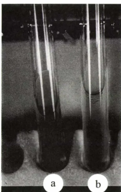
일반적인 관찰 - 간섬유화(경화)를 유도한 Op-2군, Op-4군그리고 DW 경구투여군인 OpDW-2군, OpDW- 4군의 랫드 귀에서 황달이 줄현하였고, proximal bile duct에서는 담즙 울체가 관찰되었으며,OpDW-2군, OpDW-4군의 담즙양은 op-22-, Op-4군보다 약간 적 었고(1~3 m/), 또한 담즙색은약간 맑았다(Fig. 1).
체중,장기 무게 및장기/체중변화 - 정싱군에 비해
서 Op군과 OpDW군에서 간종대,비종대가관찰 되었
다(Table I). 간 무게는 Op-4군,OpDW-4군에서 모두 정싱군보다유의성 있게 높게나타났고(p<0.005), 간/ 체중의 무게비 또한 Op-4군, OpDW-4군 뿐만 아니라
Op-2군,OpDW-2군에서도유의성 있게높게 관찰되었
다(Table I, p<0.005). OpDW-4군에서 간무게(18.8%, p<0.05), 간/체중비 (p<0.05)는 Op-4군과 비교했을 때 유의성 있게낮았다. 신장, 비장의무게와비장/체중,신 장/체중은 정상군보다 Op-2, 4군과 OpDW-2, 4군에서 모두 높았으며,비장/체중 비에서는 Op-2군보다 OpDW-2군에서 유의성있게 낮았다(p<0.05, Table I).
혈청생화학적 변호t - 정싱군에 비해서 Op-2군, Op-
Fig. 1 - The bile juice of expanded proximal bile duct from a rat in op-4 group and OpDW-4 group, a: bile juice from a rat in Op-4 group, b: bile juice from a rat in OpDW-4 group
Table I - The weight of organ and the ratio of organ/body weight in normal, fibrotic and DW treated rats
Group normal Op-2 Op-4 OpDW-2 OpDW-4
Liver weight(g) Kidney weight(g) Spleen weight(g) Ratio of LW/BW(%) Ratio of KW/BW(%) Ratio of SW/BW(%) Number of animal
9.66 ± 1.30 1.92 ± 0.21 0.60 ± 0.10 3.75 ± 0.24 0.75 ± 0.05 0.24 ± 0.05
6
11.66 ± 0.98 2.23 ± 0.46 1.13 ± 0.31*
5.89 ± 0.45**
1.13 ± 0.29*
0.58 ± 0.19*
6
21.10 ± 1.90**
2.34 ± 0.18*
1.42 ± 0.21*
7.66 ± 0.69**
0.85 ± 0.06 0.52 ± 0.08*
11
12.09 ± 2.26 2.12 ± 0.49 0.78 ± 0.29#
6.11 ± 1.17**
1.08 ± 0.31*
0.40 ± 0.16*,# 5
17.05 ± 3.95** # 2.11 ± 0.31
1.36 ± 0.42*
6.56 ± 1.31** # 0.82 ± 0.08 0.48 ± 0.13*,#
---:*The significant value different from normal (p<0.05), **The significant value different from normal (p<0.005), The --:--- --- 3---6 significant value different from Op-4 group(p<0.05)
Op-2: The 2 weeks observed rats group after BDL/S Operation Op-4: The 4 weeks observed rats group after BDL/S Operation
OpDW-2: The 2 weeks observed rats group after BDL/S Operation and treatment with DW OpDW-4: The 4 weeks observed rats group after BDL/S Operation and treatment with DW
Table II - The value of clinical parameters in sera of normal, fibrotic and DW treated rats
Group normal Op-2 Op-4 OpDW-2 OpDW-4
GOT 44.0 ± 13.2 238.5 ± 44.8* 317.3 ± 72.3* 154.7 ± 35.8*’# 264.5 ± 100.0*
GTP 21.8 ± 6.7 32.3 ± 9.3 30.4 ± 4.5 24.7 ± 4.9 33.5 ± 9.5
BUN 17.7 ± 1.5 19.5 ± 4.2 21.7 ± 4.8 19.8 ± 4.2 17.1 ± 5.4
ALP 161.4 ± 95.1 261.0 ± 65.5* 291.1 ± 38.2* 231.5 ± 82.1* 214.1 ± 57.9*’##
T-bilirubin 0.09 ± 0.18 8.4 ± 1.3* 6.5 ± 0.56* 6.7 ± 2.8* 6.4 ± 1.7*
Number of animal 6 6 11 5 6
---3---1--- ;--- ;--- T S ---
*The significant value different from normal (p<0.01), The significant value different from Op-2 group (p<0.005), The significant value different from Op-4 group (p<0.05)
4군, OpDW-2군,OpDW-4군에서 GOT, GTR ALR
t-bilirubin의 수치가 유의성 있게 높게 나타났고,
OpDW-2, OpDW4군과 Op-2군, Op>4군을 비교했을 때 GOT, ALR t-bili의 수치가낮았으나 유의성은 관 찰되지 않았으며, 단지 OpDW-2군의 GOT 수치가
Op-2군과 비교했을 때 유의성 있게 낮았다(Table H,
p<0.01). 그리고 OpDW-4군의 ALP수치는 Op>4군과 비교했을 때유의성 있게 낮았다(p<0.05).
MDA 농도의 변화-혈청중 MDA 농도는 Op군과 OpDW군 모두에서 정상군보다 유의성 있게 높았다 (p<0.05). 그러나 Op-2군, Op-4군보다 MDA농도가 OpDW-2군, OpDW-4군에서 낮았으며특히 OpDW-4군
에서는 Op-4군보다 유의성있게 낮았다(Fig. 2a,
p < 0.001).
Hydroxyproline양의 변화 - 정상군과 비교했을때 Op군과 OpDW군에서모두유의성 있게높은 hyp양이 관찰되었으나(p<0.05~<0.005), OpDW-4군의 hyp 양
은 Op-4군과 비교했을 때 유의성 있게 낮았다(Fig.
2b, p<0.05).
MDA와 hyp, GOT, ALE T-bili와의상관성 - 정상 군에서는 MDA와 GOT, t-bili와상관성이 있었고(Table
N orm a l O p - 2 O p - 4 O p D W - 2 O p D W - 4
Group
Fig. 2 - The diagrams of (a) MDA in sera and (b) hyp in liver tissue of rats. *The significant value different from normal (p<0.05), **The significant value different from normal (p <0.005), #The significant value different from Op-4 group (p<0.05)
J. Pharm. Soc. Korea
실험동물에서 복합천연물 (DW) 의 지질 과산화 억제와 간섬유화 저해 효과 검색 517
0.014 0.509 0.656
0.251 0.673 0.540
0.252 0.385 0.398
Normal Op-group OpDWgroup
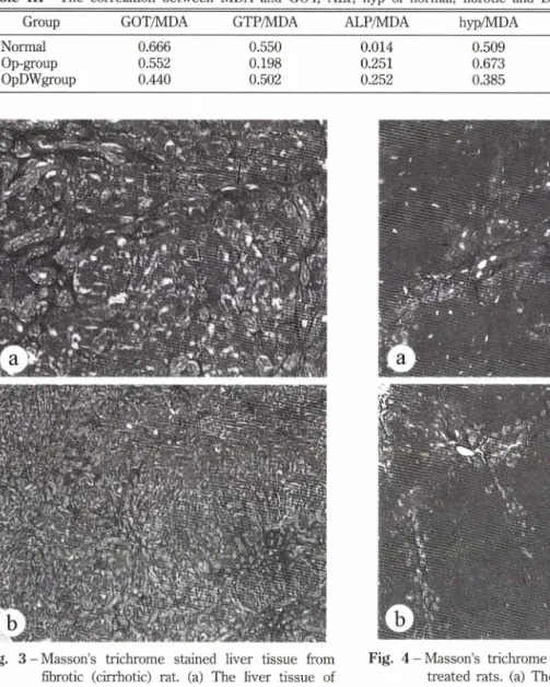
Fig. 3 - Masson's trichrome stained liver tissue from fibrotic (cirrhotic) rat. (a) The liver tissue of BDL/S operated rat ( x 80), (b) The liver tissue of BDL/S operated rat ( x 40)
<~ : collagen fiber, : bile duct proliferation
HI, r=0.666, r= 0.656), Op군에서는 MDA와 hyp가
상관성이 있으며(r=0.673), OpDW군에서는 다른
parameter와상관성은전혀나타나지않았다(Table IH).
조직학적변화 - 정상군보다는 Op-2, 4군과 OpDW- 2,4군 모두에서 담관증식(++〜 + + +), 섬유화 (+H h + +), portal triads에서의 과도한 collagen
fiber 축적, 간세포의 팽대와핵의붕괴 등이 관찰되었
다(Fig. 3a, 3b). 그러나 OpDW-4군에서 담관증식이 주로 portal triads에편재되어 있고 stroma로진전된 것이 많지 않았으나, Op-4군에서는 portal triads와
Fig. 4 - Masson's trichrome stained liver tissue of DW treated rats, (a) The liver tissue of in 4 weeks DW treated rat after BDL/S operation ( x 80), (b) The liver tissue of in 4 weeks DW treated rat after BDL/S operation ( x 40)
<~ : collagen fiber, : bile duct proliferation
stroma에서도 담관증식이 관찰되었다. OpDW-4군에서
간세포의종창은 Op-4군보다 익하고, 세포질의 파괴와
핵붕괴 등은 Op군보다는 상태가 좀더 약하게 관찰되 었다(Fig. 4a, 4b).
고 찰
각종 화학물질의 대사과정에서 생성된 free radical(NO- OH_, 02!은 지질과산화물의 개시물질로 Table III - The correlation between MDA and GOT, ALR hyp of normal, fibrotic and DW treated rats
Group GOT/MDA GTP/MDA ALP/MDA hyp/MDA t-bili/MDA n
6
17 12
0.551 0.19;
0.50:
.666.552
•440
서 나타나는데 이는 분자상 산소와 직접 반응하여 organic peroxy free radical(유기과산화)을 형성하고9’385 독특한세포장해를 일으켜 간괴사세까지 이르게 된다. In vivo 실험에서 지질과산화는간,신장, 심장 뇌등 에서 장기손상을 일으킨다고알려져 있으며,39’40) 특히 지질과산화분해물이 담즙울체에 기인하는실험적 간 섬유화의 간, 신장,심장의 조직에서 중가되었다는보
고가있고,2> 지질과산화를억제할수 있는물질은항
산화 효과가 있는것으로 간주되고있으며 여러가지 천연물에서 항 화 효과가검색되고 있다.33’34) 간섬유 화는간구성 세포의 정량적인변화와형태학적 변화를 수반하고, 특히 ECM(extracellular matrix protein) 의 간조직내 합성과 분해의 불균형이 일어나서 과도한
ECM의축적을일으킨다. 따라서 항섬유화효과는 과
도한 EMC의조직내 축적을 방지하거나합성을 억제
하고분해를촉진하는 것이다.
간질환과 관련하여 지금까지 penicillamine,41'42*
16,16-dimethyl prostaglandin E》43) biphenyl dimethyl dicarboxylate,441 colchicine,45) glucocorticoid,^ malo- tilate,47) interferon-Y,483 pentoxifylline49' 및 prolyl 4- hydroxylase inhibitor 인 pyridine-2,4-dicarboxylic-die thylamide(SO 885) 와 pyridine-2,4-dicarboxylic-di(2- methoxyethyl) amide(HOE OO7)5®등의 간섬유화 억 제효과에 대한 많은 연구가 진행되고 있으나 임상에 적용시 항섬유화작'§써 미약하거나또는그부작용이 심하여 뚜렷한항섬유화효과가있는물질이 아직까지 없는실정이다. 따라서 지질과산화분해물의 정량적인 측정과간섬유화의 지표(간조직중 collagen 축적량)의 측정은항산화및항섬유화효괴를동시에 검색할수 있다는 점에서 큰의미가있다.
본연구에서는천연물건류추출액 (DW)의항섬유화
효과와항산화효과를검색하기 위해간조직중 hyp(총
collagen)양과 지질과산화 분해물인 MDA(malondial-
dehyde)를 측정하며 그 효과를 검색하였다. 일차적으
로본시험에 앞서 이미 BDL/S 수술방법으로간섬유
화(경화)를 유도한 랫드에서 MDA와다른 parameter 에대한결과36>를확인하여 천연물의 투여기간을 2주,
유 로 결정하였으며,데이터 해석에도많이참고하였다. 본 실험에서 간섬유화(경화)를 유도한 수술군과 DW(천연물)투여군 모두에서 정상군동물과 비교했을 때 귀와 눈주위에 황달, expanded proximal bile duct에담f울체가나타났으나, DW투여군(OpDW-2, 4
군)의 담즙색이 수술군(Op-2,4군)보다 맑고 투명한
것으로미루어 보아 DW의투여가황달을약화시키는 효과가있음이 육간적인관찰에서 과악되었다(Fig. 1).
장기의 무게와장기/체중의 비는정상군보다 Op-2, 4군과 OpDW-2, 4군에서 모두높게 나타났으며,Op군 과 OpDW군을서로 비교했을때 OpDW-2군에서 비 장/체중비가 17.2%(p<0.05), OpDW-4군에서간무게와 간/체중비가 19.2%(p<0.05)로유의성 있게낮게 나타 남으로서 (Table I) DW의 4주간투여는담즙울체에 기 인하는비종대와간종대를약화시킬수 있는가능성이 나타났다. 이는영지균사체추출물을 4주간투여 후간 종대가약화되었다는박등29>의 연구결과와유사하다.
본실험의 Op군과 OpDW군모두에서는 정상군보다
혈청중 GOT, ALR t-bilirubin의 수치가 유의성 있게 높게 나타났고(p<0.05~<0.005), OpDW-2군에서는 GOT, GTR BUN, t-bilirubin 이 Op-2군보다 수치가
낮았으나 유의성은 검증되지 않았으며 (Table II),
OpDW-4군에서 ALP의수치가 Op-4군보다유의성 있 게 낮았다(p<0.05). 간섬유화의 지표인 hyp수치는 opDW-2군에서 op-2군보다높게 나타났지만, opDW-4
군에서는 op-4군보다 유의성 있게 낮게 관찰되었다
(p<0.05, Fig. 2b). 조직학적 관찰에서 약간 경미한 간세포종창, 섬유화와육안적인 관찰에서 경미한황 달출현,간조직중 유의성 있게 낮은 hyp 수치, 혈청 진단지표에서 낮은수치가 나타남으로서 간기능 개선, 간보호 및항섬유화효과의 /[능성이 높게 나타났다.
항산화 효과와 관련하여 Shimizu등26>은 DMN (dimethylnitrosamine)으로유도한간섬유화동물의 간 조직중 지질과산화분해물인 MDA 수치의 유의성 없 는 감소를 보고하였으나, 본 연구에서는 MDA가 Op
군과 OpDW군에서 정상군보다약 3~8.5배정도유의
성 있게 높게 나타났고 Op-2군과 OpDW-2군에서 거 의비슷한양상을보였다(Fig. 2a). 그러나 OpDW-4군 에서 59.4%(p<0.005, Fig. 2a) 유의성 있게 낮은 것 으로미루어 보아항산화효과있다고사료된다. 이와 유사한 결과에 대해서 Ljubumcic둥51}은 common bile duct ligation ᄋ-?- 담즙울체성간손상을유도한 랫
드에서 UDCA를 24일간투여하였을 때혈청 간기능
지표의 개선효과는없으나지질과산화는 억제되었다고 보고하였다.
또한 최근에 Yasuda등18〕은 lipid peroxide인 MDA 와 hepatic collagen양 사이에서 유사한 변화양상을
J. Pharm. Soc. Korea
실험동물에서 복합천연물 (DW) 의 지질 과산화 억제와 간섬유화 저해 효과 검색 519
보고하고있으나, 아직은뚜렷한조직중 hyp와혈청중
MDA와의 상관관계에 대한 보고는 없다. 그러나 본 실험에서 측정된 hyp와 MDA는수술후 4주군의 비교 에서 두지표사미에 중간정도의 상관관계를 나타내고 있고(r=0.673, Table IH) 항섬유화와 관련하여 간조직 중 hyp가 OpDW4군에서 Op-4군보다 24.5%(p<0.05) 유의성 있게 낮음으로서 항섬유화효과가있으며, Op 군에서는 MDA와 hyp가중간정도의 상관성 (r=0.673)
이있었으나 OpDW군에서는상관성이 관찰되지 않았
다. 이러한 결과는 BDIVS로간섬유화(경화)를유도한 랫드에서 간조직중 hyp는 혈청중 PNniP(procollagen in peptide)와상관성이 있으나, 항섬유화효과가있는 경우에는상관성이 관찰되지 않는다는김등52}의보고 와일치한다.
따라서 천연물또는 약물이 항산화및 항섬유화효 능이 있는경우에는각지표들사이에 상관성이 없을 가능성이 있으며,간섬유화가유도되었을경우에는 지
질과산화 분해물인 MDA의 농도가 간조직중 hyp와
상관성이 있다고생각된다. 혈청중 MDA의측정과간
조직 중 hyp의측정은 항산화와항섬유화효과의 검색
에좋은지표로사용할수있고혈청과간기능진단지
표인 MDA와 hyp의상관관계를 조사하는 것은항산
화, 항섬유화효과 검색에 유용하리라사료된다. 또한 DW는 4주간의 투여에서 간,간/체중비, ALR MDA,
hyp가유의성 있게 비투여군(수술군) 보다 낮은 수치
를나타냄으로서 확실한항산화, 항섬유화 효과가 있 다고사료된다.
감사의 말씀
본 연구는원광대학교 한의학전문대학원 BK21 사업
비, 2000년교비에 의해 수행된 것으로 지원에 감•사
드립니다.
문 헌
1) Szuster-Ciesielska, A., Daniluk, J. and Kandefr- Szerszen, M. : Alcohol-related cirrhosis with pancreatitis. The role of oxidative stress in the progression of the disease. Arch. Immunol Ther.
Exp. (Warsz) 49(2), 139 (2001).
2) Ljubuncic, R, Tanne, Z. and Bomzon, A. : Evidence of
a systemic phenomena for oxidative stress in cholestatic liver disease. GUT 47(5), 710 (2000).
3) Maria-Liisa, S., Heikki, A., Anni, H., Fulvio, U., Tom, M., Antonella, R., Dontscho, K.,and Harry, H. : Lipid peroxidation in human proteinuric disease. Cell Biol.
Immunol. Pathol Intern. Soc. Nephrol. 59(2), 481
(2001).
4) Diamond, J. R .: The role of reactive oxygen species in animal models of glomerular disease. Am. J.
Kidney Dis. 19,292 (1992).
5) Cracoski, J. L., Marpeau, C., Carpentier,R H., lmbert, B., Hunt, M., Stanke-Labesque, E and Bessard, G .: Enhanced in vivo lipid peroxidation in scleroderma spectrum disorders. Arthritis Rheum.
44(5),1143 (2001).
6) Stanely Mainzen Prince, R and Menon, V R : Antioxidant action of Tinospora cordifolia root extract in alloxan diabetic rats. Phytother. Res. 15(3),
213 (2001).
7) Young, I. S. and McEneny, J. : Lipoprotein oxidation and atherosclerosis. Biochem. Soc. Trans. 29(2), 358 (2001)
8) Miller, E. R.,Appel, L. J. and Risby, T. H. : Effect of dietary patterns on measures of lipid peroxidation;
Results from a randomized clinical trial. Circulation 98(22), 2390 (1998).
9) Radi, R.,Beckman, J. S., Bush, K. M. and Freeman, B. A .: Peroxynitrite-induced membrane lipid peroxi
dation; The cytotoxic potential of superoxide and nitric oxide. Arch. Biochem. Biophys. 288,481 (1991).
10) Lyon, B. L., Schwarz, R. I . : Ascorbate stimulation of PAT cells causes an increase in transcription rates and a decrease in degradation rates of procollagen mRNA. Nucleic Acids Res. 12,2569 (1984).
11) Lee, KS., Bruck, M.,Houglum, K.,Chojkier, M. : Activation of hepatic stellate cells by TGFa and collagen typells mediated by oxidative stress through c-myb expression. J. Clin. Invest 96,2461 (1995).
12) Ohkawa, H., Ohishi, N., and Yagi, K. : Assay for lipid peroxides in animal tissues by thiobarbituric acid reaction. Anal. Biochem. 95, 351 (1979).
13) Bacon, B. R., Tavill, A. S., Brittenham, G. M., Park, C. H. and Recknagel, R. 0 .: Hepatic lipid peroxidation
in vivo in rats with chronic iron overload. /• Clin.
Invest 71, 429 (1983).
14) Chance, B., Sies, H., and Boveris, A. : Hydroperoxide metabolism in mammalian organs. Physiol. Rev. 59, 527 (1979).
15) Tasi, L. Y., Lee, K. T, Tsai, S. M., Lee, S. C. and Yu, S. H .: Changes of lipid levels in blood and liver tissues of patients with obstructive jaundice. Clin.
Chim. Acta. 215, 41 (1993).
16) Cutrin, J. C., Zingaro, B.,Camandola, S., Boveris, A., Pompella, A., Poli, G .: Contribution of gamma glutamyl transpeptidase to oxidative damage of ischemic rat kidney. Kidney International. 57(2), 526 (2000).
17) Leclercq, I., Horsmans, Y., Desager, J-F, Delzenne, N. and Geubel, A .: Reduction in hepatic cytochrome P-450 is correlated to the degree of liver fat content in animal models of steatosis in the absence of inflammation. J. Hepatol 28(3), 410 (1998).
18) Yasuda, M., Shimizu, I., Shiba, M., Ito, S. and Itᄋ. S.
: Suppressive effects of estradiol on dimethylnitro- samine-induced fibrosis of the liver in rats, Hepatology 29, 719 (1999).
19) Clement, B., Grimaud,J.A., Campion,J. R, Deugnier, Y.
and Guillouzo, A .: Cell types involved in collagen and fibronectin production in normal and fibrotic human liver. Hepatology 6, 225 (1986).
20) Maher, J. J., Bissell, D. M., Friedmann, S. L. and Roll, E J. : Collagen measured in primary cultures of normal rat hepatocytes derives from lipocytes within the monolayer. J. Clin. Invest. 82,450 (1989).
21) Thumham, D. I .: Carotenoids; functions and fallacies.
Proc. Nutr. Soc. 53, 77 (1994).
22) Riemersma, R. A., Wood, D. A., Macintyre, CCA., Elton, R. A., Gey, K. F. and Oliver, M. E : Risk of angina pectoris and plasma concentrations of vitamin A, C, and E and carotene. Lancet 337,1 (1991).
23) Gey, K. E, Moser, U. K., Jordan, R, Stahelin, H. B.,
Eichholzer, M. and Ludin, E. : Increased risk of cardiovascular disease at suboptimal plasma concen
trations of essential antioxidants: An epidemiological update with special attention to carotene and vitamin C. Am. J. Clin. Nutr. 57,787S (1993).
24) Lin, C. C., Hsu, Y. E, Lin, T. C. and Hsu, H. Y. : Antioxidant and hepatoprotective effects of punicalagin
and punicalin on acetaminophen induced liver damage in rats. Phytother. Res. 15(3),206 (2001).
25) Trivedi, N. R and Rawal, U. M. : Hepatoprotective and antitoxidant property of andrographis paniculata (Nees) in BHC induced liver damage in mice. Indian J. Exp. Biol 39(1), 41 (2001).
26) Shimizu, I., Ma, Y. R.,Mizobuchi, Y., Liu, E, Miura, T, Nakai, Y., Yasuda, M., Shiba, M., Hone, T, Amagaya, S., Kawada, N., Hori, H. and Ito, S. : Effects of Sho-saiko-to, a Japanese herbal medicine, on hepatic fibrosis in rats. Hepatology 29,149 (1999).
27) Locher, R., Suter, R M., Weyhenmeyer R. and Vetter W. : Inhibitory action of silybinin on low density lipoprotein oxidation. Arzneim-Forsch 48(1), 236 (1998).
28) 林 藤 : egfe藤 湯 S K가 CC14 賴 臓 胞 讓 에 미치는틜#게 關한 職 . 利# #紙, 1(1), 70 (1976).
29) 박은전, 김기영, 김재백, 김수웅, 이승룡, 손동환: 영지 로부터추출한다당체의실험적간경화에대한섬유화 억제효과. 약학회지 38(3), 338 (1994).
30) Schuppan, D. : Chronische Hepatitis-Antifibrotische Therapieausaetze Z. Allg. Med 71,11531 (1995).
31) Batta, A. K.,Saken, G., Arora, R. and Sheffer, S. : Effect of ursodeoxycholic acid on bile add metabolism in primary biliary cirrhosis. Hepatology 10,414 (1998).
32) Jafri M, M. A., Jalis, S., Kalim, J. and Surender, S. : Hepatoprotective activity of leaves Cassia occidentalis against paracetamaol and ethyl alcohol intoxication in rats. J. Ethnopharmacol 66,355 (1999).
33) Inoue, T. and Jackson, E. K. : Strong antiproliferative effects of baicalein in culures rat hepatic stellate cells. Eur. J. Pharmacol 378(1), 129 (1999).
34) Xiao-Ming, D., Ning-Yi, S., Yang, C., Nobuta, I, and Yukihiro, S .: Hepatoprotective aliphatic glycosides from three Goodyera species. Biol Pharm. Bull.
23(6), 731 (2000).
35) Kim, K. Y., Suh, E. S., Park, S. Y. and Lim, J. A.: The bile duct obstruction stimulated lipid peroxidation and liver fibrosis (cirrhosis) in animal. J. Toxicology in press.
36) Kountras, J., Billing, B. H. and Scheuder, R J. : Prolonged bile duct obstruction: a new experimental
J. Pharm. Soc. Korea
실험동물에서 복합천연물 (DW) 의 지질 과산화 억제와 간섬유화 저해 효파 검색 521
model for cirrhosis in the rat. Br. J. Exp. Path. 65, 305 (1984).
37) Jamall, I. S. and Finelli, V N. : A single method to determine nanogram levels of 4-hydroxyproline in biological tissue. Anal. Biochem. 112,70 (1981).
38) Beckman, J. S., Beckman, T. W., Chen, J., Marshall, R A. and Freeman, B. A .: Apparent hydroxyl radical production by peroxynitrite; Implications for endothelial injury iron nitric oxide and superoxide.
Proc. Natl. Acad. Sci., 87,1620 (1990).
39) Solin, M. L., Ahola, H., Hatia, A., Ursini, E, Montine, T., Roveri, A., Keijaschki, D. and Holthoefer, H. : Lipid peroxidation in human proteinuric disease.
Kidney Int. 59(2),481 (2001).
40) Johnson, R. J., Lovett, D.,Lehrer, R. I. Couser, W. G., Klebanoff, S., J. : Role of oxidants and proteases in glomerular injury. Kidney Int. 45,352 (1994).
41) Neubauer,J., Cristensen, E.,Portmann, B., Caballeri, J., Rodes, J. and Ranek, L. : Double blind controlled trial of d-penicillamine in patients with primary biliary cirrhosis. GUT 26,114 (1985).
42) Wiesner, R. M.,Dickson, E. R., Lindor, D., Jorgensen, R.,LaRusso, N. E and Baldus, W : A controlled clinical trial evaluating cyclosporin in the treatment of primary biliary cirrhosis: A preliminary report.
Hepatology 7,1025 (1987).
43) Ruwart, M. J., Wilkinson, K. E, Rus, B. D., Vidmazr, T. J., Hahn, E. G., Peters, K. M.,Hendley, K. S., Appelman, H. D., Kim, K. Y. and Schuppan, D. : The integrated value of serum procollagen HI peptide over time predicts hepatic hydroxyproline content and stainable collagen in a model of dietary cirrhosis in the rat. Hepatology 10,801 (1989).
44) Nan, J. X., Park, E. J., Kim, H. J., Ko. G. Sohn, D. H.
: Antifibrotic effects of the methanol extract of Polygonum aviculare in fibrotic rats induced by bile
duct ligation and scission. Biolog & Pharmaceut.
Bulletin. 23(2), 240 (2000).
45) Kershenobich, H. D., Vargas., Garcia-Tsao, G., Perez- Tamayo, R. and Rojkind, M. : Effectiveness of colchichine in patients with cirrhosis. Hepatology 7, 1104 (1987).
46) Guzelian, D, S., Lindblad, W. J. and Diegelmann, R. E : Glucocorticoids suppress formation of collagen by the hepatocyte: Studies in primary monolayer cultures of paraenchymal cells prepared from rat liver. Gastroenterology. 86,897 (1984).
47) Ala-Kokko, L.,Stenback, E and Ryhanen, L.:
Preventive effect of malotilate on dimethylnitxo- samine-induced liver fibrosis in the rat. /. Lab. Clin.
Med. 113,177 (1989).
48) Svegliati Beroni, G., Curto, R, Recchioni, A., Mancini, R., Benedetti, A., Jezequel, A. M. and Oralan, E : Interferon-gamma reduces liver injury and collagen deposition in dimethylnitrosamine- induced cirrhosis. Hepatology 18,209A (1993).
49) Peterson, T. C. : Pentoxifylline prevents fibrosis in an animal model and inhibits platelet-derived growth factor-driven proliferation of fibroblasts. Hepatology 17,486 (1993).
50) Boker, K., Schwarting, G., Kaule, G., Gunzler,V and Schmidt, E .: Fibrosis of the liver in rats induced by bile duct ligation: Effects of inhibition of prolyl 4- hydroxylase. J. Hepatol 13,35 (1991).
51) Ljubuncic, E,Tanne, Z.,Bomzon, A .: Ursodeoxycholic acid suppresses extent of lipid peroxidation in diseased liver in experimental cholestatic liver disease. Dig. Dis. Sci 45(10),1921 (2000).
52) Kim, K. Y.: Pharmakologische Pruefung potentieller Antifibrotika in einem Ratten-Leberfibrose-Modell sowie Ermittlung der Stoffwechselwege zirkulierender Prokollagen-Peptide. Dissertation, Berlin (1991).