Immunomodulatory Effects of β-1,3/1,6-glucan and Lactobacillus plantarum LM1004 on Atopic Dermatitis Models
In Sung Kim
1†, Sung Hak Kim
2†, Jeong A Kim
1, Da Yoon Yu
1, Gwang Il Kim
1, Dong-Chan Park
3, Jong Min Lim
3, Sang Suk Lee
4, In Soon Choi
5and Kwang Keun Cho
1*
1Department of Animal Resources Technology, Gyeongnam National University of Science and Technology, Jinju 52725, Korea
2Department of Animal Science, Chonnam National University, Gwangju 61186, Korea
3Glucan Corporation, Busan 46048, Korea
4Department of Animal Science and Technology, Sunchon National University, Suncheon 57922, Korea
5Departmnet of Life Science, Silla University, Busan 46958, Korea
Received September 29, 2017 /Revised January 5, 2018 /Accepted January 8, 2018
In this study, we examined the efficacy of the immune regulation of β-1,3/1,6-glucan and Lactobacillus plantarum LM1004 on atopic dermatitis models. The oral administration of β-1,3/1,6-glucan and L.
plantarum LM1004 on mice significantly decreased the amount of scratching, leakage to evans blue, and concentrations of serum immunoglobulin E (IgE) and histamine compared with the atopic derma- titis–induced group. When atopic dermatitis was induced, the transcription factors (GATA-3, retinoic acid-related orphan receptor γ T [RORγT]) and cytokines (interleukin-4 [IL-4], IL-17) of Th2 and Th17 cells were overexpressed at the transcriptional level, and they significantly decreased with oral admin- istration of β-1,3/1,6-glucan and L. plantarum LM1004. In addition, β-1,3/1,6-glucan and L. plantarum LM1004 were shown to modulate the immune balance by increasing the expression of Th1 and Treg transcription (T-bet, forkhead box p3 [Foxp3]) and cytokines (interferon-γ [IFN-γ], transforming growth factor-β [TGF-β]). Galectin-9 and filaggrin were significantly lower in the atopic dermatitis–
induced group and significantly higher in the β-1,3/1,6-glucan-treated group. In contrast, thymic stro- mal lymphopoietin (TSLP) was highest in the atopic dermatitis–induced group, while mice that were orally administered β-1,3/1,6-glucan and L. plantarum LM1004 showed similar TSLP levels to the con- trol group. These results indicate that β-1,3/1,6-glucan and L. plantarum LM1004 have immunomodu- latory effects and atopic dermatitis improvement effects in an animal model of atopic dermatitis.
Therefore, it is expected that β-1,3/1,6-glucan and L. plantarum LM1004 can be used as natural materi- als in the treatment of atopic dermatitis.
Key words : Atopic dermatitis, β-Glucan, immunomodulatory, Lactobacillus plantrum LM1004
†Authors contributed equally.
*Corresponding author
*Tel : +82-55-751-3286, Fax : +82-55-751-3689
*E-mail : [email protected]
This is an Open-Access article distributed under the terms of the Creative Commons Attribution Non-Commercial License (http://creativecommons.org/licenses/by-nc/3.0) which permits unrestricted non-commercial use, distribution, and reproduction in any medium, provided the original work is properly cited.
Journal of Life Science 2018 Vol. 28. No. 1. 17~25 DOI : https://doi.org/10.5352/JLS.2018.28.1.17
서 론
아토피 피부염은 주로 영유아기에 발병되는 대표적인 난치 성 질환으로 알려져 있으며 일반적으로 피부 건조(xeroder- ma), 가려움(pruritus), 염증(inflammation)을 동반한 아토피 성 습진(eczema)을 말한다[28]. 아토피성 피부염의 면역학적 기전으로는 T helper cell type 1 (Th1)/Th2 세포간의 불균형, cytokine 체계 이상(dysfunction), Langerhans cell의 활성 증 가 등이 있다[30, 31]. 또한 Nograles 등[39]은 Th17 세포의
생산(produce)과 기능 조절의 불균형으로 아토피성 피부염이 발생된다고 보고하였다.
아토피성 피부염의 원인 물질이 체내에 들어오면 Th2 세포 는 IL-4, IL-5, IL-13 등의 cytokine을 과잉 분비하여 IgE의 생성 을 증가시키고 비만 세포(mast cell)와 호산구(eosinophil)의 분화(differentiation)를 유도시킴으로써 histamine, leuko- triene 등의 화학물질 탈과립(degranulation)이 일어난다[21, 34, 35]. 이러한 탈과립 물질들에 의해 피부에 붉은 반점(spot), 부종(edema), 가려움증 등을 동반한 아토피성 피부염이 유발 된다. 아토피성 피부염 발병에 관여하는 중요한 분자로 TSLP 는 급성 및 만성 아토피성 피부염 환자와 생쥐 모델의 각질형 성세포(keratinocyte)로부터 과발현 되며[47, 51], IL-3, IL-13, TNF-α mRNA의 발현을 증가시켜 수지상세포로부터 Th2 세 포 반응을 유도한다[38]. 이로 인해 표피단백질(epidermis protein)인 filaggrin mRNA의 발현이 감소되어 피부 장벽 손 상이 일어나 아토피 피부염이 유발된다[54].
Galectin은 체내의 여러 장기(viscera)에 분포하면서 특히
면역계 반응(immune system reaction)에 관여하는 다양한 세포 들의 기능을 조절하는데, Galectin-9은 Treg 세포를 활성화하 여 자가면역질환(autoimmune disease)을 완화시킨다[29, 44].
아토피성 피부염 완하제로는 항히스타민과 스테로이드 제 제가 많이 사용되고 있으나 장기간 사용할 경우 부작용이 발 생하여, 최근에는 부작용이 적은 천연소재에 대한 연구가 활 발히 이루어지고 있다[1].
Aureobasidium pullulans 균주에 의해 생산되는 풀루란 중에 서 가장 잘 알려진 것은 α-glucan이지만[24], β-1,3/1,6- 결합의 분자 구조를 가짐으로써 생리활성 촉진 능력이 우수한 β-glu- can을 생산한다[26]. β-glucan은 다당류의 일종으로 효모의 세 포벽, 버섯류, 곡류를 비롯하여 효모, 조류, 박테리아 등에 존 재하는 생리활성 물질이다[2, 11, 32]. 낮은 농도에서 높은 점성 을 나타내는 수용성 식이 섬유의 형태로서 항산화, 항암, 항콜 레스테롤, 면역증강 및 피부 재생 효과와 같은 다양한 생리활 성 촉진 효과가 밝혀져 있어 건강 식품 소재 및 식품 첨가물로 많이 사용되고 있다[5, 12, 16, 37, 52]. β-glucan은 Dectin-1, Toll-like receptor (TLR)-2, TLR-6 등의 세포 수용체를 통해 단핵구, 대식세포, 수지상세포, 자연살생세포, 호중구 내로 들 어가서 cytokine 분비를 유발하고 T 세포 및 B 세포를 자극시 켜 면역 반응을 조절하게 된다[6].
프로바이오틱스의 대표적인 유산균은 체내에서 유익한 효 과를 나타내며, 대부분 식품이나 체내에서 유래한 균주로서 부작용이 거의 없고 비교적 안전하다[25, 36, 53]. 알레르기 및 아토피성 피부염에 대한 유산균의 효과에 대하여 많은 임상 연구가 이루어졌으며[33], 특히 L. plantarum은 비병원성, 그람 양성 미생물로서 생쥐와 사람에게 있어 알레르기 증상을 완화 시키고[14, 20, 42, 43]. L. plantarum ATCC 8014는 CD4
+/ CD25
+/foxp3
+T 세포를 증가시킴으로써 면역반응을 자극시 키는 것으로 보고 되었다[19]. 하지만 아토피성 피부염에 대한 β-1,3/1,6-glucan과 L. plantarum의 면역조절 기전 연구는 현재 미흡한 부분이 많다.
본 연구에서는 비교적 소재가 풍부하지만 아토피성 피부염에 대한 연구가 미흡한 β-1,3/1,6-glucan과 김치 유래 L. planta- rum LM1004를 이용하여 아토피 피부염을 유발한 흰쥐와 생 쥐의 가려움증, 유출된 evans blue, 혈청 IgE, histamine, 면역 세포의 전사인자와 cytokine, galectin-9, filaggrin 및 TSLP의 발현을 분석하였으며, 이러한 결과를 통해 β-1,3/1,6-glucan 과 L. plantarum LM1004의 면역조절 효과를 규명하고자 하였다.
재료 및 방법
시료
본 실험에서 사용한 β-glucan은 흑효모 Aureobasidium pul- lulans SM-2001 균주에서 체외로 분비되는 β-1,3/1,6-glucan (β - 1,3 linkage 68: β-1,6 linkage 32)이며, 평균 분자량은 2.6×10
5Da, 순도는 15%이며[50], ㈜글루칸(Busan, Korea)으로부터 제 공받았다. L. plantarum LM1004 (KCCM 43246) 농도는 2×10
12cell/g이며, 배추김치에서 분리된 프로바이오틱스이다.
실험동물
본 실험에서 사용한 6주령의 Sprague-Dawley (SD) 수컷 흰쥐는 ㈜샘타코(Osan, Korea)에서 구입하여 아토피 피부염 유발에 이용하였으며, 6주령의 ddY 수컷 생쥐는 ㈜중앙실험 동물(Seoul, Korea)에서 구입하여 가려움증 유발에 이용하였 다. 모든 동물은 일정한 온도(22±1℃), 습도(60±10%)가 유지되 며 12시간의 명암주기가 조절되는 동물실에서 사양관리 되었 다. 또한 동물들은 1주일간 동물사양실에서 적응 과정을 거친 후 5개의 그룹으로 분리되었고, 전 실험기간 동안 고형사료 AIN- 76 (Seoul, Korea)와 물을 자유롭게 섭취할 수 있도록 하였다. 동물실험은 경남과학기술대학교 동물실험윤리위원 회의 인준을 받아 수행하였다(승인번호 2017-2).
아토피 피부염 유발 및 약물 투여
아토피 피부염 유발을 위하여 Ishiguro 등[18]의 방법에 따 라 히스타민 분비 촉진제인 Compound 48/48 (COM, Sigma, USA)을 사용하였다. β-1,3/1,6-glucan과 L. plantarum LM1004 는 7일 동안 매일 1회 0.2 ml(250 mg/60 kg)를 경구투여 하였 으며, 7일차에는 SD 흰쥐의 등 부위를 제모하고 COM 50 μl (10 μg/ml)를 제모 부위에 피내주사 하였다. 그리고 30분 후에 0.5%의 Evans blue (Sigma, USA) 200 μl를 미정맥으로 투여하 였다.
가려움증 판정 시험
가려움증 판정 시험을 위하여 β-1,3/1,6-glucan과 L. planta- rum LM1004는 7일 동안 매일 1회 0.2 ml (250 mg/60 kg)를 경구투여 하였으며, 7일차에는 ddY 생쥐 등 부위를 제모하고 COM 10 ml/kg (3 mg/kg)를 제모 부위에 피하주사 하였다.
그리고 COM 투여 30분 후부터 Ohtsuka 등[40]의 방법에 따라 20분간 ddY 생쥐 몸 전체에서의 가려움증이 나타나는 횟수를 관찰하였다.
유출된 evans blue 반점 직경의 측정
Evans blue가 함유된 생리식염수 투여 30분 후, 모세혈관의 확장에 의해 evans blue의 유출이 일어난 등 피부를 잘라내어 digital caliper (Bluebird, NA500-150S)로 직경을 측정하여 mm로 나타내었다.
유출된 evans blue의 정량
Katayama 등[22]의 방법에 따라 감작부위인 등 피부를 직
경 10 mm의 크기로 잘라내어 1.0 N KOH (1.0 ml) 용액에
37℃, 48시간 동안 반응시킨 다음, 0.6 N H
3PO
4(2.5 ml)와
Table 1. Primers for the target genes and GAPDH used in RT-PCR
Target gene Primer
GAPDH Forward
Reverse
5’ CCACCCAGAAGACTGTGGAT 3’
5’ CACATTGGGGGTAGGAACAC 3’
T-bet Forward
Reverse
5’ TCAACCAGCACCAGACAGAG 3’
5’ AAACATCCTGTAATGGCTTGTG 3’
GATA-3 Forward
Reverse
5’ CATTACCACCTATCCGCCCTATG 3’
5’ CACACACTCCCTGCCTTCTGT 3’
RORγT Forward
Reverse
5’ TTCACCCCACCTCCACTG 3’
5’ TGCAAGGGATCACTTCAATTT 3’
Foxp3 Forward
Reverse
5’ CCCATCCCCAGGAGTCTTG 3’
5’ CCATGACTAGGGGCACTGTA 3’
INF-γ Forward
Reverse
5’ TCAAGTGGCATAGATGTGGAAGAA 3’
5’ TGGCTCTGCAGGATTTTCATG 3’
IL-4 Forward
Reverse
5’ ACAGGAGAAGGGACGCCAT 3’
5’ GAAGCCCTACAGACGAGCTCA 3’
IL-17 Forward
Reverse
5’ TTC ATC TGT GTC TCT GAT GCT 3’
5’ TTG ACC TTC ACA TTC TGG AG 3’
TGF-β Forward
Reverse
5’ GAAGGCAGAGTTCAGGGTCTT 3’
5’ GGTTCCTGTCTTTGTGGTGAA 3’
Galectin-9 Forward
Reverse
5’ GAGAGGAAGACACACATGCCTTTC 3’
5’ GACCACAGCATTCTCATCAAAACG 3’
Filaggrin Forward
Reverse
5’ CACTGAGCAAAGAAGAGCTGAA 3’
5’ CGATGTCTTGGTCATCTGGA 3’
TSLP Forward
Reverse
5’ AGAGAAGCCCTCAATGACCAT 3’
5’ GGACTTCTGTGCCATTTCC 3’
아세톤(6.5 ml)을 첨가하여 균질화 시키고 원심분리(900× g, 10분)하여, 그 상층액을 620 nm에서 흡광도를 측정하여, 유출 된 evans blue의 양을 mg/site로 나타내었다.
혈청 IgE 및 histamine의 측정
Evans blue 투여 30분 후, 동물 희생시 대정맥에서 혈액을 채혈하여 1,500 rpm에서 15분간 원심분리하여 혈청을 분리하 였다. 분리된 혈청에서 각각의 Hitachi optigen Allergen spe- cific IgE assay system (Hitachi Chemical Diagnostics, USA) 과 Histamine EIA Kit (LDN, Germany)를 사용하여 IgE 및 histamine의 양을 측정하였다.
전사인자 및 cytokine의 측정
β-1,3/1,6-glucan과 L. plantarum LM1004의 면역조절 효과 를 분석하기 위해 혈관확장 SD 흰쥐를 희생시킨 후 장간막 림프절을 분리하였다. 장간막 림프절은 Trizol® Reagent (ambion, USA)에 넣고 Silent Crusher M (Heiodlph, Ger- many)을 사용하여 균질화하였다. Chomczynski와 Sacch[8]의 방법에 따라 RNA를 분리하였으며, cDNA 합성할 때까지 -20
℃에 보관하였다. cDNA 합성은 reverse transcription poly- merase chain reaction (RT-PCR) Kit (TaKaRa, Japan)를 사용
하였으며 50℃에서 30분간 수행하였다. PCR cycling의 조건은 95℃에서 10분 동안 initial denaturation 후 95℃에서 30초간 denaturation, 55℃에서 30초간 annealing 및 72℃에서 1분간 elongation의 조건으로 35cycle을 진행하였고 최종적으로 7 2℃에서 5분 동안 final elongation을 수행하였다. 실험에 사용 한 primer는 Table 1과 같다. Glyceraldehyde- 3-phosphate- dehydrogenase (GAPDH)를 reference gene으로 사용하여 Th1, Th2, Th17 및 Treg 세포의 전사인자 및 cytokine의 발현 을 분석하였으며, 또한 Galectin-9, Filaggrin, TSLP의 발현을 분석하였다.
통계처리
반복실험을 통해 얻은 결과는 IBM SPSS Statistics 20 (IBM, USA)을 사용하여 분산분석 하였으며, 평균±표준편차로 나타 내었다. 분석 결과에 대한 유의성 검정은 분산분석을 한 후 p<
0.05 수준에서 Duncan’s multiple range test를 실시하였다[13].
결과 및 고찰
가려움증 판정 시험
COM을 피하주사하여 ddY 생쥐의 20분간 긁는 행동의 횟
Fig. 1. Effects of β-1,3/1,6-glucan and L. plantarum LM1004 on the incidences of scratching behaviors in the acute pruri- tus induced by COM. C: Control, N: Negative control (induced AD), T1: β-1,3/1,6-glucan, T2: L. plantarum LM1004, T3: β-1,3/1,6-glucan and L. plantarum LM1004.
a-bMeans are significantly different in each group (p<
0.05). Data represent means±SD of 6 replicates.
A B
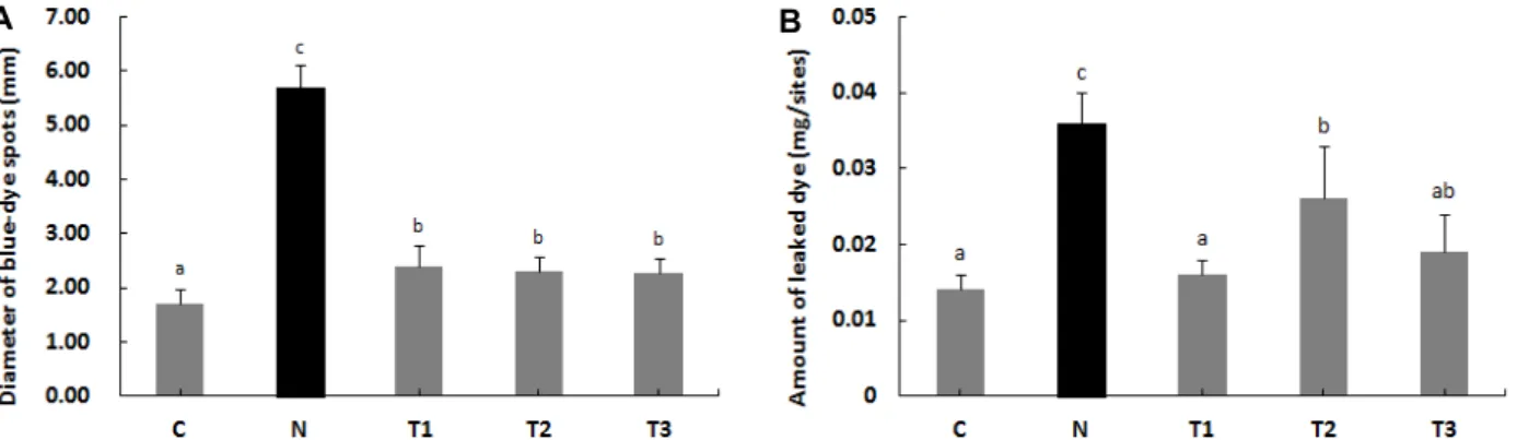
Fig. 2. Effects of β-1,3/1,6-glucan and L. plantarum LM1004 on the diameter of blue-dye spots and amount of leaked dye. (A) The diameter of blue-dye spots in the vasodilation induced by COM. (B) The amount of leaked dye in the vasodilation induced by COM. C: Control, N: Negative control (induced AD), T1: β-1,3/1,6-glucan, T2: L. plantarum LM1004, T3: β-1,3/1,6-glucan and L. plantarum LM1004. a-cMeans are significantly different in each group (p<0.05). Data represent means ± SD of 5 replicates.
수를 관찰한 결과 Fig. 1과 같이 아토피 피부염 유발그룹(N)에 서 39.17±9.95회의 긁는 행동이 관찰되었다. β-1,3/1,6-glucan 처리군(T1), L. plantarum LM1004 처리군(T2) 및 복합 처리군 (T3)에서 긁는 횟수는 각각 19.83±4.92회, 19.67±4.97회 및 18.67±5.68회로 나타나 아토피 피부염 유발그룹(N) 보다 가려 움증의 횟수가 유의적으로 적었다.
유출된 evans blue 반점의 직경 및 염색량의 변화 COM으로 유발된 모세혈관확장 아토피피부염 모델에 대한 β-1,3/1,6-glucan과 L. plantarum LM1004의 효과를 분석하기 위해 SD 흰쥐를 희생시킨 후 유출된 evans blue 반점 직경을 측정한 결과는 Fig. 2A와 같다. 대조군(C)은 유출된 evans blue 반점의 크기가 유의적으로 가장 작았으며, 아토피 피부염 유발그룹(N)은 5.7 mm로 유의적으로 가장 크게 나타났다. β- 1,3/1,6-glucan 처리군(T1), L. plantarum LM1004 처리군(T2)
및 복합 처리군(T3)의 유출된 evans blue 반점의 크기는 서로 유의적인 차이가 없었지만 아토피 피부염 유발그룹(N) 보다 유의적으로 낮은 값을 나타내어 COM으로 유발된 모세혈관확 장을 억제하는 효과를 보였다.
유출된 evans blue의 양을 분석하기 위해 SD 흰쥐의 등 피 부를 잘라내어 알칼리성 용액으로 용해한 후 상층액의 흡광도 를 측정한 결과는 Fig. 2B와 같다. 아토피 피부염 유발그룹(N) 은 유출된 evans blue가 0.04 mg/sites로 유의적으로 가장 많 았으며, β-1,3/1,6-glucan 처리군(T1)과 복합 처리군(T3)은 대 조군(C)과 유사한 수준으로 나타났다.
Histamine 분비 촉진제인 COM은 아토피 피부염 모델의 모세혈관확장을 유발시켜 evans blue의 유출을 유도한다[18].
따라서 유출된 evans blue의 감소는 투여한 실험재료가 모세 혈관확장을 완화시켜 아토피 피부염에 효과가 있다는 것을 의미한다[17, 46]. 본 연구에서 SD 흰쥐에 아토피 피부염을 유 발시킨 후 β-1,3/1,6-glucan과 L. plantarum LM1004를 투여하 였을 때 유출되는 evans blue의 크기와 양이 감소하는 것으로 나타났다. 따라서 β-1,3/1,6-glucan과 L. plantarum LM1004는 아토피 피부염에서 초래되는 급성 염증성 변화인 모세혈관확 장을 억제하여 아토피 피부염을 완화하는 데 도움이 되는 것 으로 판단된다.
혈청 IgE 및 histamine의 변화
아토피 피부염의 면역학적 지표로 알려진 혈청 IgE의 함량 을 측정한 결과는 Fig. 3A와 같다. 혈청 IgE의 함량은 아토피 피부염 유발그룹(N)에서 7.44 ng/ml로 유의적으로 가장 높게 나타났으며 β-1,3/1,6-glucan 처리군(T1), L. plantarum LM1004 처리군(T2) 및 복합 처리군(T3)은 대조군(C)과 같은 수준으로 나타내어 혈청 IgE의 생성은 β-1,3/1,6-glucan과 L. plantarum LM1004에 의해 유의적으로 억제된 것을 확인하였다.
Histamine은 주로 비만세포와 호염기성 세포에서 분비되
며, 아토피 피부염에서 모세혈관확장의 주요인이 된다. COM
A B
Fig. 3. Effects of β-1,3/1,6-glucan and L. plantarum LM1004 on the serum IgE and histamine levels. (A) The serum histamine level in the vasodilation induced by COM. (B) The serum IgE level in the vasodilation induced by COM. C: Control, N: Negative control (induced AD), T1: β-1,3/1,6-glucan, T2: L. plantarum LM1004, T3: β-1,3/1,6-glucan and L. plantarum LM1004. a-bMeans are significantly different in each group (p<0.05). Data represent means ± SD of 6 replicates.
으로 유발된 모세혈관확장을 조사하기 위하여 histamine을 분 석한 결과는 Fig. 3B와 같다. 아토피 피부염 유발그룹(N)에서 41.43 ng/ml로 유의적으로 가장 높았으며, β-1,3/1,6-glucan 처리군(T1), L. plantarum LM1004 처리군(T2) 및 복합 처리군 (T3)은 아토피 피부염 유발그룹(N) 보다 histamine의 함량이 유의적으로 낮아 모세혈관확장을 완화하는 효과를 보였다.
IgE는 대부분의 아토피 피부염 환자들에게서 발현되는 im- munoglobulin이며 비만세포, 호산구 및 호중구의 탈과립화를 유발시켜 histamine과 같은 염증성 물질의 분비를 유도한다 [7]. Histamine은 급성 과민반응과 소양감을 유발하여 아토피 피부염을 유도한다[48]. 본 연구에서 SD 흰쥐에 아토피 피부염 을 유발시킨 후 β-1,3/1,6-glucan과 L. plantarum LM1004를 투 여하였을 때 혈청 IgE 및 histamine은 정상수준으로 나타났다.
이러한 결과로 볼 때 β-1,3/1,6-glucan과 L. plantarum LM1004 는 염증성 물질의 분비를 감소시킴으로써 아토피 피부염을 완화하는 효과가 있는 것으로 사료된다.
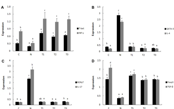
전사인자 및 cytokine의 변화
β-1,3/1,6-glucan과 L. plantarum LM1004가 아토피 피부염 을 완화하는 기전을 분석하기 위해 Th1, Th2, Th17 및 Treg 반응의 전사인자로서 T-bet, GATA-3, RORγT 및 Foxp3의 발 현과 cytokine으로 알려진 IFN-γ, IL-4, IL-17 및 TGF-β의 발현 을 나타낸 결과는 Fig. 3과 같다.
Th1 반응의 전사인자 T-bet (Fig. 4A)와 cytokine IFN-γ (Fig. 4A), Treg 반응의 전사인자 Foxp3 (Fig. 4D)와 cytokine TGF-β (Fig. 4D)는 아토피 피부염 유발그룹(N)에서 유의적으 로 가장 낮았으며, β-1,3/1,6-glucan 처리군(T1), L. plantarum LM1004 처리군(T2) 및 복합 처리군(T3)에서 유의적으로 높게 나타났다. Th2 반응의 전사인자 GATA-3 (Fig. 4B)와 cytokine IL-4 (Fig. 4B), Th17 반응의 전사인자 RORγT (Fig. 4C)와 cyto- kine IL-17 (Fig. 4C)은 아토피 피부염 유발그룹(N)에서 유의적
으로 가장 높게 나타났으며, β-1,3/1,6-glucan 처리군(T1), L.
plantarum LM1004 처리군(T2) 및 복합 처리군(T3)에서는 대조 군(C)과 유사한 수준으로 나타났다.
Th1 세포는 IL-2, IFN-γ, TNF-α 등의 cytokine을 분비하여 대식세포의 기능을 향상시켜 세포성 면역반응을 증진시키며, Th2 세포는 IL-4, IL-5, IL-13 등의 cytokine을 분비하여 B세포 의 항체 생산을 증가시키고 이로써 체액성 면역반응을 활성화 시킨다[4, 45]. 정상적인 상태에서 Th1과 Th2 세포는 상호작용 으로 균형을 이루어 면역반응을 유지하지만, 아토피 피부염은 Th2 세포가 다량의 염증성 cytokine을 분비시킴으로써 면역 학적 불균형으로 인해 발생한다[27]. 또한 Th17 세포가 분비하 는 IL-17도 염증반응을 증가시켜 아토피 피부염을 유발하는 것으로 알려져 있다[3]. Treg 세포는 IL-10, TGF-β와 같은 면역 억제 cytokine을 분비하여 과한 염증반응을 억제할 뿐만 아니 라 무해한 항원이나 자가항원에 대한 면역 관용을 유도한다 [41]. 본 연구에서 SD 흰쥐에 아토피 피부염을 유발시킨 후 β-1,3/1,6-glucan과 L. plantarum LM1004를 투여하였을 때 Th1 및 Treg 세포의 전사인자 및 cytokine의 발현은 정상 수준 이거나 유의적으로 높게 나타났다. 그와 반대로 Th2 및 Th17 세포의 전사인자 및 cytokine의 발현은 아토피 피부염 유발 그룹보다 유의적으로 낮게 나타났다. 이러한 결과로 볼 때 β- 1,3/1,6-glucan과 L. plantarum LM1004는 과발현 된 Th2와 Th17 세포의 발현을 감소시키고 Th1과 Treg 세포의 발현을 증가시킴으로써 면역 균형을 조절하여 아토피 피부염의 증상 을 완화하는 것으로 관찰되었다.
Galectin-9, Filaggrin 및 TSLP의 변화
Galectin-9, filaggrin 그리고 TSLP의 발현을 나타낸 결과는
Fig. 5와 같다. Galectin-9과 filaggrin은 아토피 피부염 유발그
룹(N)에서 유의적으로 가장 낮은 값이었으며, β-1,3/1,6-glu-
can 처리군(T1), L. plantarum LM1004 처리군(T2) 및 복합 처리
A B
C D
Fig. 4. Effects of β-1,3/1,6-glucan and L. plantarum LM1004 on the transcription factor and cytokine expression. T-cell polarization in mesenteric lymph nodes (MLN) was evaluated by analyzing the expression of T-bet, IFN-γ (Th1, A), GATA-3, IL-4 (Th2, B), RORγT, IL-17 (Th17, C) and Foxp3, TGF-β (Treg, D). C: Control, N: Negative control (induced AD), T1: β-1,3/1,6-glucan, T2: L. plantarum LM1004, T3: β-1,3/1,6-glucan and L. plantarum LM1004. a-eMeans are significantly different in each group (p<0.05). Data represent means ± SD of 5 replicates.
Fig. 5. Effects of β-1,3/1,6-glucan and L. plantarum LM1004 on the galectin-9, filaggrin, TSLP expression. C: Control, N:
Negative control (induced AD), T1: β-1,3/1,6-glucan, T2:
L. plantarum LM1004, T3: β-1,3/1,6-glucan and L. planta- rum LM1004. a-dMeans are significantly different in each group (p<0.05). Data represent means±SD of 5 replicates.
군(T3)은 대조군(C)과 유사한 수준이었으며, 그 중에서도 β- 1,3/1,6-glucan 처리군(T1)은 유의적으로 가장 높은 값을 나타 내었다. Galectin-9은 활성화된 장상피세포 및 면역세포로부 터 생성되며 CD4
+T cell을 자극시켜 Th1 면역 반응을 증진시
키고[9], Th17 면역 반응은 억제시키며 Treg 세포 유도는 증진 시킨다[49]. 또한 아토피 피부염이 유발된 상태에서 synbiotics 를 섭취하였을 때 장상피세포에서 galectin-9의 분비가 증가되 면서 Th1과 Treg의 반응이 유도된다는 보고가 있으며[10], fi- laggrin의 발현 증가는 Th2 cytokine을 감소시키며 IgE의 저하 및 피부장벽의 기능 완화와 관련이 있다[15].
아토피 피부염의 주요한 생물학적 마커로 사용되는 TSLP 는 아토피 피부염 유발그룹(N)에서 유의적으로 가장 높은 값 을 나타내었으며 β-1,3/1,6-glucan 처리군(T1), L. plantarum LM1004 처리군(T2) 및 복합 처리군(T3)은 대조군(C)과 유사한 수준이었으며, 특히 L. plantarum LM1004 처리군(T2) 및 복합 처리군(T3)에서 유의적으로 가장 낮은 값을 나타내었다. TSLP 는 피부의 각질형성세포, 상피세포, 평활근세포 및 폐의 섬유 모세포 등에서 발현되며, 비만세포와 NK-T 세포를 유도하여 과도한 Th2와 Th17 면역반응을 일으킨다[23].
본 연구에서 β-1,3/1,6-glucan과 L. plantarum LM1004는 아
토피 피부염 유발 그룹보다 galectin-9과 filaggrin의 발현을
유의적으로 증가시켰으며 TSLP는 유의적으로 감소시켰다. 이
러한 결과로 볼 때 β-1,3/1,6-glucan과 L. plantarum LM1004는
Th1과 Treg 반응을 증가시키며 Th2와 Th17 반응을 감소시키
는 것으로 나타나 전사인사 및 cytokine의 결과와 일치하는
경향이었다. 따라서 β-1,3/1,6-glucan과 L. plantarum LM1004 는 아토피 피부염의 증상을 개선하는 synbiotics로 개발될 수 있음을 나타내고 있다.
감사의 글
본 연구는 산업통상자원부에서 수행된 산업핵심기술개발 사업(No. 10049026)의 지원을 받아 수행되었음.
References
1. Allerano, F. M., Wetworth, C. E., Arana, A., Fernandez, C.
and Paul, C. F. 2007. Risk of lymphoma following exposure to calcineurin inhibitors and topical steroids in patients with atopic dermatitis. J. Invest. Dermatol. 127, 808-816.
2. Babicek, K., Cechova, I., Simon, R. R., Harwood, M. and Cox, D. J. 2007. Toxicological assessment of a particulate yeast (1,3/1,6)-beta-D-glucan in rats. Food Chem. Toxicol. 45, 1719-1730.
3. Bedoya, S. K., Lam, B., Lau, K. and Larkin, J., 3rd. 2013. Th17 cells in immunity and autoimmunity. Clin. Dev. Immunol.
2013, 1-16.
4. Belardelli, F. 1995. Role of interferons and other cytokines in the regulation of the immune response. APMIS 103, 161-179.
5. Bobek, P. and Galbavy, S. 2001. Effect of pleuran (beta-glu- can from Pleurotus ostreatus) on the antioxidant status of the organism and on dimethylhydrazine-induced precancerous lesions in rat colon. Br. J. Biomed. Sci. 58, 164-168.
6. Chan, G. C., Chan, W. K. and Sze, D. M. 2009. The effects of beta-glucan on human immune and cancer cells. J. Hema- tol. Oncol. 2, doi: 10.1186/1756-8722-2-25.
7. Choi, Y., Kim, M. S. and Hwang, J. K. 2012. Inhibitory effects of panduratin A on allergy-related mediator production in rat basophilic leukemia mast cells. Inflammation 35, 1904- 1915.
8. Chomczynski, P. and Sacchi, N. 1987. Single-step method of RNA isolation by acid guanidinium thiocyanate-phenol- chloroform extraction. Anal. Biochem. 162, 156-159.
9. Dai, S. Y., Nakagawa, R., Itoh, A., Murakami, H., Kashio, Y., Abe, H., Katoh, S., Kontani, K., Kihara, M., Zhang, S.
L., Hata, T., Nakamura, T., Yamauchi, A. and Hirashima, M. 2005. Galectin-9 induces maturation of human mono- cyte-derived dendritic cells. J. Immunol. 175, 2974-2981.
10. de Kivit, S., Saeland, E., Kraneveld, A. D., vande Kant, H.
J., Schouten, B., van Esch, B. C., Knol, J., Sprikkelman, A.
B., van der Aa, L. B., Knippels, L. M., Garssen, J., van Kooyk, Y. and Willemsen, L. E. 2012. Galectin-9 induced by dietary synbiotics is involved in suppression of allergic symptoms in mice and humans. Allergy 67, 343-352.
11. Delaney, B., Nicolosi, R. J., Wilson, T. A., Carlson, T., Frazer, S., Zheng, G. H., Hess, R., Ostergren, K., Haworth, J. and Knutson, N. 2003. Beta-glucan fractions from barley and oats are similarly antiatherogenic in hypercholesterolemic
syrian golden hamsters. J. Nutr. 133, 468-475.
12. Delatte, S. J., Evans, J., Hebra, A., Adamson, W., Othersen, H. B. and Tagge, E. P. 2001. Effectiveness of beta-glucan collagen for treatment of partical-thickness burns in children.
J. Pediatr. Surg. 36, 113-118.
13. Duncan, D. B. 1955. Multiple range and multiple F tests.
Biometrics 11, 1-42.
14. Fujiwara, D., Inoue, S., Wakabayashi, H. and Fujii, T. 2004.
The anti-allergic effects of lactic acid bacteria are strain de- pendent and mediated by effects on both Th1/Th2 cytokine expression and balance. Int. Arch. Allergy Immunol. 135, 205- 215.
15. Furue, M., Chiba, T., Tsuji, G., Ulzii, D., Kido-Nakahara, M., Nakahara, T. and Kadono, T. 2017. Atopic dermatitis:
immune deviation, barrier dysfunction, IgE autoreactivity and new therapies. Allergol. Int. 66, 1-6.
16. Hetland, G., Ohno, N., Aaberge, I. S. and Lovik, M. 2000.
Protective effect of β-glucan against systemic Streptococcus pneumonia infection in mice. FEMS Immunol. Med. Microbiol.
27, 111-116.
17. Inagaki, N., Miura, T., Nagai, H., Ono, Y. and Koda, A. 1988.
Inhibition of vascular permeability increase in mice. An ad- ditional anti-allergic mechanism of glucocorticoids. Int.
Arch. Allergy Appl. Immunol. 87, 254-259.
18. Ishiguro, K., Oku, H., Suitani, A. and Yamamoto, Y. 2002.
Effects of conjugated linoleic acid on anaphylaxis and aller- gic pruritus. Biol. Pharm. Bull. 25, 1655-1657.
19. Jo, Y. R. and Kang, S. M. 2012. Lactococcus lactis culture meth- od for enhanced depression of inducer in atopic diseases.
Kor. J. Microbiol. Biotechnol. 40, 310-318.
20. Kalliomaki, M. and Isolauri, E. 2003. Role of intestinal flora in the development of allergy. Curr. Opin. Allergy Clin.
Immunol. 3, 15-20.
21. Kang, J. S., Lee, K., Han, S. B., Ahn, J. M., Lee, H., Han, M. H., Yoon, Y. D., Yoon, W. K., Park, S. K. and Kim, H.
M. 2006. Induction of atopic eczema/dermatitis syndrome- like skin lesions by repeated topical application of a crude extract of Dermatophagoides pteronyssinus in NC/Nga mice.
Int. Immunopharmacol. 6, 1616-1622.
22. Katayama, S., Shionoya, H. and Ohtake, S. 1978. A new method for extraction of extravasated dye in the skin and the influence of fasting stress on passive cutaneous anaphy- laxis in guinea pigs and rate. Microbiol. Immunol. 22, 89-101.
23. Kim, H. J., Kim, Y. J., Lee, S. H., Yu, J., Jeong, S. K. and Hong, S. J. 2014. Effects of Lactobacillus rhamnosus on allergic march model by suppressing Th2, Th17, and TSLP re- sponses via CD4+ CD25+ Foxp3+ Tregs. Clin. Immunol. 153, 178-186.
24. Kim, J. H., Kim, M. R., Lee, J. H., Lee, J. W. and Kim, S.
K. 2000. Production of high molecular weight pullulan by Aureobasidium pullulans using glucosamine. Biotechnol.
Lett. 22, 987-990.
25. Klein, G., Pack, A., Bonaparte, C. and Reuter, G. 1998.
Taxonomy and phsiology of probiotic lactic acid bacteria.
Int. J. Food Microbiol. 41, 103-125.
26. Kraus, J. Blaschek, W., Schutz, M. and Franz, G. 1992.
Antitumor activity of cell wall β-1,3/1,6-glucans from Phytophthora species. Planta. Medica. 58, 39-42.
27. Lee, G. S., Jung, H. M., Oh, S. K., Cheong, J. H. and Kang, T. J. 2012. Effects of herbal complex on atopic dermatitis in BALB/c mice. Kor. J. Pharmacogn. 43, 59-65.
28. Leung, D. Y., Boguniewicz, M., Howell, M. D., Nomura, I.
and Hamid, Q. A. 2004. New insights into atopic dermatitis.
J. Clin. Invest. 113, 651-657.
29. Liu, F. T., Patterson, R. J. and Wang, J. L. 2002. Intracellular functions of galectins. Biochim. Biophys. Acta. 1572, 263-273.
30. Lugovic, L., Lipozencic, J. and Jakic-Razumovic, J. 2005.
Prominent involvement of activated Th1-subset of T-cells and increased expression of receptor for IFN-gamma on ker- atinocytes in atopic dermatitis acute skin lesions. Int. Arch.
Allergy Immunol. 137, 125-133.
31. Matsui, K. and Nishikawa, A. 2013. Peptidoglycan-induced T helper 2 immune response in mice involves interleukin-10 secretion from langerhans cells. Microbiol. Immunol. 57, 130- 138.
32. Mclntosh, M., Stone, B. A. and Stanisich, V. A. 2005. Curdlan and other bacterial (1→3)-beta-D-glucans. Appl. Microbiol.
Biotechnol. 68, 163-173.
33. Mercenier, A., Pavan, S. and Pot, B. 2003. Probiotics as bio- therapeutic agents: present knowledge and future prospects.
Curr. Pharm. Des. 9, 175-191.
34. Metcalfe, D. D., Kaliner, M. and Donlon, M. A. 1981. The mast cell. Crit. Rev. Immunol. 3, 23-74.
35. Miyajima, I., Dombrowicz, D., Martin, T. R., Ravetch, J. V., Kinet, J. P. and Galli, S. J. 1997. Systemic anaphylaxis in the mouse can be mediated largely through IgG1 and Fc gammaRⅢ. Assessment of the cardiopulmonary changes, mast cell degranulation, and death associated with active or IgE- or IgG1-dependent passive anaphylaxis. J. Clin.
Invest. 99, 901-914.
36. Naidu, A. S., Bidlack, W. R. and Clemens, R. A. 1999.
Probiotic spectra of lactic acid bacteria (LAB). Crit. Rev. Food Sci. Nutr. 39, 13-126.
37. Nicolosi, R., Bell, S. J., Bistrian, B. R., Greenberg, I., Forse, R. A. and Blackburn, G. L. 1999. Plasma lipid changes after supplementation with beta-glucan fiber from yeast. Am. J.
Clin. Nutr. 70, 208-212.
38. Noda, S., Krueger, J. G. and Guttman-Yassky, E. 2015. The translational revolution and use of biologics in patients with inflammatory skin diseases. J. Allergy Clin. Immunol. 135, 324-336.
39. Nograles, K. E., Zaba, L. C., Guttman-Yassky, E., Fuentes- Duculan, J., Suarez-Farinas, M., Cardinale, I., Khatcherian, A., Gonzalez, J., Pierson, K. C., White, T. R., Pensabene, C., Coats, I., Novitskaya, I., Lowes, M. A. and Krueger, J. G.
2008. Th17 cytokines interleukin (IL)-17 and IL-22 modulate distinct inflammatory and keratinocyte-response pathways.
Br. J. Dermatol. 159, 1092-1102.
40. Ohtsuka, E., Kawai, S., Ichikawa, T., Nojima, H., Kitagawa, K., Shirai, Y., Kamimura, K. and Kuraishi, Y. 2001. Roles of mast cells and histamine in mosquito bite-induced aller- gic itch-associated response in mice. Jpn. J. Pharmacol. 86,
97-105.
41. Palomeres, O., Yaman, G., Azkur, A. K., Akkoc, T., Akdis, M. and Akdis, C. A. 2010. Role of Treg in immune regu- lation of allergic diseases. Eur. J. Immunol. 40, 1232-1240.
42. Peng, G. C. and Hsu, C. H. 2005. The efficacy and safety of heat-killed Lactobacillus paracasie for treatment of per- ennial allergic rhinitis induced by house-dust mite. Pediatr.
Allergy Immunol. 16, 433-438.
43. Prescott, S. L., Dunstan, J. A., Hale, J., Breckler, L., Lehmann, H., Weston, S. and Richmond, P. 2005. Clinical effects of probiotics are associated with increased interferon-gamma responses in very young children with atopic dermatitis.
Clin. Exp. Allergy 35, 1557-1564.
44. Rabinovich, G. A., Liu, F. T., Hirashima, M. and Anderson, A. 2007. An emerging role for galectins in tuning the im- mune response: lessons from experimental models of in- flammatory disease, autoimmunity and cancer. Scand J.
Immunol. 66, 143-158.
45. Romagnani, S. 2000. T-cell subsets (Th1 versus Th2). Ann.
Allergy Asthma. Immunol. 85, 9-18.
46. Rothschild, A. M., Gomes, E. L. and Rossi, M. A. 1991.
Reversible rat mesenteric mast cell swelling caused by vagal stimulation or sham-feeding. Agents Actions 34, 295-301.
47. Sano, Y., Masuda, K., Tamagawa-Mineoka, R., Matsunaka, H., Murakami, Y., Yamashita, R., Morita, E. and Katoh, N.
2013. Thymic stromal lymphopoietin expression is increased in the horny layer of patients with atopic dermatitis. Clin.
Exp. Immunol. 171, 330-337.
48. Schroeder, J. T. 2011. Basophils: emerging roles in the patho- genesis of allergic disease. Immunol. Rev. 242, 144-160.
49. Seki, M., Oomizu, S., Sakata, K. M., Sakata, A., Arikawa, T., Watanabe, K., Ito, K., Takeshita, K., Niki, T., Saita, N., Nishi, N., Yamauchi, A., Katoh, S., Matsukawa, A., Kuchroo, V. and Hirashima, M. 2008. Galectin-9 suppresses the gen- eration of Th17, promotes the induction of regulatory T cells, and regulates experimental autoimmune arthritis. Clin.
Immunol. 127, 78-88.
50. Seo, H. P., Kim, J. M., Shin, H. D., Kim, T. K., Chang, H.
J., Park, B. R. and Lee, J. W. 2002. Production of β- 1,3/1,6-glucan by Aureobasidium pullulans SM-2001. Kor. J.
Biotechnol. Bioeng. 17, 376-380.
51. Soumelis, V., Reche, P. A., Kanzler, H., Yuan, W., Edward, G., Homey, B., Gilliet, M., Ho, S., Antonenko, S., Lauerma, A., Smith, K., Gorman, D., Zurawski, S., Abrams, J., Menon, S., McClanahan, T., de Waal-Malefyt, Rd. R., Bazan, F., Kastelein, R. A. and Liu, Y. J. 2002. Human epithelial cells trigger dendritic cell mediated allergic inflammation by pro- ducing TSLP. Nat. Immunol. 3, 673-680.
52. Tokunaka, K., Ohno, N., Adachi, Y., Miura, N. N. and Yadomae, T. 2002. Application of Candida solubilized cell wall β-glucan in antitumor immunotherapy against P815 mastocytoma in mice. Int. Immunopharmacol. 2, 59-67.
53. Vitaliti, G., Pavone, P., Guglielmo, F., Spataro, G. and Falsa- perla, R. 2014. The immunomodulatory effect of probiotics beyond atopy: an update. J. Asthma. 51, 320-332.
54. Young, C. A., Rorke, E. A., Adhikary, G., Xu, W. and Eckert,
초록:아토피 피부염 모델에 대한 β-1,3/1,6-glucan과
Lactobacillus plantarumLM1004의 면역 조절 효과
김인성
1†․김성학
2†․김정아
1․유다윤
1․김광일
1․박동찬
3․임종민
3․이상석
4․최인순
5․조광근
1*
(1경남과학기술대학교 동물소재공학과, 2전남대학교 동물자원학부, 3㈜글루칸, 4순천대학교 동물자원과학과, 5신라
대학교 생명과학과)
본 연구에서는 아토피 피부염 동물 모델에 대한 β-1,3/1,6-glucan과 L. plantarum LM1004의 면역조절 효과를 확인하고자 하였다. 가려움증의 횟수와 유출된 evans blue, 그리고 혈청 IgE와 histamine의 농도는 β-1,3/1,6-glu- can과 L. plantarum LM1004를 섭취한 그룹에서 아토피 피부염 유발그룹에 비해 유의적으로 감소하는 결과를 나타 내었다. 아토피 피부염이 유발되면 전사 수준에서 Th2 및 Th17 세포의 전사인자 및 cytokine은 과발현되며, β- 1,3/1,6-glucan과 L. plantarum LM1004를 섭취하였을 때 이를 유의적으로 감소되었다. 또한 β-1,3/1,6-glucan과 L.
plantarum LM1004는 Th1 및 Treg 세포의 전사인자(T-bet, GATA-3, RORγT, Foxp3) 및 cytokine (INF-γ, IL-4, IL-17, TGF-β)의 발현을 증가시킴으로써 면역 균형을 조절하는 것으로 나타났다. Galectin-9과 filaggrin은 아토피 피부염 유발 처리군에서 유의적으로 가장 낮았으며, β-1,3/1,6-glucan 처리군에서 유의적으로 가장 높게 나타났다.
이와 반대로 TSLP는 아토피 피부염 유발그룹에서 유의적으로 가장 높았으며 β-1,3/1,6-glucan과 L. plantarum LM1004를 섭취한 그룹은 대조군과 유사한 수준이었다. 이러한 결과를 통해 β-1,3/1,6-glucan과 L. plantarum LM1004는 아토피 피부염 동물 모델에서 면역조절 작용 및 아토피 피부염의 개선 효과를 가짐을 알 수 있었다.
따라서 β-1,3/1,6-glucan과 L. plantarum LM1004는 아토피 피부염에 유용한 천연소재로서 사용될 것으로 기대된다.
R. L. 2017. Loss of epidermal AP1 transcription factor func- tion reduces filaggrin level, alters chemokine expression and
produces an ichthyosis-related phenotype. Cell Death Dis.
8, e2840.