Overexpression of Clast4 Reduces Cell Proliferation
Minkook Kang and Seung Jin Han*
Department of Biological Sciences, Inje University, 197 Inje-ro, Gimhae, Gyeongnam 621-749, Korea Received July 8, 2014 /Revised August 12, 2014 /Accepted August 18, 2014
The eIF4E protein is the key regulator of translation initiation. The interaction of eIF4E with eIF4G triggers the translation of mRNA, and several proteins interrupt this association to modulate translation. Human 4E-T is one of the eIF4E-binding partners that represses the translation of bound mRNAs, and it is involved in the transport of eIF4E to processing bodies (P-bodies). Although Clast4, the mouse homolog of human 4E-T, might play critical roles in the regulation of translation, its prop- erties are not well known. In this report, we deciphered the properties of Clast4 by determining its phosphorylation state, binding to eIF4E, and effects of overexpression on cell proliferation. Clast4 was phosphorylated by protein kinase A (PKA) in vivo on several residues of its amino terminus.
Nevertheless, the PKA phosphorylation of Clast4 appeared to have no effect on either its eIF4E-bind- ing ability or localization. Clast4 interacted with eIF4E1 and CPEB. The conserved eIF4E-binding se- quence in Clast4, YXXXXLϕ, was important for binding eIF4E1A but not eIF4E1B. Similar to that of another well-known eIF4E regulator, the eIF4E binding protein (4E-BP), the overexpression of Clast4 decreased cell proliferation. These results suggest that Clast4 acts as a global translation regulator in cells.
Key words :
4E-T, Clast4, CPEB, eIF4E1, translation initiation
*Corresponding author
*Tel : +82-55-320-3787, Fax : +82-55-336-7706
*E-mail : [email protected]
This is an Open-Access article distributed under the terms of the Creative Commons Attribution Non-Commercial License (http://creativecommons.org/licenses/by-nc/3.0) which permits unrestricted non-commercial use, distribution, and reproduction in any medium, provided the original work is properly cited.
Journal of Life Science 2014 Vol. 24. No. 10. 1144~1150 DOI : http://dx.doi.org/10.5352/JLS.2014.24.10.1144
Introduction
In eukaryotes, the translation of mRNA is tightly regu- lated to orchestrate many cellular mechanisms such as cell proliferation, differentiation, neural cell plasticity, embryo development, and oocyte maturation [12, 19]. Therefore, mis- regulation of these steps is related with cell malfunction, such as the creation of cancer cell [1]. Translation starts with the formation of eukaryotic initiation factor 4F (eIF4F) com- plex, which recruits ribosomes and other initiation factors [6]. The eIF4F complex consists of eIF4E, eIF4G, and eIF4A.
The eIF4E interacts with the 5’cap structure, m
7GpppN, which is present on all nuclear-encoded mRNAs. The eIF4G, a scaffold protein, binds the eIF4E and recruits an eIF4A, ATP-dependent RNA helicase, to resolve the mRNA secon- dary structure [6]. Because eIF4E is the first protein in- corporated into the translation initiation complex in mRNA among eIF4F complex factors, it is a major target for regulation. The eIF4G subsequently binds eIF4E for the basal
translation initiation complex, and this interaction is regu- lated by factors such as the 4E-BP. Hypophosphorylated 4E-BP competes with eIF4G to bind with eIF4E through its YXXXXLϕ (ϕ is the hydrophobic residue) motif, which is also found in the eIF4G protein [11]. By hyperphosphorylation through the mTOR signaling pathway, 4E-BP dissociates from eIF4E, resulting in the docking of eIF4G to eIF4E to initiate translation [7]. Mutation or unregulated hyper- phosphorylation of 4E-BP induces an increase of pro- liferation due to weakening or loss of interaction with eIF4E in cancer cells [17]. Translation initiation is regulated not only by proteins on the 5’ untranslated region (UTR) but also by the factors on the 3’UTR. Cytoplasmic poly- adenylation element-binding protein (CPEB) is loaded on the cytoplasmic polyadenylation element (CPE) of 3’UTR [12].
CPEB is implicated in the repression of translation by inter-
acting poly(A)-specific ribonuclease (PARN) to shorten the
poly(A) tail or by binding one of the proteins such as
Maskin, Neuroguidin, and eIF4E transporter (4E-T) to ob-
struct the association of eIF4E with eIF4G [2, 9]. Following
a translation initiation signal (e.g., growth factors or nu-
trients), CPEB is phosphorylated, and it facilitates poly(A)
tail elongation on the 3’UTR. The poly(A)-binding protein
(PABP) is placed on the poly(A) tail to generate a closed-loop
structure by the interaction with eIF4G on the 5’UTR. This
structure stabilizes the initiation complex and enhances the
- Note -
initiation of translation [12].
Clast4 is a mouse homolog of human 4E-T [20], a protein responsible for the transport of eIF4E from cytoplasm to nu- cleus or P-body [4]. It is believed that mRNA decay and/or storage take place in the P-body, because they contain some proteins related to RNA degradation such as PAT1, Dcp1 and Xrn1 [14]. Human 4E-T is also a P-body component pro- tein, and the depletion of 4E-T induces a remarkable de- crease of P-body assembly in HeLa cells [5]. Given that Clast4 also possesses the eIF4E-binding motif, YXXXXLϕ, it appears to be a translation regulator interrupting the inter- action between eIF4E and eIF4G. This is consistent with the fact that the Drosophila ortholog of Clast4, Cup, interacts with eIF4E and 3’UTR-bound factors to repress translation during ovary development and growth [15]. It has been reported that Xenopus 4E-T can associate with CPEB similar to how Maskin does in the Xenopus oocyte, and Xenopus 4E-T also binds eIF4E1B, a germinal-specific isoform of eIF4E, in an YXXXXLϕ-independent manner, unlike eIF4E1A, which is a canonical form of eIF4E1 [13]. However, the regulation mechanism of the mouse Clast4 protein is still unclear.
Therefore, in this report we deciphered the properties of Clast4 by examining its phosphorylation, binding properties, and overexpression effect on cell proliferation.
Materials & Methods Materials
The anti-4E-T and anti-phospho (Ser/Thr) PKA substrate antibodies were purchased from Bethyl (Montgomery, AL) and Cell Signaling (Danvers, CO), respectively. FLAG epit- ope antibody and other chemicals were purchased from Sigma Aldrich (St. Louis, MO). Oligonucleotide synthesis and DNA sequencing was performed by Cosmo Genetech (Seoul, Korea). The eIF4E1B gene was isolated from mouse ovaries by RT-PCR with a forward primer of T A G C C C G G G G T A C C A T G A A- CAAAGTTGAGGGTGGAGGGC and a reverse primer of TTAACGCGGCCGCCACCACAAACTTGTTTGCTAAG.
Construction of Clast4 mutants
The Clast4 cDNA was obtained from Open Biosystems (MMM1013-7512592, Clone ID: 5359290) and cloned into the pcDNA 3.1 topo (Invitrogen Corp., Carlsbad, CA) or pCMV Tag2 (Clontech, Mountain View, CA) vectors. To determine the localization of Clast4, the Clast4-GFP fusion gene was
generated by inserting the GFP gene into the Clast4-pcDNA 3.1 construct. The truncated forms of Clast4 (Fig. 1D) were made by cutting with restriction enzymes or by PCR reaction. To generate the non-phosphorylated form of Clast4, indicated serine or threonine sites of Clast4 (Ser212, Thr256, Ser258, Ser352) were mutated using the QuikChange Site-Directed Mutagenesis Kit (Stratagene, La Jolla, CA) with specific primers.
Immunoprecipitation and in vitro kinase assay HEK293T cells were transfected with JetPEI (Polyplus-transfection Inc., NY) at 60% confluence, and har- vested with lysis buffer (50 mM Tris-HCl pH 7.4, 150 mM NaCl, 0.1% NP40, 1 mM EDTA, 1 μg/ml leupeptin, 1 mM aprotinin, 1 mM PMSF, 1 mM sodium orthovadate, 1 mM NaF) 36 h after transfection. Harvested cell lysate was in- cubated with anti-FLAG antibody at 4°C and then pre- cipitated using protein G sepharose (GE Healthcare Bio-Sciences, Piscataway, NJ). The immunoprecipitates were washed with lysis buffer and kinase buffer (25 mM Tris-HCl pH 7.5, 5 mM β-glycophosphate, 0.2 mM dithiothreitol, 1 mM sodium orthovadate, 1 mM magnesium chloride) three times, consecutively. The immunoprecipitates were in- cubated with kinase buffer containing 250 μM ATP and 10 μCi/ml [γ-
32P] ATP (3000 Ci/mmol, PerkinElmer) for 30 min at 37°C with or without 5 units of PKA catalytic subunit (Promega, Madison, Wisconsin), Plk1, Aurora A kinase (SignalChem, Richmond, CA), and Cdk1, respectively. The samples were resolved on the 8% SDS- PAGE gel and trans- ferred to PVDF membranes. Radiolabeled Clast4 was de- tected with the X-ray film (Kodak, Rochester, NY) or by a phosphor image analyzer (Typhoon 9400, GE Healthcare Bio-Sciences).
In vivo phosphorylation
For the in vivo phosphorylation test, Clast4-transfected cells were cultured in the DMEM with 0.2% FBS for 16 hr.
To activate in vivo PKA, 10 μM Forskolin and 0.1 mM IBMX were treated for 30 min with or without 10 μM H89. The immunoprecipitated Clast4 was detected with anti-phospho (Ser/Thr) PKA substrate antibody following SDS-PAGE and transferred to a PVDF membrane.
Co-immunoprecipitation
HA-tagged eIF4E1B-transfected cells were divided into 60
mm dishes, and FLAG-tagged Clast4 fragments were sub-
Fig. 1. PKA phosphorylates Clast4 in vivo. (A) Immunoprecipitated FLAG-Clast4 was incubated with PKA, Plk1, Aurora A kinase, and Cdk1/cyclin B complex with 5 μCi [γ-32P] ATP. The phosphorylation state and the expression of Clast4 were checked by autoradiography (upper panel) and FLAG-specific antibody (lower panel) after SDS-PAGE. (B) Clast4-transfected HEK293T cells were treated with forskolin and IBMX. Immunoprecipitated Clast4 was detected with anti-phospho (Ser/Thr) PKA sub- strate antibody (upper panel) and FLAG-specific antibody (lower panel). The phosphorylation of Clast4 was completely dimin- ished by the addition of 10 μM of H89. (C) Clast4-transfected cells were co-transfected with either wild-type (WT) or kin- ase-dead mutant (KD) of PKA catalytic subunit. Western blot analysis for immunoprecipitates using anti-phospho (Ser/Thr) PKA substrate antibodies indicated that Clast4 is phosphorylated by WT PKA overexpression (upper blot). The expression of Clast4 and PKA constructs was confirmed using anti-FLAG or HA antibodies, respectively (lower two blots). (D) The amino terminus of Clast4 is the major PKA phosphorylation site. A schematic diagram of the FLAG-tagged full-length or truncated form of Clast4 is shown (upper image). The putative PKA phosphorylation sites (S212, RRRNDSYT; T256, S258, RTRRRTASVK; S352, GSRSSSLG) were indicated. The in vitro kinase assay with PKA catalytic subunits in [γ-32P] ATP showed that F1 and F2 fragments were phosphorylated by PKA (left panel). The immunoprecipitated proteins were monitored using FLAG antibody (right panel). (E) The putative PKA-phosphorylated serine or threonine amino acids were changed to alanine.
The phosphorylation states of mutated F1 or F2 constructs were checked by autoradiography after an in vitro kinase assay.
(F) The localization of EGFP-tagged wild-type or phosphorylation sites mutated Clast4 were checked after transfection into HEK293T cells. The localization of Clast4 to P-body was increased by PKA inhibitor treatment but not by the mutation of phosphorylation sites. Dcp1 and Hoechst were used as P-body and nucleus indicators, respectively.
sequently transfected. After immunoprecipitation with FLAG antibody, the western blotting was performed with HA antibody.
Immunocytochemistry
HEK293T cells were cultured on the poly-L-lysine-coated slide glass and transfected with GFP-tagged Clast4. After se-
rum starvation for 16 hr, forskolin and IBMX were treated
with or without H89. The cells were fixed in 4% form-
aldehyde in PBS for 1 hr and were permeabilized in blocking
buffer (1% BSA, 0.3% Triton-X-100 in PBS). The Dcp1 anti-
body in blocking buffer was added for 2 hr followed by the
treatment of CY3-conjugated anti-rabbit (PA43004, GE
Healthcare Bio-Sciences) antibody. The samples were ob-
served under a confocal laser-scanning microscope (LSM 510 META, Carl Zeiss, Germany).
Proliferation test
The GFP, Clast4, or 4E-BP-transfected cells (2,000/well) were plated in 96-well culture plates, and 20 μl of MTT (3-[4,5-dimethylthiazol-2-yl]-2,5-diphenyltetrazolium bromide, 0.5 mg/ml in PBS) was added at the indicated time point to check the number of viable cells. After incubation for 4 h at 37°C, generated formazan was dissolved in MTT assay solution (4 mM HCL, 0.1% NP40 in isopropanol). The ab- sorbance of the colored solution was quantified by measur- ing at 590 nm with a reference filter of 620 nm using a micro- plate reader (MQX 200R, BioTek Instruments, Winooski, VT, USA).
Results and Discussion Clast4 is phosphorylated by PKA
It has been reported that Clast4 is phosphorylated by an unknown kinase during oocyte maturation [20]. To identify the kinase(s) that phosphorylates Clast4 during cell cycle progression, we performed in vitro kinase assays with sev- eral of the kinases involved in cell cycle regulation. Among the kinases tested, PKA could phosphorylate Clast4 much more strongly than the other kinases (i.e., polo-like kinase 1 (plk1), Aurora A kinase, and cyclin-dependent kinase 1 (Cdk1)) (Fig. 1A). Because human 4E-T is shuttled to the P-body, and PKA is related to the P-body formation in yeast [18], we focused on the role of PKA in Clast4 phosphorylation. To confirm the phosphorylation of Clast4
in vivo, PKA was activated by the treatment of forskolin,an adenylyl cyclase activator, and IBMX, a phosphodiester- ase inhibitor. After the immunoprecipitation of Clast4, the phosphorylation state of Clast4 was detected with anti-phos- pho (Ser/Thr) PKA substrate antibody that could detect the phosphorylated conserved site (RXXpS/T). Clast4 was phos- phorylated in vivo by the activation of PKA, and this effect was completely removed by treatment of H89, a PKA in- hibitor (Fig. 1B). In addition, the phosphorylation state of Clast4 in the cell was increased by the overexpression of PKA catalytic subunit, not by that of the kinase-dead form (Fig. 1C). These results show that Clast4 is per se a PKA sub- strate in the cell.
Using web-based programs (http://scansite.mit.edu/ and http://www.phosphosite.org/homeAction.do), the S212,
S256, S258, and S352 sites were found to be potential PKA phosphorylation sites. To delineate PKA phosphorylation sites in Clast4, several truncated forms of Clast4 were generated. F1 and F2 (but not F3 or F4) fragments were phos- phorylated by PKA catalytic subunit in [γ-
32P] ATP-contain- ing reaction buffer (Fig. 1D), indicating that the amino-termi- nal half of Clast4 contains the major PKA phosphorylation sites. To define the phosphorylated residues in the F1 and F2 fragments, S212 of F1 and T256, S258, and S352 of F2 residues were mutated to alanine, a non-phosphorylated amino acid residue, and in vitro phosphorylation was con- ducted (Fig. 1E). The degree of [γ-
32P] incorporation into the F1 fragment was reduced by the mutation of S212A. In the F2 fragment, the signal intensity was not significantly re- duced in the single amino acid mutants, but it was dramati- cally decreased by the mutation of all putative phosphor- ylation sites. These results strongly suggest that multiple sites of Clast4 can be phosphorylated by PKA.
The depletion of human 4E-T induces a significant reduc- tion in the size of the P-body [5], and the oxidative stress promotes multisite human 4E-T phosphorylation by c-Jun N-terminal kinase (JNK) to facilitate the formation of larger P-body [3]. In addition, PKA catalytic subunit Tpk associates with the translation initiation factors Pab1 and Rps3 in ex- ponentially growing cells and possibly controls cap-depend- ent translation by regulating Rpg1 and eIF4G
1proteins in yeast. Glucose starvation promotes the loss of interaction be- tween Tpk and initiation factors followed by their accumu- lation into P-bodies [18]. Therefore, we checked the effect of PKA phosphorylation on the P-body localization of Clast4 by the transfection of GFP-fused wild-type or phosphor- ylation site mutant Clast4. After transfection into HEK293T cells, Clast4 was found in both the P-body and cytoplasm (Fig. 1F). Although treatment of PKA inhibitor (H89) led to an increase of P-body size, mutation of the PKA site on Clast4 had no effect on either its localization or the size of the P-body (Fig. 1F). It would be very interesting to inves- tigate the relationship between Clast4 phosphorylation by other kinases and P-body formation.
Clast4 interacts with eIF4E1 and CPEB
It has been proposed that human 4E-T plays a similar role
with Maskin, another eIF4E-binding protein, modulating the
initiation of translation by linking eIF4E on the Cap structure
and CPEB on the 3’UTR of mRNA. However, to our knowl-
edge, no report has described the interaction of Clast4 with
A B
C D
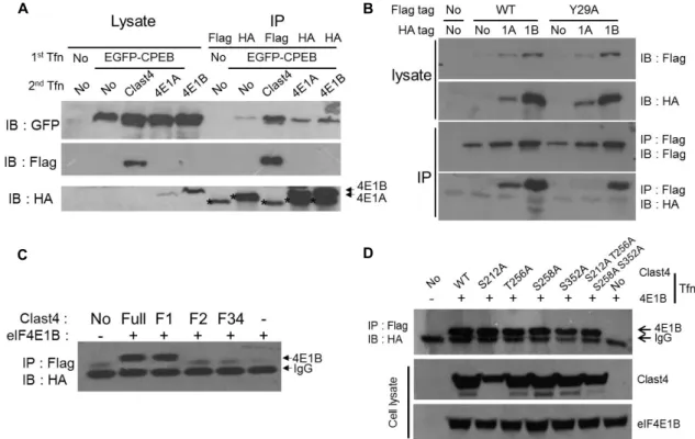
Fig. 2. Clast4 interacts with eIF4E1s and CPEB (A) After co-transfection of EGFP-CPEB and one of FLAG-Clast4, HA-eIF4E1A, or HA-eIF4E1B, the interaction between two proteins was checked by co-immunoprecipitation. Clast4 bound CPEB directly, while eIF4E1A and eIF4E1B were not able to bind directly with CPEB. Asterisks indicate the IgG light chain. (B) After either the wild-type or Y29A mutant form of Clast4 was transfected, either eIF4E1A or eIF4E1B was consecutively transfected.
The association with Clast4 and eIF4E was monitored by co-immunoprecipitation. While the wild-type of Clast4 was shown to bind with eIF4E1A and eIF4E1B, the binding affinity of Clast4 with eIF4E1A (but not with eIF4E1B) was abolished by the mutation of tyrosine 29 to alanine. (C) eIF4E1B associated with the amino-terminus of Clast4. After the co-transfection of HA-eIF4E1B and one of the truncated forms of FLAG-Clast4, the co-immunoprecipitates were detected using HA antibody.
(D) None of the putative PKA phosphorylation sites was involved in its ability to bind eIF4E. One of the FLAG-tagged Clast4 phosphorylation site mutants and HA-tagged eIF4E1B were transfected into HEK293T cells, and the co-immun- oprecipitation of Clast4 and eIF4E1B was monitored.
CPEB and eIF4E in the mammalian system, which led us to investigate the binding properties of Clast4 with these proposed partners. Co-immunoprecipitation results show that Clast4 associates with CPEB, eIF4E1A, and eIF4E1B (Fig.
2A). Because eIF4E1A can bind with the protein that contains the eIF4E-binding motif, YXXXXLϕ, the corresponding ty- rosine residue of Clast4 was changed to alanine, and the binding specificity between Clast4 and either eIF4E1A or eIF4E1B was monitored. The binding affinity between Clast4 and eIF4E1A was dramatically reduced by this mutation, but it was not for eIF4E1B (Fig. 2B). These results suggest that Clast4 associates with both eIF4E1s in a different manner of eIF4E-binding motif dependency. By co-immun- oprecipitation with several truncated forms of Clast4, the amino terminus of Clast4 was identified as a binding part to eIF4E1B (Fig. 2C). Given that the Clast4 can bind with
eIF4E1s and CPEB, Clast4 could be involved in mRNA trans- lation regulation. However, the way in which the binding of Clast4 with other factors is regulated and the kinds of mRNA that are modulated by the action of Clast4 are cur- rently unclear.
The binding of 4E-BP with eIF4E relies on phosphor-
ylation by the mTOR signal pathway, and the hyper-phos-
phorylated human 4E-T has a weak affinity for eIF4E in mi-
tosis [16]. Clast4 is phosphorylated by an unknown kinase
in the MI and MII phases during mouse oocyte maturation
[20]. Therefore, the effect of Clast4 phosphorylation on the
binding properties with eIF4E was monitored. The co-im-
munoprecipitation result demonstrated that the mutant of
the putative PKA phosphorylation sites of Clast4 does not
have an effect on the binding capacity between Clast4 and
eIF4E (Fig. 2D). Although this result is somewhat dis-
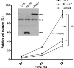
Fig. 3. Overexpression of Clast4 decreases the proliferation rate of the cell. (A) The GFP, 4E-BP, or Clast4 construct was introduced into 4 × 10³ HEK293T cells, and the cell num- bers were counted at the indicated times. Statistically sig- nificant differences of cell proliferation in Clast4 or 4E-BP expression cells compared to GFP expression cells are represented by asterisks (***p<0.001). Results represent three separate experiments. The inset panel indicates the expression of transfected proteins (NS: nonspecific band).
appointing, we think that ruling out one candidate kinase that might be able to regulate Clast4 is also informative for further studies. As several kinases such as Aurora A kinase, Plk1, Greatwall kinase, and MAP kinase are activated during the MI–MII transition in oocyte maturation, these kinases could be involved in Clast4 phosphorylation. In fact, several kinases ―including PKA, Plk1, Aurora A kinase, and cdc2/cyclin B― can phosphorylate Clast4 at least in vitro (Fig. 1). To elucidate the role of Clast4 in translation regu- lation, the kinases responsible for Clast4 phosphorylation must be revealed.
Overexpression of Clast4 delays cell proliferation Because 4E-BP binds eIF4E to hinder translation initiation, and the expression level of 4E-BP correlates with oncogenic activation [17], we checked whether the overexpression of Clast4 influences cell proliferation. The overexpression of 4E-BP decreased cell proliferation by up to 30% compared to GFP overexpression. Interestingly, Clast4 overexpression also reduced cell proliferation as much as 4E-BP did (Fig.
3). This result demonstrated that Clast4 might act as an mRNA translation regulator similar to 4E-BP. Although 4E-BP has received attention as an important regulator of translation initiation, Clast4 seems to be a new candidate
for the performance of a similar function. Recently, another result showed that treatment of human 4E-T siRNA in HeLa cells increases global protein synthesis by about 55%, which reinforces this idea [10]. Since the Drosophila homolog, CUP, promotes deadenylation and inhibits the decapping of mRNA targets [8], it is possible that Clast4 inhibits trans- lation using a different mechanism from that of 4E-BP. It would be very interesting to determine the new function of Clast4 during inhibition of translation.
In this study, we found that Clast4 is phosphorylated by PKA in vivo on several residues of its amino terminus and Clast4 can associate with CPEB and eIF4E1. The over- expression of Clast4 decreases cell proliferation. Collectively, all of these results suggest that Clast4 acts as a novel global translation regulator in the cell.
Acknowledgements
This work was supported by the Korea Research Founda- tion Grant funded by the Korean Government (MOEHRD, Basic Research Promotion Fund) (KRF-2007-331-C00196).
References
1. Audic, Y. and Hartley, R. S. 2004. Post-transcriptional regu- lation in cancer. Biol Cell 96, 479-498.
2. Cao, Q. and Richter, J. D. 2002. Dissolution of the mas- kin-eIF4E complex by cytoplasmic polyadenylation and poly(A)-binding protein controls cyclin B1 mRNA trans- lation and oocyte maturation. EMBO J 21, 3852-3862.
3. Cargnello, M., Tcherkezian, J., Dorn, J. F., Huttlin, E. L., Maddox, P. S., Gygi, S. P. and Roux, P. P. 2012.
Phosphorylation of the eukaryotic translation initiation fac- tor 4E-transporter (4E-T) by c-Jun N-terminal kinase pro- motes stress-dependent P-body assembly. Mol Cell Biol 32, 4572-4584.
4. Dostie, J., Ferraiuolo, M., Pause, A., Adam, S. A. and Sonenberg, N. 2000. A novel shuttling protein, 4E-T, medi- ates the nuclear import of the mRNA 5' cap-binding protein, eIF4E. EMBO J 19, 3142-3156.
5. Ferraiuolo, M. A., Basak, S., Dostie, J., Murray, E. L., Schoen- berg, D. R. and Sonenberg, N. 2005. A role for the eIF4E- binding protein 4E-T in P-body formation and mRNA decay. J cell biol 170, 913-924.
6. Gingras, A. C ., Raught, B. and Sonenberg, N. 1999. eIF4 initiation factors: effectors of mRNA recruitment to ribo- somes and regulators of translation. Annu Rev Biochem 68, 913-963.
7. Heesom, K. J. and Denton, R. M. 1999. Dissociation of the eukaryotic initiation factor-4E/4E-BP1 complex involves phosphorylation of 4E-BP1 by an mTOR-associated kinase.
초록:Clast4의 과발현에 의한 세포 증식의 감소 강민국 · 한승진
(인제대학교 생명과학과)
eIF4E는 번역개시과정에서 중심조절자 역할을 한다. eIF4E와 eIF4G의 결합이 mRNA의 번역을 촉발하기 때문 에, 여러 단백질들이 이 결합을 저해함으로써 번역과정을 조절한다. 인간 4E-T는 eIF4E 결합단백질 중의 하나로, 결합한 mRNA의 번역을 저해할 뿐 아니라, eIF4E를 processing body (P-body)로 이동시키는 기능을 가지고 있다.
Clast4는 인간의 4E-T와 상동성을 가지는 생쥐 단백질로 번역 조절에 중요한 기능을 할 것으로 추측되지만, 그 특징은 아직 잘 알려져 있지 않다. 본 연구에서는 Clast4의 인산화된 상태와 eIF4E와의 결합력, Clast4 과발현시 세포증식의 변화에 대한 특징을 관찰하였다. Clast4는 PKA에 의해 in vivo에서 아미노말단의 몇몇 잔기가 인산화 되는 것으로 확인되었다. 그러나 PKA에 의해 인산화된 Clast4는 eIF4E와의 결합력이나 Clast4의 세포 내 위치에 는 큰 변화가 없었다. Clast4는 eIF4E1과 CPEB와 결합하며, Clast4의 보존된 eIF4E 결합 서열인 YXXXXLϕ가 eIF4E1A와의 결합에서는 중요하지만 eIF4E1B와의 결합에서는 큰 영향이 없는 것으로 관찰되었다. 잘 알려져 있 는 eIF4E 조절자인 4E-BP의 경우와 유사하게 Clast4를 과발현하였을 때 세포의 증식이 감소되었다. 이러한 결과는 Clast4가 세포 내에서 전반적인 번역 조절에 관여하고 있다는 것을 시사한다.
FEBS Lett 457, 489-493.
8. Igreja, C. and Izaurralde, E. 2011. CUP promotes dead- enylation and inhibits decapping of mRNA targets. Genes Dev 25, 1955-1967.
9. Jung, M. Y., Lorenz, L. and Richter, J. D. 2006. Translational control by neuroguidin, a eukaryotic initiation factor 4E and CPEB binding protein. Mol Cell Biol 26, 4277-4287.
10. Kamenska, A., Lu, W. T., Kubacka, D., Broomhead, H., Minshall, N., Bushell, M. and Standart, N. 2014. Human 4E-T represses translation of bound mRNAs and enhances microRNA-mediated silencing. Nucleic Acids Res 42, 3298-3313.
11. Mader, S., Lee, H., Pause, A. and Sonenberg, N. 1995. The translation initiation factor eIF-4E binds to a common motif shared by the translation factor eIF-4 gamma and the trans- lational repressors 4E-binding proteins. Mol Cell Biol 15, 4990-4997.
12. Mendez, R. and Richter, J. D. 2001. Translational control by CPEB: a means to the end. Nat Rev Mol Cell Biol 2, 521-529.
13. Minshall, N., Reiter, M. H., Weil, D. and Standart, N. 2007.
CPEB interacts with an ovary-specific eIF4E and 4E-T in ear- ly Xenopus oocytes. J Biol Chem 282, 37389-37401.
14. Mir, M. A., Duran, W. A., Hjelle, B. L., Ye, C. and Pangani- ban, A. T. 2008. Storage of cellular 5' mRNA caps in P bod- ies for viral cap-snatching. Proc Natl Acad Sci USA 105,
19294-19299.
15. Piccioni, F., Zappavigna, V. and Verrotti, A. C. 2005. A cup full of functions. RNA Biol 2, 125-128.
16. Pyronnet, S., Dostie, J. and Sonenberg, N. 2001. Suppression of cap-dependent translation in mitosis. Genes Dev 15, 2083-2093.
17. She, Q. B., Halilovic, E., Ye, Q., Zhen, W., Shirasawa, S., Sasazuki, T., Solit, D. B. and Rosen, N. 2010. 4E-BP1 is a key effector of the oncogenic activation of the AKT and ERK signaling pathways that integrates their function in tumors.
Cancer Cell 18, 39-51.
18. Tudisca, V., Simpson, C., Castelli, L., Lui, J., Hoyle, N., Moreno, S., Ashe, M. and Portela, P. 2012. PKA isoforms coordinate mRNA fate during nutrient starvation. J Cell Sci 125, 5221-5232.
19. Ule, J. and Darnell, R. B. 2006. RNA binding proteins and the regulation of neuronal synaptic plasticity. Curr Opin Neurobiol 16, 102-110.
20. Villaescusa, J. C., Allard, P., Carminati, E., Kontogiannea, M., Talarico, D., Blasi, F., Farookhi, R. and Verrotti, A. C.
2006. Clast4, the murine homologue of human eIF4E-Transporter, is highly expressed in developing oo- cytes and post-translationally modified at meiotic maturation. Gene 367, 101-109.
![Fig. 1. PKA phosphorylates Clast4 in vivo. (A) Immunoprecipitated FLAG-Clast4 was incubated with PKA, Plk1, Aurora A kinase, and Cdk1/cyclin B complex with 5 μCi [γ- 32 P] ATP](https://thumb-ap.123doks.com/thumbv2/123dokinfo/5012681.549579/3.892.107.792.123.652/phosphorylates-clast-immunoprecipitated-clast-incubated-aurora-kinase-complex.webp)