Inhibition of Cell Invasion by Indole-3-Carbinol in OVCAR-3 Human Ovarian Cancer Cells
Yung Hyun Choi
1,2* and Sung Ok Kim
11
Department of Biomaterial Control (BK21 program) and Blue-Bio Industry RIC, Dongeui University, Busan 614-714, Korea
2
Department of Biochemistry and Research Institute of Oriental Medicine, Dongeui University College of Oriental Medicine, Busan 614-052, Korea
Received March 18, 2011 /Revised May 3, 2011 /Accepted May 3, 2011
In the present study, we investigated the effect of indole-3-carbinol (I3C), a natural compound pres- ent in vegetables, on the cell migration and invasion of OVCAR-3 ovarian cancer cells. Our results indicated that I3C inhibited the proliferation of OVCAR-3 cells, a process which was associated with inhibition of cell motility as determined by wound healing experiments and cell invasion studies.
I3C treatment increased the tightness of the tight junctions (TJs), which was demonstrated by an in- crease in transepithelial electrical resistance and a decrease in paracellular permeability. The RT-PCR and immunoblotting results indicated that I3C repressed the levels of claudin-3 as well as claudin-4, proteins that comprise a major part of TJs and play a key role in the control and selectivity of para- cellular transport. Furthermore, the activities of matrix metalloproteinase (MMP)-2 and MMP-9 were also decreased by treatment with I3C, which was connected with the down-regulation of their mRNAs and protein expression. The results suggest that I3C may be expected to inhibit cancer cell metastasis and invasion by restoring TJs and decreasing MMP activity in ovarian cancer cell line OVCAR-3.
Key words : Indole-3-carbinol, invasion, tight junctions, claudin, matrix metalloproteinase
*Corresponding author
*Tel:+82-51-850-7413, Fax:+82-51-853-4036
*E-mail : [email protected]
Introduction
Epithelial ovarian cancer is the leading cause of death among female genital malignancies due to difficulties in both diagnosis and therapy. Despite recent efforts in the search for molecular mechanisms responsible for the devel- opment of this cancer, the pathways important for the ini- tiation and development of ovarian malignant trans- formation have remained elusive [40].
Indole-3-carbinol (I3C) is a hydrolysis product of gluco- brassicin, an indolylmethyl glucosinolate, found in crucif- erous vegetables such as cabbage, cauliflower, broccoli and Brussels sprouts [21,39]. In vitro, I3C was found to suppress the proliferation of various tumor cells, including breast, prostate, colon and endometrial cancer cells, which was as- sociated with G1 arrest of the cell cycle and apoptosis in- duction [4-8,30]. In vivo, I3C also has been shown to suppress tumorigenesis of the colon, lung, breast, cervix and liver [10,14,18,26,41]. Although this wide range of activities has been assigned to I3C, its basic mechanisms of anti-invasive
activity in ovarian cancer remain poorly understood.
Recent findings indicate that tight junction (TJ) dysfunc- tion can contribute to cancer progression and metastasis [27,32]. Claudins, which are the major integral membrane proteins that form the backbone of TJs, are overexpressed in cancer tissues, possibly suggesting that claudins have cel- lular functions distinct from their roles at TJ-complexes. For example, De Oliveira et al. [12] reported that the levels of claudin-1, claudin-3 and -4 were upregulated in colorectal tumors which correlated with disorganized TJ strands and increased paracellular permeability. Rangel et al. [29] and Kominsky [19] also reported claudin-3 and -4 were overex- pressed in many types of cancers including ovarian cancer cells and in particular, metastatic cells. When claudin-3 or -4 is expressed in an ovarian cancer cell that normally do not express these proteins, these cells show a greater ten- dency to spread to other sites. Conversely, the inhibition of claudin-3 and -4 in ovarian cancer cells reduced their in- vasive potential [3].
In addition, matrix metalloproteinases (MMPs) have been
implicated as possible mediators of invasion and metastasis
in some cancers [1,22,24]. Since MMPs have many physio-
logical functions in metastasis, inhibition of the activity of
MMPs holds great promise for the prevention or inhibition of metastasis. In this study, to determine whether I3C has the ability to enhance TJ and suppress MMPs activities, we examined the effects of I3C on invasive parameters included:
matrigel membrane invasion; zymography for MMP activ- ities and gene expression; and the levels of TJ-associated pro- teins including claudins, ZO-1 and E-cadherin using a hu- man ovarian carcinoma cell line OVCAR-3.
Materials and Methods Cell culture
OVCAR-3 human ovarian cancer cells were obtained from the American Type Culture Collection (Rockville, MD) and cultured in RPMI-1640 (Gibco-BRL, Gaithersburg, MD) sup- plemented with 10% of fetal bovine serum (FBS), 2 mM of L-glutamine, and 100 μg/ml of streptomycin. The cells were maintained in a humidified atmosphere containing 5% CO
2at 37
oC. I3C (LKT Labs, St Paul, MN) was dissolved in di- methyl sulfoxide (DMSO) as a stock solution at a 100 mM concentration and was stored in aliquots at -20
oC.
MTT assay
Cell viability was analyzed by an MTT [3-(4,5-dimethylth- iazol-2-yl)-2,5-diphenyltetrazolium bromide] assay. In brief, the cells were seeded at 2x10
5cells in 6-well plates, respectively. After 24 hr of culturing, the cells were treated with the various concentrations of I3C for the indicated times, and were incubated with 0.5 mg/ml of MTT (Chemicon, Temecula, CA) for 4 hr. The culture supernatant was removed from the wells, and DMSO was added to dis- solve the formazan crystals. The absorbance of each well was measured at 540 nm with an ELISA reader (Molecular Devices, Sunnyvale, CA).
Measurement of transepithelial electrical resistance
OVCAR-3 cells were grown as confluent monolayer at a density of 1X10
5per well on 12-well polycarbonate mem- branes (1 cm
2surface area, 0.4 mm pore size, Costar, Corning, NY). The cells were treated with I3C or vehicle (DMSO) for 72 hr. Transepithelial electrical resistance (TEER) was measured with an EVOM Epithelial Tissue Voltohmmeter (World Precision Instruments, Sarasota, FL) at four different areas of each transwell and the values aver- aged [16].
Transport studies
Transepithelial transport studies were performed using cells that were grown in the transwells (Costar, Corning, NY) for 48 hr. In brief, the cells were treated with I3C or DMSO for 48 hr, after which the media from the apical and basolateral compartments was removed by aspiration, and the monolayers were washed twice with pre-warmed PBS.
At time 0 hr, [
14C] D-mannitol (Sigma) was added to the apical compartment and the movement across the cell mono- layers was determined. During the transport experiments, the monolayers were continuously agitated on a shaker and the amount of mannitol transported across the monolayers was quantitated using a LS 6500TA liquid scintillation coun- ter (Beckman Coulter, Fullerton, CA). The apparent perme- ability coefficient, P
app(cm/sec), for mannitol was then de- termined using the following equation: P
app=(Vr/A·D
0)·
(dQ/dt), where dQ/dt is the flux across the monolayer, Vr is the volume of the receiver compartment (1.5 ml), A is the surface area (1 cm
2) of the transwell membrane and D
0is the initial concentration of mannitol in the donor compart- ment [20].
Wound healing migration assay
To test the effect of I3C on motility of OVCAR-3 cells, the wound healing experiment was performed. This assay measures the rate of motility of cells to fill in an empty area of the tissue culture plate created by scratching a confluent monolayer of cells. For this study, OVCAR-3 cells were grown to confluency on 30-mm cell culture dishes coated with rat tail collagen (BD Biosciences, Bedford, MA) and then treated with vehicle or I3C for 24 hr. A scratch was made through the cell monolayer using a pipette tip. After washing with PBS, serum free media (to prevent cell proliferation) containing either vehicle (DMSO) or I3C (100 μM) was added. Photographs of the wounded area were taken right after the scratch was made (0 hr) 48 hr and 72 hr later to monitor cell move- ment into the wounded area.
Matrigel invasion assay
We then measured the effect of I3C on OVCAR-3 cell
invasiveness using the Boyden chamber (BD Biosciences)
invasion assay. Cells were exposed to 100 μM of I3C for
6 hr and the treated cells were plated onto the apical
side of the matrigel coated filters in serum-free medium
containing either I3C or DMSO. Medium containing 20%
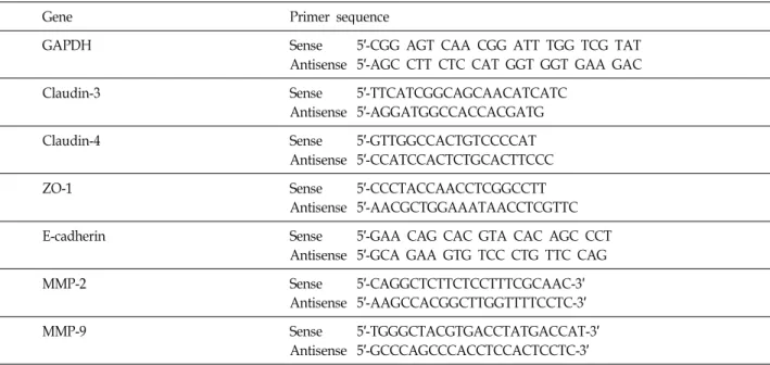
Table 1. Oligonucleotides used in RT-PCR
Gene Primer sequence
GAPDH Sense 5′-CGG AGT CAA CGG ATT TGG TCG TAT
Antisense 5′-AGC CTT CTC CAT GGT GGT GAA GAC
Claudin-3 Sense 5′-TTCATCGGCAGCAACATCATC
Antisense 5′-AGGATGGCCACCACGATG
Claudin-4 Sense 5′-GTTGGCCACTGTCCCCAT
Antisense 5′-CCATCCACTCTGCACTTCCC
ZO-1 Sense 5′-CCCTACCAACCTCGGCCTT
Antisense 5′-AACGCTGGAAATAACCTCGTTC
E-cadherin Sense 5′-GAA CAG CAC GTA CAC AGC CCT
Antisense 5′-GCA GAA GTG TCC CTG TTC CAG
MMP-2 Sense 5′-CAGGCTCTTCTCCTTTCGCAAC-3′
Antisense 5′-AAGCCACGGCTTGGTTTTCCTC-3′
MMP-9 Sense 5′-TGGGCTACGTGACCTATGACCAT-3′
Antisense 5′-GCCCAGCCCACCTCCACTCCTC-3′
FBS was placed in the basolateral chamber to act as a chemoattractant. After 72 hr, cells on the apical side were wiped off using a Q-tip. The cells on the bottom of the filter were stained with hematoxylin and Eosin Y and then counted (three fields of each triplicate filter) using an inverted microscope.
Matrix metalloproteinase activity using gelatin zymography
Following incubation with I3C for 48 hr, cell culture supernatants were collected and centrifuged at 400× g for 5 min. Cell-free supernatant was mixed with 2× sample buffer (Invitrogen) and zymography was performed using precast gels (10% polyacrylamide and 0.1% gelatin).
Following electrophoresis, the gels were washed twice at room temperature for 30 min in 2.5% Triton X-100, sub- sequently washed in buffer containing 50 mM Tris-HCl, 150 mM NaCl, 5 mM CaCl
2,1 μM ZnCl
2, and 0.02% NaN
3at pH 7.5 and incubated in this buffer at 37
oC for 24 hr.
Thereafter, gels were stained with 0.5% (w/v) Coomassie brilliant blue G-250 (Bio-Rad Laboratoties, Hercules, CA) for 1 hr, then lightly destained in methanol:acetic acid:wa- ter (3:1:6). Clear bands appear on the Coomassie stained blue background in areas of gelatinolytic activity. Gels were scanned and images were processed by extraction of the blue channel signal, converting it to black and white, and inverting it for quantification of gelatinolytic activities from the integrated optical density.
RNA isolation and reverse transcription-polymerase chain reaction (RT-PCR)
Total RNA was prepared using an RNeasy kit (Qiagen, La Jolla, CA) and primed with random hexamers for syn- thesis of complementary DNA using AMV reverse tran- scriptase (Amersham Corp., Arlington Heights, IL), accord- ing to the manufacturer's instructions. Polymerase chain re- action (PCR) was carried out in a Mastercycler (Eppendorf, Hamburg, Germany) using the primers indicated in Table 1. Conditions for PCR reactions were 1× (94
oC for 3 min), 35× (94
oC for 45 sec; 58
oC for 45 sec; and 72
oC for 1 min), and 1× (72
oC for 10 min). Amplification products obtained by PCR were electrophoretically separated on 1% agarose gel and visualized by ethidium bromide (EtBr) staining.
Immunoblot analysis
After treatment with I3C for the indicated amounts of time, total cell lysates were prepared in an extraction buffer [25 mM Tris-Cl (pH 7.5), 250 mM NaCl, 5 mM ethyl- endiaminetetra acetic acid, 1% nonidet P-40, 0.1 mM sodium orthovanadate, 2 μg/ml leupeptin, and 100 μg/ml phenyl- methylsulfonyl fluoride]. Protein concentration was de- termined using a Bio-Rad protein assay kit (Bio-Rad, Laboratories, Hercules, CA). For Western blot analysis, pro- teins (30~50 μg) were separated by 8~13% sodium dodecyl sulfate (SDS)-polyacrylamide gel electrophoresis and then electrotransferred to a nitrocellulose membrane (Schleicher
& Schuell, Keene, NH). Membranes were blocked with 5%
skim milk for 1 hr, and were then subjected to immunoblot analysis using the desired antibodies (MMP-2 and MMP-9 from NeoMarkers, Fremont, CA; claudin-3, -4, ZO-1 and E-cadherin from Zymed, San Francisco, CA; and β-actin from Sigma). Proteins were then visualized by the enhanced chemiluminescence (ECL) method, according to the recom- mended procedure (Amersham Co.). Peroxidase-labeled donkey anti-rabbit immunoglobulin and peroxidase-labeled sheep anti-mouse immunoglobulin were purchased from Amersham.
Statistical analysis
All data are presented as mean±SD. Significant differ- ences among the groups were determined using the un- paired Student’s t-test. A value of *p<0.05 was accepted as an indication of statistical significance. All of the figures shown in this article were obtained from at least three in- dependent experiments.
Results
I3C inhibited cell growth, motility and invasion of OVCAR-3 cells
Initially, we determined the cytotoxic effect of I3C on the growth of OVCAR-3 cells. As shown in Fig. 1, I3C had in- hibitory effects on this cell proliferation in a dose- and time-dependent manner. When compared with the control, treatment with 150 μM and 200 μM of I3C for 72 hr caused approximately 45% and 70% inhibition of cell growth, respectively. To determine whether I3C inhibits the cell mo- tility OVCAR-3 cells, the wound healing experiment was performed. The results, as shown in Fig. 2, demonstrated that 100 μM of I3C, which was not significant cytotoxic, as shown by MTT assay, time-dependently delayed the cell mo- tility of OVCAR-3 cells as compared to that of control cells.
Using a Boyden chamber invasion assay, we next examined the question of whether or not I3C decrease the activity of cell invasion. As shown in Fig. 3, I3C treatment reduced cell invasion through the Matrigel chamber in a concen- tration-dependent manner.
I3C down-regulated expression and activities of MMPs in OVCAR-3 cells
Because cell migration plays an important role in the metastasis process and since invasion of the basement mem- brane is primarily mediated by gelatinase MMPs, we tested
Fig. 1. Inhibition of cell growth of OVCAR-3 cells by I3C.
OVCAR-3 cells were treated with the indicated concen- trations of I3C for the desired times, and cell viability was estimated by the MTT assay. Each bar represents the mean±S.D. calculated from three independent experiments. Significance was determined by the Student’s
t
-test (*p
<0.05 versus untreated control).Fig. 2. Inhibition of cell migration of OVCAR-3 cells by I3C.
Cells were grown to confluency on 30-mm cell culture dishes coated with rat tail collagen (20 μg/ml) and then treated with vehicle (control) or 100 μM I3C for 24 hr.
A scratch was made through the cell layer using a pip- ette tip. After washing with PBS, serum free media (to prevent cell proliferation) containing either vehicle (DMSO) or I3C (100 μM) was added. Photographs of the wounded area were taken immediately after the scratch was made (0 hr) 48 and 72 hr later to evaluate cell movement into the wounded area.
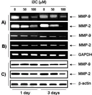
the effects of I3C on MMPs expression and their activ- ities by RT-PCR, Western blot and gelatin zymography.
As shown in Fig. 4, I3C decreased MMP-2 and -9 activ-
ities in time- and concentration-dependent manner,
which was connected with a concurrent down-regulation
Fig. 3. Decrease of cell invasion by I3C in OVCAR-3 cells.
(A) Cells pretreated with I3C for 6 hr were plated onto the apical side of matrigel coated filters in se- rum-free medium containing either vehicle or I3C.
Medium containing 20% FBS was placed in the baso- lateral chamber to act as a chemoattractant. After 48 hr, the cells on the apical side were wiped off using a Q-tip. Next, cells on the bottom of the filter were stained using hematoxylin and Eosin Y, and then counted using an inverted microscope (three fields of each triplicate filter). (B) Data are shown as the mean of triplicate samples (error bars, ±SD) and represent invasive cell numbers compared with those of control cells. Significance was determined using a Student’s t-test (*,
p
<0.05 versus untreated control).Fig. 4. Effects of I3C on MMP-2 and MMP-9 activities, pro- tein and mRNA expressions in OVCAR-3 cells. (A) The cells were incubated in the absence or presence of 50 μM and 100 μM of I3C for 24 hr and 72 hr.
Medium was collected and the activities of MMP-2 and MMP-9 were measured by zymography.
Photograph of the MMP bands, which is representa- tive of three independent experiments, is shown. (B) For RT-PCR analysis, the cells were cultured under the same conditions as those of (A) and total RNAs were isolated and RT-PCR was performed to inves- tigate the mRNA expression of MMP-2 and MMP-9.
Photographs of ethidium bromide-stained gel, which were representative of three independent experiments, are shown. (C) Cells under the same conditions as those of (A) were lysed, and proteins were separated by electrophoresis on SDS-polyacrylamide gels.
Western blotting was then performed using an- ti-MMP-2 and anti-MMP-9 antibodies, and an ECL detection system. Actin was used as an internal control.
of their mRNA and protein levels. These results suggest that the antiinvasive effect of I3C is associated with in- hibition of MMP-2 and-9 expression, and activity in OVCAR-3 cells.
I3C increased the TEER and decreased the paracellular permeability of OVCAR-3 cells
In order to examine the relationship between TJ tighten- ing and anti-invasive activity of I3C, the values of TEER were determined. As shown in Fig. 5A, incubation of OVCAR-3 cells with I3C substantially increased their TEER (a measure of tight junction formation) levels in a concen-
tration- and time dependent manner. To further characterize the TJ changes induced by I3C, the effect of I3C on the changes of paracellular permeabilities were determined.
After 72 hr of treatment with 50 μM and 100 μM of I3C, the apparent permeability of mannitol (P
appmannitol), a measure of the paracellular flux, decreased by approx- imately 39% and 84%, respectively, when compared to that of the untreated control cells (Fig. 5B).
I3C repressed the TJ-regulatory gene products in OVCAR-3 cells
Because the increase in TEER and decrease in paracellular
Fig. 5. Effects of I3C on the values of TEER and paracellular permeability in OVCAR-3 cells. (A) Cells were treated with either 50 μM or 100 μM I3C for the indicated times and the TEER was then measured as described in the materials and methods section. (B) The apparent perme- ability coefficient, Papp (cm/sec), for mannitol of cells treated for two days with either 50 μM or 100 μM I3C or the vehicle, DMSO, was determined as described in the materials and methods section. Results are shown as the mean±S.D. of three independent experiments.
The significance was determined using a Student’s
t
-test. *,p
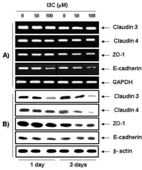
<0.05 vs. untreated control cells.permeabilitiy by I3C may result from the "tightening" of pre-existing TJs or alternatively, by increasing the total num- ber of TJs, we further elucidated the question of whether or not I3C reduces levels of TJ components using RT-PCR and Western blot analyses. As shown in Fig. 6, I3C repressed the levels of claudin-3 and -4 proteins, the most important components of the TJ [17,25], beginning by 24 hr and sup- pressing further by 72 hr, but there were no significant changes in the claudin-3 and -4 mRNA levels by I3C treat- ment, suggesting that the reduction of claudin-3 and -4 pro- tein levels occurs post-transcription. Since, the ZO-1 protein serves as a link between the integral TJ proteins, such as claudins, and the actin cytoskeleton as well as an adapter
Fig. 6. Effect of I3C on expressions of TJ-related proteins and mRNAs in OVCAR-3 cells. (A) The cells were treated with 50 μM or 100 μM I3C for the indicated times. Total RNAs were then isolated and reverse-transcribed. The resulting cDNAs were subjected to PCR with the in- dicated primers and the reaction products were sub- jected to electrophoresis in a 1% agarose gel and vi- sualized by EtBr staining. GAPDH was used as an in- ternal control. (B) The cells grown under the same con- ditions as (A) were lysed and the cellular proteins were then separated by electrophoresis on SDS-poly- acrylamide gels and transferred onto nitrocellulose membranes. Next, the membranes were probed with the indicated antibodies and the proteins were visualized us- ing an ECL detection system. Actin was used as an in- ternal control.
for cytosolic signaling proteins, and E-cadherin, as an adher- ent junction protein, is also known to regulate TJ formation [36], their expression levels were also investigated to see whether or not the modulation of ZO-1 and E-cadherin was connected with the anti-invasive action of I3C. As indicated in Fig. 6, the levels of both mRNA and protein of ZO-1 were decreased on 72 hr but there was not significantly different compared with change of claudin levels, and the levels of E-cadherin mRNA and protein also similarly decreased with those of ZO-1.
Discussion
Ovarian cancer is the most lethal of all gynecological
cancers. Most deaths from ovarian cancer are due to wide- spread intraperitoneal metastases and malignant ascites [11, 40]. However, mechanisms of metastasis in ovarian cancer remain poorly understood. Because cell migration and in- vasion are critical steps in metastasis, inhibition of tumor cell migration and invasion are important mechanisms in the anti-metastatic properties of anti-cancer drugs. Many re- cent studies have shown that chemopreventive and/or che- motherapeutic agents can inhibit tumor cell invasion and metastasis. About 10 years ago, Meng et al. [23] first men- tioned that I3C can activate the function of invasion sup- pressor molecules associated with the suppression of in- vasion and migration in breast cancer cells in vitro. More recently, Takada et al. [33] showed that I3C inhibited cell proliferation and invasion of leukemic cells through in- hibition of MMP-9 activity. And, Aggarwal and Ichikawa [2] reported that I3C treatment resulted in the regression of cervical intraepithelial neoplasia and recurrent respiratory papillomatosis in phase II clinical trials. However, the molec- ular mechanisms of anti-metastasis and anti-invasive activ- ities by I3C are not well known in human ovarian cancer cells. Therefore, this investigation attempted to address the role of I3C on cell migration and invasion using human ovarian cancer OVCAR-3 cell line, and we found that I3C significantly inhibited cell motility and invasive activity by decreasing MMPs activity and tightening TJs.
MMPs are important proteolytic enzymes during organ development and tissue regeneration, but they also play im- portant roles in cancer invasion and metastasis [24].
Particularly, MMP-2 and MMP-9 play important roles in tu- mor invasion and angiogenesis, and Wang et al. [38] found cervical cancerous tissues had higher expressions of MMP-2 mRNA and protein than their normal counterparts. MMPs are also collectively capable of cleaving virtually all ex- tracellular matrix (ECM) substrates, and degradation of ma- trix is a key event in progression, invasion, and metastasis of potentially malignant and malignant lesions [13,37], there- fore tumor metastasis can be inhibited by blocking MMP synthesis and activity. Our results indicated marked in- hibition of MMP-2 and -9 mRNA and protein levels and ac- tivities following I3C treatment (Fig. 4), suggesting that the anti-invasive activity of I3C in OVCAR-3 cells was asso- ciated with inhibition of MMP-2 and -9 activities.
Changes in permeability properties and loss of cell polar- ity are other hallmarks of epithelial cell tumorigenesis.
Modulation of TJs, which are structures critical for main- tenance of these functions in epithelial cells, in a number of epithelial cancers has been demonstrated [25,27,28]. Thus,
TJ disruption and dysregulation of its composite proteins play critical roles in cancer progression, invasion, and meta- stasis, particularly epithelial cancers [36]. For example, Soler et al. [32] first demonstrated that TEER of colon carcinoma tissue was significantly lower than that of normal colon tis- sues but showed higher transepithelial paracellular perme- ability, which confirmed the loss of TJs. Other studies also have shown that many anti-cancer drugs are inhibitory to motility and invasiveness and that they act by reduction of transepithelial paracellular permeability [9,15,35]. These ob- servations indicated that TJ leakiness was associated with cancer progression and TJ tightening might have anti-cancer activity [25,27]. In this study, I3C treatment increased the TEER and increased the paracellular permeability of OVCAR-3 cells, which was associated with lower cell mo- tility and invasiveness (Fig. 5), indicating that the anti-in- vasive activity of I3C may be due, in part, to its ability to enhance TJ activity.
Many components of TJs have recently been identified.
Among these, members of the claudin family, which are transmembrane proteins with extracellular domains, interact with other claudins associated with adjacent cells for regu- lation of paracellular permeability [25]. Emerging evidence indicates that disruption of TJs, with concomitant dysregula- tion of TJ proteins, is an early event in cancer cell invasion and metastasis. In particular, overexpression of claudin-3 and -4 has been demonstrated in several tumors, including breast and ovarian cancers [29]. Conversely, "knockdown"
of these two claudins inhibited the invasiveness of cancer cells [1]. The observations indicate that claudins are dysregu- lated in many types of cancers and claudin proteins may prove to be useful biomarkers for detection and diagnosis of certain cancers. In the present study, I3C treatment mark- edly altered the levels of claudin-3 and -4 proteins (Fig. 6), indicating that down-regulation of claudin expression by I3C relate to increased TJ tightening. In addition, ZO-1 levels in the cells are strictly regulated to correlate closely to the number of TJs, and ZO-1 is usually located only at TJ-com- plexes and E-cadherin, the adherens junction protein, medi- ate critical cell-cell interactions and regulate TJ formation [31,34], suggesting these two proteins may regulate in the tightening of TJs. As indicated in Fig. 6, the levels of ZO-1 and E-cadherin were gradually down-regulated by I3C treatment. Although we need to validate this study, we ten- tatively suggest that I3C, through effects on expression of these proteins, may mediate anti-metastasis and anti-in- vasiveness in OVCAR-3 cells.
Although this study will require validation, the present
results suggest that I3C inhibits cell migration and invasion in OVCAR-3 cells while concurrently repressing the MMPs activities, as well as tightening of TJs through inhibition of the levels of claudin expression. Taken together, the data indicate that I3C may be a promising new dietary source for decreasing the risk of cancer cell metastasis.
Acknowledgments
This research was supported by Technology Development Program for Agriculture and Forestry (610003-03-1-SU000), Ministry for Food, Agriculture, Forestry and Fisheries, and Blue-Bio Industry RIC at Dong-Eui University as a RIC (08-06-07) program of KIAT under Ministry of Knowledge Economy, Republic of Korea.
References
1. Agarwal, R., T. D'Souza, and P. J. Morin. 2005. Claudin-3 and claudin-4 expression in ovarian epithelial cells enhances invasion and is associated with increased matrix metal- loproteinase-2 activity.
Cancer Res.
65, 7378-73852. Aggarwal, B. B. and H. Ichikawa. 2005. Molecular targets and anticancer potential of indole-3-carbinol and its derivatives.
Cell Cycle
4, 1201-1215.3. Anderson, J. M. 2006. Molecular structure of tight junctions and their role in epithelial transport.
News Physiol. Sci.
16, 126-130.4. Bonnesen, C., I. M. Eggleston, and J. D. Hayes. 2001. Dietary indoles and isothiocyanates that are generated from crucif- erous vegetables can both stimulate apoptosis and confer protection against DNA damage in human colon cell lines.
Cancer Res.
61, 6120-6130.5. Brandi, G., M. Paiardini, B. Cervasi, C. Fiorucci, P. Filippone, C. De Marco, N. Zaffaroni, and M. Magnani. 2003. A new indole-3-carbinol tetrameric derivative inhibits cyclin-de- pendent kinase 6 expression, and induces G1 cell cycle ar- rest in both estrogen-dependent and estrogen-independent breast cancer cell lines.
Cancer Res.
63, 4028-4036.6. Chinni, S. R., Y. Li, S. Upadhyay, P. K. Koppolu, and F.
H. Sarkar. 2001. Indole-3-carbinol (I3C) induced cell growth inhibition, G1 cell cycle arrest and apoptosis in prostate can- cer cells.
Oncogene
20, 2927-2936.7. Cho, H. J., S. Y, Park, E. J. Kim, J. K. Kim, and J. H. Park.
2011. 3,3'-Diindolylmethane inhibits prostate cancer devel- opment in the transgenic adenocarcinoma mouse prostate model.
Mol. Carcinog.
50, 100-112.8. Choi, H. S., M. C. Cho, H. G. Lee, and D. Y. Yoon. 2010.
Indole-3-carbinol induces apoptosis through p53 and activa- tion of caspase-8 pathway in lung cancer A549 cells.
Food Chem. Toxicol.
48, 883-890.9. Choi, Y. H., W. Y. Choi, S. H. Hong, S. O. Kim, G. Y. Kim,
W. H. Lee, and Y. H. Yoo. 2009. Anti-invasive activity of sanguinarine through modulation of tight junctions and ma- trix metalloproteinase activities in MDA-MB-231 human breast carcinoma cells.
Chem. Biol. Interact.
179, 185-191.10. Dashwood, R. H., A. T. Fong, D. N. Arbogast, L. F. Bjeldanes, J. D. Hendricks, and G. S. Bailey. 1994. Anticarcinogenic ac- tivity of indole-3-carbinol acid products: ultra-sensitive bio- assay by trout embryo microinjection.
Cancer Res.
54, 3617-3619.11. Davidson, B., R. Reich, C. G. Trope, T. L. Wang, and IeM.
Shih. 2010. New determinates of disease progression and outcome in metastatic ovarian carcinoma.
Histol. Histopathol.
25, 1591-1609.
12. De Oliveira, S. S., I. M. De Oliveira, W. De Souza, and J.
A. Morgado-Diaz. 2005. Claudins upregulation in human colorectal cancer.
FEBS Lett.
579, 6179-6185.13. Duffy, M. I., T. M. Maguire, A. Hill, E. McDermott, and N. O'Higgins. 2000. Metalloproteinases: role in breast carci- nogenesis, invasion and metastasis.
Breast Cancer Res.
2, 252-257.14. Exon, J. H., E. H. South, B. A. Magnuson, and K. Hendrix.
2001. Effects of indole-3-carbinol on immune responses, aberrant crypt foci, and colonic crypt cell proliferation in rats.
J. Toxicol. Environ. Health A
62, 561-573.15. Gitter, A. H., K. Bendfeldt, K. Schmitz, J. D. Schulzke, C.
J. Bentzel, and M. Fromm. 2000. Epithelial barrier defects in HT-29/B6 colonic cell monolayers induced by tumor ne- crosis factor-α.
Ann. N. Y. Acad. Sci.
915,193-203.16. Goskonda, V. R., M. A. Khan, C. M. Hutak, and I. K. Reddy.
1999. Permeability characteristics of novel mydriatic agents using an
in vitro
cell culture model that utilizes SIRC rabbit corneal cells.J. Pharm. Sci.
88, 180-184.17. Hewitt, K. J., R. Agarwal, and P. J. Morin. 2006. The claudin gene family: expression in normal and neoplastic tissues.
BMC Cancer
6, 186-193.18. Kojima, T., T. Tanaka, and H. Mori. 1994. Chemoprevention of spontaneous endometrial cancer in female Donryu rats by dietary indole-3-carbinol.
Cancer Res.
54, 1446-1449.19. Kominsky, S. L. 2006. Claudins: emerging targets for cancer therapy.
Expert Rev. Mol. Med.
8, 1-11.20. Liang, M., C. R. Ramsey, and F. G. Knox. 1999. The para- cellular permeability of opossum kidney cells, a proximal tubule cell line.
Kidney Int.
56, 2304-2308.21. Loub, W. D., L. W. Wattenberg, and D. W. Davis. 1975. Aryl hydrocarbon hydroxylase induction in rat tissues by natu- rally occurring indoles of cruciferous plants.
J. Natl. Cancer Inst.
54, 985-988.22. Matrisian, L. M. 1992. The matrix-degrading metalloproteinases.
Bioessays
14, 455-463.23. Meng, Q., M. Qi, D. Z. Chen, R. Yuan, I. D. Goldberg, E.
M. Rosen, K. Auborn, and S. Fan. 2000. Suppression of breast cancer invasion and migration by indole-3-carbinol:
associated with up-regulation of BRCA1 and E-cadher- in/catenin complexes.
J. Mol. Med.
78, 155-165.24. Mook, O. R., W. M. Frederiks, and C. J. Van Noorden. 2004.
The role of gelatinases in colorectal cancer progression and
초록:Indole-3-carbinol에 의한 OVCAR-3 인체 난소암세포의 침윤 억제 최영현
1,2*․김성옥
1(동의대학교
1대학원 바이오물질제어학과 및 블루바이오 소재 개발 센터,
2한의과대학 생화학교실 및 한의학
연구소)
본 연구에서는 식물체에 널리 분포하는 indole-3-carbinol (I3C)에 의한 OVCAR-3 인체 난소암세포의 이동성 및 침윤성 억제 가능성과 이와 연관된 기전을 조사하였다. 본 연구의 결과에 의하면 I3C에 의한 OVCAR-3 세포 의 증식억제는 세포의 이동성 억제와 연관이 있었으며, 이를 wound healing 및 matrigel invasion assay로 확인 하였다. 아울러 I3C 처리에 의하여 transepithelial electrical resistance가 증가되었으며, cellular paracellular per- meability는 감소되었는데, 이는 I3C 처리에 의해 세포 내 치밀결합(tight junctions, TJs)의 tightness가 증가되었 음을 의미한다. RT-PCR 및 immunoblotting 결과에 의하면, I3C는 TJs의 구성 성분이면서 paracellular transport 의 선택적 투과성을 조절하는 주요 인자인 claudin-3 및 -4의 발현을 유의적으로 억제하였다. 또한 matrix metal- loproteinase (MMP)-2 및 -9의 활성이 I3C 처리에 의하여 매우 억제되었는데, 이는 그들의 mRNA 및 단백질 수준 에서의 발현 감소와 연관성이 있었다. 따라서 I3C에 의한 OVCAR-3 난소암세포의 침윤성 억제는 TJs 기능의 강 화와 MMP 활성의 저하가 주요 인자로 작용함을 알 수 있었다.
metastasis.
Biochim. Biophys. Acta.
1705, 69-89.25. Morin, P. J. 2005. Claudin proteins in human cancer: promis- ing new targets for diagnosis and therapy.
Cancer Res.
65, 9603-9606.26. Morse, M. A., S. D. LaGreca, S. G. Amin, and F. L. Chung.
1990. Effects of indole-3-carbinol on lung tumorigenesis and DNA methylation induced by 4-(methylnitros-ami- no)-1-(3-pyridyl)-1-butanone (NNK) and on the metabolism and disposition of NNK in A/J mice.
Cancer Res.
50, 2613-2617.27. Mullin, J. M., N. Agostino, E. Rendon-Huerta, and J. J.
Thornton. 2005. Keynote review: epithelial and endothelial barriers in human disease.
Drug Discov. Today
10, 395-408.28. Ouban, A. and A. A. Ahmed. 2010. Claudins in human can- cer: a review.
Histol. Histopathol.
25, 83-90.29. Rangel, L. B., R. Agarwal, T. D'Souza, E. S. Pizer, P. L. Alò, W. D. Lancaster, L. Gregoire, D. R. Schwartz, K. R. Cho, and P. J. Morin. 2003. Tight junction proteins claudin-3 and claudin-4 are frequently overexpressed in ovarian cancer but not in ovarian cystadenomas.
Clin. Cancer Res.
9, 2567-2575.30. Rogan, E. G. 2006. The natural chemopreventive compound indole-3-carbinol: state of the science.
In Vivo
20, 221-228.31. Schneeberger, E. E. and R. D. Lynch. 2004. The tight junction:
a multifunctional complex.
Am. J. Physiol. Cell. Physiol.
286, 1213-1228.32. Soler, A. P., R. D. Miller, K. V. Laughlin, N. Z. Carp, D.
M. Klurfeld, and J. M. Mullin. 1999. Increased tight junc- tional permeability is associated with the development of colon cancer.
Carcinogenesis
20, 1425-1431.33. Takada, Y., M. Andreeff, and B. B. Aggarwal. 2005.
Indole-3-carbinol suppresses NF-κB and IκBα kinase activa- tion, causing inhibition of expression of NF-κB-regulated antiapoptotic and metastatic gene products and enhance-
ment of apoptosis in myeloid and leukemia cells.
Blood
106, 641-649.34. Tunggal, J. A., I. Helfrich, A. Schmitz, H. Schwarz, D.
Günzel, M. Fromm, R. Kemler, T. Krieg, and C. M. Niessen.
2005. E-cadherin is essential for
in vivo
epidermal barrier function by regulating tight junctions.EMBO J.
24, 1146-1156.35. Van Deun, K., F. Pasmans, F. Van Immerseel, R. Ducatelle, and F. Haesebrouck. 2008. Butyrate protects Caco-2 cells from Campylobacter jejuni invasion and translocation.
Br.
J. Nutr.
100, 480-484.36. Van Itallie, C. M. and J. M. Anderson. 2006. Claudins and epithelial paracellular transport.
Annu. Rev. Physiol.
68, 403-429.37. Vihinen, P. R. Ala-aho, and V. M. Kähäri. 2005. Matrix metal- loproteinases as therapeutic targets in cancer.
Curr. Cancer Drug Targets
5, 203-220.38. Wang, P. H., J. L. Ko, H. T. Tsai, S. F. Yang, C. P. Han, L. Y. Lin, and G. D. Chen. 2008. Clinical significance of ma- trix metalloproteinase-2 in cancer of uterine cervix: a semi- quantitative study of immunoreactivities using tissue array.
Gynecol. Oncol.
108, 533-542.39. Wattenberg, L. W. and W. D. Loub. 1978. Inhibition of poly- cyclic aromatic hydrocarbon-induced neoplasia by naturally occurring indoles.
Cancer Res.
38, 1410-1413.40. Williams, T. I., K. L. Toups, D. A. Saggese, K. R. Kalli, W.
A. Cliby, and D. C. Muddiman. 2007. Epithelial ovarian can- cer: disease etiology, treatment, detection, and investiga- tional gene, metabolite, and protein biomarkers.
J. Proteome Res.
6, 2936-2962.41. Zhang, X. and D. Malejka-Giganti. 2003. Effects of treatment of rats with indole-3-carbinol on apoptosis in the mammary gland and mammary adenocarcinomas.