• 검색 결과가 없습니다.

A Textiloma on the Pterion : A Rarely Occurred Craniotomy Complication

N/A
N/A
Protected

Academic year: 2021

Share "A Textiloma on the Pterion : A Rarely Occurred Craniotomy Complication"

Copied!
3
0
0

로드 중.... (전체 텍스트 보기)

전체 글

(1)

252

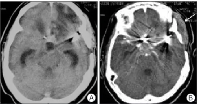
tomb-like isodense lesion having a moderate enhancement un- der the musculo-cutaneous flap (Fig. 1). Thus, a second surgery was performed. A piece of gauze encapsulated by a large amount of granulation tissue was found at the layer between temporalis muscle and dura through surgical defect of calvarium (Fig. 2).

Histopathological study confirmed cotton fibers surrounded by a granulomatous foreign body reaction (Fig. 3). She continued to do well after wound revision.

DISCUSSION

Retention of surgical materials within wounds has been re- ported to occur with a frequency of one in 5000 for all opera- tions

3)

. Among them, textilomas in the fields of neurosurgery have been found to appear after spinal surgery

7)

, craniotomies for trauma

6)

, tumor resection

1)

, clot evacuation

9)

, cotton wrap- ping of aneurysms

5)

, and microvascular decompression

2)

. The vast majority of reported postcraniotomy textilomas were sub- dural lesions

10)

.

Intraoperatively applied foreign materials can cause infection or abscess formation in the early stage, whereas others mostly remain asymptomatic for many years

8)

. Chronic cases of fibrin- ous textiloma like the present one involves slow adhesion for- mation such as encapsulation and granuloma. They are often overlooked or can be confused with other soft-tissue masses.

INTRODUCTION

Textiloma is a term used to describe a foreign-body reaction to the retained material routinely used during operations such as cotton or gauze. Textilomas are inflammatory granulomas that often mimic residual or recurrent tumors or abscesses clin- ically or radiologically. A study involving 254 cases of textilo- mas showed common occurrences in the order of abdomen (56%), pelvis (18%), thorax (11%), and limb (7%), spine (4%), and head and neck (4%)

10)

. In contrast, the existence of textilo- mas after craniotomies has been observed in a limited number of studies. The author reports the first case of textiloma due to retained gauze that was left under the temporalis muscle eight years ago in a patient who underwent pterional craniotomy for ruptured aneurysm.

CASE REPORT

A 64-year-old woman complained of a painful expansion in the temporal region that she had first noticed five months pre- viously. This patient underwent left pterional craniotomy 8 years earlier for a ruptured aneurysm. There were no complica- tions at the time of her operation. On examination, a well-cir- cumscribed, firm, raised scalp mass at the area of previous cra- niotomy was defined. Computed tomography (CT) showed a

A Textiloma on the Pterion : A Rarely Occurred Craniotomy Complication

Ealmaan Kim, M.D., Ph.D.

Department of Neurosurgery, Dongsan Medical Center, Keimyung University School of Medicine, Daegu, Korea

Textiloma is an inflammatory mass containing surgical sponges that are unintentionally left behind in a surgical wound. This complication has been most commonly described by abdominal and gynecologic surgeons. However, the occurrence of textiloma after intracranial procedures especially under the temporalis muscle has not been documented. The author reports a rare case of textiloma of the pterion in a patient who presented with a subcutaneous tumor developed eight years after frontotemporal craniotomy for aneurysm clipping.

Key Words : Complication · Craniotomy · Pterion · Temporalis muscle · Textiloma.

Case Report

Received : July 31, 2012

Revised : February 12, 2013

Accepted : April 8, 2013

Address for reprints : Ealmaan Kim, M.D., Ph.D.

Department of Neurosurgery, Dongsan Medical Center, Keimyung University School of Medicine, 56 Dalseong-ro, Jung-gu, Daegu 700-712, Korea Tel : +82-53-250-7823, Fax : +82-53-250-7356, E-mail : [email protected]

This is an Open Access article distributed under the terms of the Creative Commons Attribution Non-Commercial License (http://creativecommons.org/licenses/by-nc/3.0) which permits unrestricted non-commercial use, distribution, and reproduction in any medium, provided the original work is properly cited.

J Korean Neurosurg Soc 53 : 252-254, 2013

http://dx.doi.org/10.3340/jkns.2013.53.4.252

Copyright © 2013 The Korean Neurosurgical Society

Print ISSN 2005-3711 On-line ISSN 1598-7876

www.jkns.or.kr

(2)

253

Postcraniotomy Textiloma | E Kim

allows for a more conservative treatment.

CONCLUSION

The author reports a case of textiloma under the temporalis muscle found in the second surgery performed 8 years after the Time to discovery in this case, 8 years, was much longer be-

cause of the high likelihood of intracranial textilomas related complications within the first 2 years after surgery

1,2,5,6,9)

. Being sterile, the surgical cottons do not initiate an infective reaction and thus remain clinically silent and missed for a long duration in the postoperative period until they are thought of. After a cer- tain period, when the patient’s resistance decreases or a bout of bacteremia ensues, organisms will lodge into the area of the for- eign body and a sterile inflammatory reaction will turn symp- tomatic. This may be associated with tooth extraction or throat infection or after a flu-like syndrome. A continuing foreign body granulomatous reaction may be speculative explanation for this case that presents late as a progressively enlarging mass in the temporalis muscle.

In contrast with other lesions, intracranial and spinal textilo- mas sometimes show early severe symptoms like neurologic defi- cit, pain or seizure. Patients with textilomas after craniotomy for tumors are at risk of unnecessary treatment because these lesions manifest as recurrent tumors. Patients with textilomas could in fact show more aggravated symptoms due to the mass effect of granuloma after microvascular decompression for cranial rhi- zopathy. Furthermore, spine surgeons may mistake textilomas for recurrent disc herniations in some cases. However, the pres- ent case showed only bulging on the surgical wound without neurological deterioration. This was quite different from most patients who underwent pterional craniotomy and experienced remarkable depression in the frontozygomatic fossa.

Imaging is essential to the workup for textilomas, and the common differential diagnosis includes primary, metastatic, re- current tumor, radiation necrosis or abscess. Suggested CT find- ings are low-density heterogeneous masses with a hyperdense rim that was further enhanced after contrast injection like the present case, and sometimes they present with a spongiform pattern containing air bubbles

6)

. The signal intensity of magnet- ic resonance imaging may vary according to aging, histologic composition, and its fluid content. Almost all lesions are hypoin- tense on T1-weighted images and hyperintense on T2-weighted images, but textilomas cannot be easily distinguished from a re- current tumor or abscess due to their less characteristics

8)

. There- fore, careful history taking about previous operations and being aware of textiomas are very important for imaging diagnosis.

An increased risk of a foreign body retainment after cranioto- my was highly correlated with emergency cases, unexpected changes in operations, and inaccurate counts of sponges and in- struments, involvement of more than one surgical team, and pa- tients with a high body-mass index

4)

. Although textilomas are rarely encountered under the temporalis muscle, these must al- ways be considered in the differential diagnosis of cranial mass- es arising after craniotomy. Early second-look surgery may be advised to ascertain the diagnosis for certain cases, especially when unusual progression of the intra- and extracranial lesion is seen during treatment or routine follow-up. This lifts the bur- den of uncertainty from both the patient and the physician and

Fig. 1. Computed tomography of a textiloma developed after pterional craniotomy for aneurysm clipping. Nonenhanced (A) and enhanced (B) scans show an isodensity of homogeneous mass with a peripheral con- trast enhancement in the temporalis muscle (arrow).

B A

Fig. 2. Intraoperative appearance of the lesion demonstrates an osseous defect and a mass of granulation tissues adherent to the temporalis muscle (arrow). The lesion is extending into the underlying dura mater.

Fig. 3. Photomicrograph reveals gauze fibers (arrowhead) surrounded by

dense fibrous connective tissue infiltrated with lymphocytes, plasma

cells, neutrophilic granulocytes and macrophages (H&E, original magnifi-

cation ×40).

(3)

254

J Korean Neurosurg Soc 53 | April 2013

factors for retained instruments and sponges after surgery. N Engl J Med 348 : 229-235, 2003

5. Goldsberry DH, Ross IB, Dhillon G, Corbett JJ : Visual dysfunction caused by gauze wrapping of an intracranial aneurysm. J Neurooph- thalmol 24 : 42-45, 2004

6. Kim AK, Lee EB, Bagley LJ, Loevner LA : Retained surgical sponges af- ter craniotomies : imaging appearances and complications. AJNR Am J Neuroradiol 30 : 1270-1272, 2009

7. Kucukyuruk B, Biceroglu H, Abuzayed B, Ulu MO, Kafadar AM : Para- spinal gossybipoma : a case report and review of the literature. J Neuro- sci Rural Pract 1 : 102-104, 2010

8. Manzella A, Filho PB, Albuquerque E, Farias F, Kaercher J : Imaging of gossypibomas : pictorial review. AJR Am J Roentgenol 193 (6 Suppl) : S94-S101, 2009

9. Vishteh AG, Apostolides PJ, Dean B, Spetzler RF : Magnetic resonance image of postcraniotomy retained cotton or rayon. Case illustration. J Neurosurg 88 : 928, 1998

10. Wan W, Le T, Riskin L, Macario A : Improving safety in the operating room : a systematic literature review of retained surgical sponges. Curr Opin Anaesthesiol 22 : 207-214, 2009

first. The rarity of postcraniotomy extracranial textilomas and their nonspecificities in the imaging features preclude preoper- ative diagnosis. The occurrence of a textiloma following crani- otomy can only be avoided by surgeons who are aware of this uncommon but clinically significant event.

References

1. Apel-Sarid L, Cochrane DD, Steinbok P, Byrne AT, Dunham C : Micro- fibrillar collagen hemostat-induced necrotizing granulomatous inflam- mation developing after craniotomy : a pediatric case series. J Neuro- surg Pediatr 6 : 385-392, 2010

2. Chen J, Lee S, Lui T, Yeh Y, Chen T, Tzaan W : Teflon granuloma after microvascular decompression for trigeminal neuralgia. Surg Neurol 53 : 281-287, 2000

3. Cima RR, Kollengode A, Garnatz J, Storsveen A, Weisbrod C, Des- champs C : Incidence and characteristics of potential and actual re- tained foreign object events in surgical patients. J Am Coll Surg 207 : 80-87, 2008

4. Gawande AA, Studdert DM, Orav EJ, Brennan TA, Zinner MJ : Risk

수치

Fig. 1. Computed tomography of a textiloma developed after pterional  craniotomy for aneurysm clipping

참조

관련 문서