大韓獸醫學會誌(2009) 第49卷 第1號
Korean J Vet Res
(2009) 49(1) : 63~6663
Cutaneous smooth muscle tumors in 3 dogs
Ji-Youl Jung
1, Sang-Chul Kang
2, Dae-Sik Park
3, Eun-Sung Lee
3, Jong-Hee Bae
1, Jae-Hoon Kim
1,*1
College of Veterinary Medicine, Jeju National University, Jeju 690-756, Korea
2
Preclinical Research Center, Chemon,Yongin 449-826, Korea
3
Saha Animal Hospital, Busan 604-010, Korea
(Accepted: March 11, 2009)
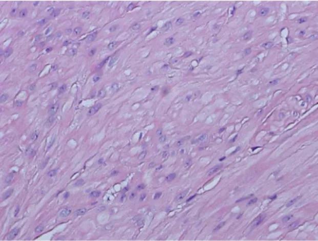
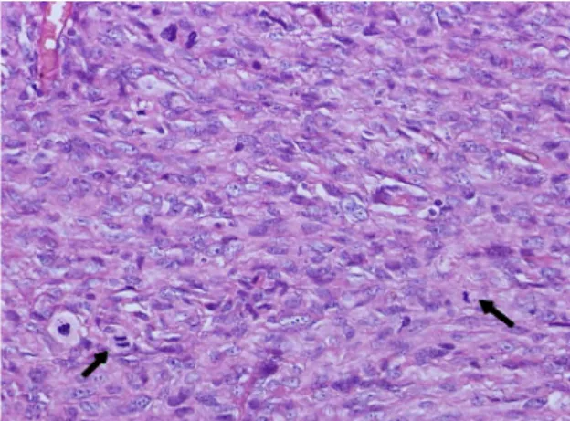
Abstract : Cutaneous leiomyomas (leiomyosarcomas) are smooth muscle tumors that occur single or as multiple lesions. They usually arise from the arrector pili muscles (piloleiomyomas) and less commonly from the muscle of veins (angioleiomyomas). This report describes histologic and immunohistochemical features of one cutaneous piloleiomyoma and two angioleiomyosarcomas. Three 7-12-year-old female dogs were presented with single or double cutaneous nodules. Histologically, the neoplastic masses were composed of densely or loosely arranged interlacing bundles. The neoplastic cells were ovoid to elongate, and had eosinophilic cytoplasms and perinuclear cytoplasmic vacuolation. Nuclei were central to eccentric, cigar shaped, oval to elongate. In two cases, high mitotic index in high power field, multifocal necrosis and local invasion were also noted. Masson’s trichrome and van Gieson staining revealed muscle origin tumors in these cases. Immunohistochemically, the tumor cells were strongly positive for smooth muscle actin. In our best knowledge, this is the first report of cutaneous smooth muscle tumors in dogs in Korea.
Keywords : dog, immunohistochemistry, leiomyoma, leiomyosarcoma, skin
Introduction
Smooth muscle is widely distributed in the body, including the gastrointestinal, respiratory, and genitourinary tracts. Cutaneous leiomyomas (leiomyosarcomas) are uncommon smooth muscle tumors of the skin that occur single or as multiple lesions. They usually arise from the muscle of veins, arrector pili muscles or genital deep dermal smooth muscles. Tumors derived from the musculature of blood vessels are termed angioleiomyomas and angioleiomyosarcomas, and those developing from arrector pili muscles are identified as piloleiomyomas and piloleiomyosarcomas. Smooth muscle tumors located in the skin of the genitalia and nipples are named genital leiomyomas and leiomyosarcomas [7, 8]. Smooth muscle tumors are rare in domestic animals. Cutaneous leiomyomas are infrequently described in dogs and cats, and there is not sufficient data to allow for determination of age, breed, or sex predilec- tions [3].
According to the data from 748 canine cutaneous neoplasms in Korea, the incidence of cutaneous leio-
myoma and leiomyosarcoma was 0.27% and 0.4%, respectively [10]. In another review of 2,616 canine and feline skin tumors from the UK, the incidence of cutaneous leiomyomas was 0.88% and 0.33%, respec- tively [2]. Distinguishing between leiomyosarcoma, rhabdomyosarcoma and fibrosarcoma may be difficult by routine histological methods, but immunohistoche- mical and ultrastructural methods are useful for accurate diagnosis of soft tissue sarcomas [3]. In the present study, we described the histopathologic and immunohi- stochemical features of 3 cutaneous smooth muscle neoplasms in dogs.
Case report
Three subcutaneous biopsies from dogs were submitted to the pathology laboratory at the college of veterinary medicine in Jeju National University. Three cases were composed of a 12-year-old mixed dog with two tumors (1.5 to 2.5 cm in diameter) on the perineal region (case 1), a 7-year-old Shih tzu with a tumor (3.5 × 3 × 2.5 cm) on the right abdominal region around 5th mammary
*Corresponding author: Jae-Hoon Kim
College of Veterinary Medicine, Jeju National University, Jeju 690-756, Korea
[Tel: +82-64-754-3387, Fax: +82-64-702-9920, E-mail: [email protected]]
64 Ji-Youl Jung, Sang-Chul Kang, Dae-Sik Park, Eun-Sung Lee, Jong-Hee Bae, Jae-Hoon Kim
teat (case 2), and a 9-year-old mixed dog with a tumor (6 × 4 cm) on the left distal area of femur (case 3), respectively. All of three dogs were female.
Submitted masses were trimmed, embedded in paraffin, sectioned at 3
µm, and stained with hematoxylin and eosin, Masson’s trichrome, and van Gieson techni- ques. Additional paraffin-embedded sections were available for immunohistochemistry. After mounting on silane coated glass slides, each section was stained by a labeled streptavidin-biotin peroxidase method. For antigen retrieval, sections were incubated with Target Retrieval solution (Dako code S3307). For the differential diagnosis, primary antibody for vimentin (1 : 100, monoclonal mouse anti-vimentin, clone V9; Abcam, UK), S-100 (1 : 1,000, rabbit polyclonal anti-S100;
Dako, Denmark) and
α-smooth muscle actin (1 : 100,
α