4)
† 주 저자 (e-mail: [email protected] call: 042)829-7566
Vol. 45, No. 3, September 2019, 255-263 http://dx.doi.org/10.15230/SCSK.2019.45.3.255
홍삼추출물에 함유된 이소말톨 글리토시드의 멜라닌 생성저해 효과
이 상 명†
목원대학교 화학화장품학부
(2019년 7월 8일 접수, 2019년 9월 6일 수정, 2019년 9월 16일 채택)
Antimelanogenic Effect of Isomaltol Glycoside from Red Ginseng Extract
Sang Myung Lee†
Department of Chemistry and Cosmetics, College of Sciences & Technology, Mokwon University, 88, Doanbuk-ro, Seo-gu, Daejeon 35349, Korea
(Received July 8, 2019; Revised September 6, 2019; Accepted September 16, 2019)
요 약: 이소말톨 글리코시드는 홍삼제조과정에서 인삼이 함유하고 있는 아미노산과 당의 열전환물질로서 홍삼 추출물에 다량 함유된 친수성 퓨란배당체이다. 현재 화장품원료로 자주 사용되고 있는 홍삼추출물의 화장품원료로서의 가치를 재고하기 위하여 홍삼추출물의 주성분인 이소말톨 글리코시드에 대하여 다양한 피부미백활성 실험을 실시 하였다. 우리는 이소말톨 글리코시드에 대한 B16-F10 세포에서의 멜라닌함량 변화, 제브라피쉬배아착색 분석, 버섯티로시나제 저해활성, 피부미백활성 기전결정을 위한 단백질 분석을 실시하였다. 제브라피쉬 멜라닌 함량 분석에서 이소말톨 글리코시드는 처리하지 않은 대조군과 비교하여 50 및 100 μg/mL 처리 후 총 멜라닌 함량을 약 20% 감소 시켰으며 제브라피쉬 속의 타이로시나제 활성을 약 10% 감소시켰다. 이소말톨 글리코시드는 또한 B16-F10 흑색 종에서 멜라닌 함량의 농도의존적인 감소를 보였으며 MITF 인산화 인자인 p-AKT 및 p-ERK의 발현을 증가시키고 MITF의 농도를 감소시켰다. 또한 이소말톨 글리코시드는 세포내 타이로시나제, TRP-1 및 TRP-2 발현을 억제 하였다. 이소말톨 글리코시드의 함량은 인삼 추출물에서 약 3%, 인삼 뿌리에서 약 1%였다. 따라서, 정량적으로 고려할 때 홍삼추출물에 다량 함유되어있는 이소말톨 글리코시드는 홍삼의 미백활성을 나타내는 주요성분 중 하나로 판단된다.
Abstract: Isomaltol glycoside is a hydrophilic furanic glycoside in which the amino acids and sugars of ginseng are thermally denatured during red ginseng production. Various skin whitening tests were conducted on isomaltol glycoside containing a lot of red ginseng extract in order to investigate the skin whitening effect as a cosmetic raw material.
We have tested melanin content assay in B16-F10 cells, zebrafish embryo pigmentation assay, mushroom tyrosinase inhibitory activity, western blot analysis to determine skin whitening activity of isomaltol glycosides. In the zebrafish melanin content assay, isomaltol glycoside decreased total melanin content by about 20% and zebrafish tyrosinase activity by about 10% after treatment with 50 and 100 μg/mL compared to the untreated control group. Isomaltol glycoside also showed a concentration-dependent decrease in melanin content in B16-F10 melanoma. Furthermore, it increased the expression of MITF phosphorylation factors p-AKT and p-ERK in B16-F10 melanoma and decreased the concentration of MITF. It also inhibited tyrosinase, TRP-1 and TRP-2 expression. The content of isomaltol glycoside was about 3%
in the ginseng extract and about 1% in the ginseng root. Thus, isomaltol glycoside is considered as one of the main components that exhibit the whitening activity of ginseng when considered quantitatively as whitening activity.
Keywords: skin whitening, antimelanogenesis, isomaltol glycoside, Red ginseng
1. Introduction
Red ginseng is steamed and dried without peeling the root of fresh ginseng (Panax ginseng meyer). Red ginseng is a herbal remedy for energy recovery and has been used in traditional folk remedies. For this reason, many pharmacological studies on ginseng have been carried out and the usefulness of red ginseng has already been clarified[1]. In recent years, red ginseng extract has been widely used as a cosmetic raw material. Ginsenosides, major components of ginseng, are known to have anti-skin-wrinkling and skin-whitening effects. Ginsenosides have been reported to inhibit the production of collagenase (MMP-2 or -9)[2], a skin-wrinkle-inducing enzyme and the melanin from melanocytes[3,4]. However, when used as a raw material for cosmetics, red ginseng is often extracted with water or alcohol. Considering the amount of ginsenosides[1]
present in a red ginseng extract and the degree of its skin- improving activity, it is difficult to evaluate the role of the ginsenosides in cosmetic raw materials on improving the skin.
Usually, an extract shows its effect, when either the phy- siological activity of the specific compound contained in the extract is highly efficient or when the chemical amount is high enough[5]. From this point of view, the ginsenosides might not be the active agent responsible for the skin improvement effect of a ginseng extract. Rather, it might be attributed to another compound contained in ginseng in a higher amount and in a higher activity than the ginsenosides. For the evaluation of ginseng in cosmetic raw materials, it is, therefore, necessary to study the compounds other than the ginsenosides.
Melanin is responsible for the pigmentation in human skin.
The two important types of melanins are deep brown eumelanin and reddish yellow pheomelanin. The melanin synthesis signalling pathway contains a variety of intracellular signalling molecules.
When skin is exposed to ultraviolet light, melanocyte cyclin adenosine monophosphate (cAMP) is increased, protein kinase (PKA) is activated[6], and CREB (cAMP response element binding protein) increases the expression of MITF (micro- phthalmia-associated transcription factor)[7]. The extracellular signal-regulated kinase (ERK) signal, which is known to be mainly involved in cell proliferation and differentiation, together with the AKT (protein kinase B) signal induces phospho-
rylation of MITF, resulting in ubiquitination of MITF and induction of tyrosinase[8], TRP-1, 2 (tyrosinase-related protein-1, -2), thereby reducing melanin synthesis[9]. Melanin synthase tyrosinase is required for the synthesis of eumelanin and pheomelanin, whereas TRP-1 and TRP-2 are known to be involved more in the synthesis of eumelanin. Many tyrosinase inhibitors which have been described include arbutin, kojic acid, and ellagic acid extracted from natural products. Since many existing pigment inhibitors show toxicity to melanocytes and have adverse effects on human cells[10,11], secondary metabolites such as flavonoids and terpenoids isolated from safe traditional herbal medicines have been developed as whitening agents[12]. While searching for skin whitening agents in traditional Chinese medicine, we have discovered that the water-soluble ingredient isomaltol glycoside (isomaltol- α -D-glycoside, IMG) in red ginseng inhibits the production of tyrosine degrading enzymes. In this paper, various charac- teristics of the skin whitening activity of IMG[13] as a water- soluble compound of red ginseng and its value as a functional raw material for cosmetic whitening activity are described.
2. Materials and Methods
2.1. Chemicals and Antibodies
Mushroom tyrosinase, L-tyrosin, kojic acid, α-melanocyte- stimulating hormone (α-MSH), 12-o-tetradecanoyl phorbol-13- acetate, 1-phenyl-2-thiourea (PTU) and 3-[4,5-dimethylthiazol- 2-yl]-2,5-diphenyltetrzolium bromide (MTT), tricaine methane- sulfonate were purchased from Sigma-Aldrich (USA). Fetal bovine serum (FBS), antibiotics (penicillin, streptomycin) and cell cure media were obtained from Gibco BRA Life Technology (USA).
Antibodies recognizing extracellular signal-regulated kinase
(ERK), phosphorylated extracellular signal-regulated kinase
(p-ERK) and protein kinase B (AKT), phosphorylated protein
kinase B (p-AKT) were obtained from Cell Signaling Technology
(USA). Tyrosinase, tyrosinase-related protein-1 (TRP-1),
tyrosinase-related protein-2 (TRP-2) and actin antibodies were
purchased from Santa Cruz Biotechnology (USA). Antibody
against microphthalmia-associated transcription factor (MITF)
was purchased from Thermo Fisher Scientific (USA). Isomatol
glycoside (IMG), a standard compound for measuring
antimelanogenesis activity and measuring the content in red ginseng samples, was purchased from Embo institute (Korea).
Red ginseng extract (Hong Sam Jeong) and Red ginseng roots were purchased from a functional food store (Korea). Commercially available red ginseng extract is prepared by adding 10 ∼ 13 times of water to red ginseng root, extracting at 85
oC for 12 h, and concentrating the extract at 50 ∼ 60
oC to 70 ∼ 73 Brix[1].
2.2. Cell Culture
B16-F10 murine melanocytes (Korean Cell Line Bank, Korea) were cultured in Dulbecco’s modified Eagle medium containing 10% fetal bovine serum, 100 U/mL penicillin, 0.1 mg/mL streptomycin and 0.25 mg/mL and cultured in a humidified 95% air / 5% CO
2atmosphere.
2.3. Cell Viability Test
B16-F10 murine melanocytes were dispensed in a 96-well plate at a density of 5×10
4cells/well (0.18 mL), and 0.02 mL of each sample (the solution adjusted to a final concentration of 10, 25, 50, 100, 200 μg/mL) was added to each well and incubated at 37
oC in a 5% CO
2incubator for 24 h. In the control group, the same amount of distilled water as that of the sample was added and the cells were cultured under the same conditions. After adding 0.02 mL of MTT solution (2.5 mg/mL) for 4 h, the culture solution was removed and 0.15 mL of DMSO was added to each well. After incubation at room temperature for 30 min, absorbance at 540 nm was measured with an ELISA reader. Cell viability was expressed as the ratio of absorbance of the sample added group when the absorbance of the non-sampl group was 100% survival and the absorbance of the cell free group was 0% survival.
2.4. Melanin Content Assay in B16-F10 Cells
B16-F10 melanoma cells were cultured in DMEM with 10% fetal bovine serum and penicillin/streptomycin (100 U/50 µg/mL) in a humidified atmosphere containing 5% CO
2in air at 37
oC. Intracellular melanin content was measured as previously described with some modifications[14]. The cells were treated with α-melanocyte stimulating hormone (α- MSH) (100 nM) for 24 h, and further treated with IMG (at final concentrations of 10, 25, 50, 100, and 200 μg/mL) for
24 h. After treatments, the cells were detached by incubation in trypsin/ethylenediamine tetraacetic acid and subsequently centrifuged at 5000 G for 5 min. Afterwards, the cell pellets were solubilized in NaOH at 60
oC for 60 min. The melanin content was assayed at 405 nm absorbance by spectro- photometric analysis. The amount of melanin in the cells was expressed as the ratio of the absorbance of the sample treated group when the absorbance of the untreated group was 100%.
2.5. Zebrafish Embryo Pigmentation Assay
Zebrafish embryos were obtained from a zebrafish organo- genesis mutant bank in Gyeongbuk National University.
Zebrafishes were kept in a 3.5 L acrylic tank. The acrylic tank was maintained at a constant temperature (28 ± 1
oC) with 14/10 h cycle of light and dark. The zebrafish were fed with alive brine shrimps (Artemia salina). The embryos were obtained from natural spawning that was induced in the morning by turning on the light. Embryo collection was completed in 30 min. Synchronized embryos were collected and arrayed using a pipette (10 embryos per well in 24-well plates containing 1 mL of egg water). Isomaltol glycoside was dissolved in water and then added to the egg water 9 - 72 h postfertilization. Effects on the pigment spots of zebrafish were observed under a stereomicroscope. In all experiments, 0.1 mM PTU was used to generate transparent zebrafish without interfering in the developmental process[15] and considered as a positive control. Phenotype-based assessment of pigmentation was carried out by dechorionation using a forceps, anesthetization in tricaine methanesulfonate solution, mounting in glycerol on a 35 mm dish, and observation under a stereoscopic microscope to take pictures[16].
2.6. Mushroom Tyrosinase Inhibitory Activity
The inhibitory activity against mushroom tyrosinase was
modified by a general tyrosinase activity assay[17]. In a
96well plate (SPL, Korea), 150 μL of 0.1 M phosphate
buffer, 38 μL of 1.5 mM tyrosine solution, and 10 μL of 2100
U/mL mushroom tyrosinase were added, and the initial values
were measured by treating 3 μL of the sample, the blank
(DMSO) and the positive control (kojic acid) solutions (the
solution adjusted to a final concentration of 10, 25, 50 μ
g/mL). Thereafter, the reaction was carried out at 37
oC for 1 h, and the amount of dopachrome generated using the microplate reader (Bio-Rad 3550) was measured at 490 nm. The tyrosinase inhibitory activity was expressed as the absorbance reduction rate of the sample solution and the non-added sample.
2.7. Western Blot Analysis
In order to study the processes related to melanogenesis, B16-F10 cells were cultured in 60 mm diameter dishes with 25, 50, and 60 μg/mL of IMG and α-MSH (200 nM). Cell pellets were harvested and lysed using a radioimmuno precipitation assay lysis buffer (EMD Millipore, USA). The samples were resolved by a 4–20% gradient sodium dodecyl sulfate-polyacrylamide gel electrophoresis (SDS-PAGE), transferred to polyvinylidene fluoride membranes, and then exposed to the appropriate primary antibodies such as tyrosinase, MITF, TRP-1, TRP-2, ERK, p-ERK, AKT, p-AKT, and β-actin. The proteins were visualized by an enhanced chemilu- minescence system (Amersham Biosciences, USA) using horseradish peroxidase-conjugated secondary antibodies.
2.8. IMG Content Analysis from Red Ginseng Samples by HPLC
The red ginseng extract was dissolved in water and adjusted to 0.02 g/mL. The red ginseng root was pulverized into a fine powder, and 1 g of the powder was extracted with 50 mL of water at 60
oC for 1 h. The samples were filtered with a membrane filter (0.4 μm), and 20 μL were injected to HPLC. Isomaltol glycoside solutions as a standard, purchased from Embo institute (Korea), were prepared at concentrations of 12, 25, 50, and 100 μg/mL in methanol and calibration curves were prepared under given HPLC conditions. The HPLC (Shimazu class VP series, Japan) base separation of isomaltol glycoside for quantitative analysis was performed using a stationary phase system. Afterwards, an NH
2column (NUCLEDUR 100-5 NH2-RP, Machrey-Nagel) with acetonitrile (70 - 100%, in water) at a flow rate 1.3 mL/min during 50 min, UV detection at 285 nm.
3. Results and Discussion
IMG, a food chemistry product of red ginseng, is a hydrophilic caramel flavor compound produced by Maillard’s reaction of maltose in ginseng[18]. We have tested various whitening experiments to see if IMG has the effect of improving human skin, and it has been confirmed that this compound has various inhibitory activities on melanin production.
3.1. Effects of IMG on Cell Viability and Melanin Biosynthesis
To investigate the effect of IMG on melanogenesis in B16- F10 cells, cell viability and melanin content experiments were performed. IMG did not show cytotoxicity on B16-F10 cells in a concentration of 10-200 μg/mL. Melanin content in B16-F10 cells was significantly decreased by IMG concentration.
As shown in Figure 1, IMG shows that the content of melanin is decreased in a concentration-dependent manner, such as 100% for 0 μg/mL, 85% for 10 μg/mL, 83% for 25 μg/mL, 65% for 50 μg/mL, 58% for 100 μg/mL and 38% for 200 μg/mL. The viability and melanin content assay were examined three times each.
3.2. Effect of IMG on Melanogenesis in Zebrafish Zebrafish is a new in vivo model for evaluating the activity of melanogenesis inhibitors[15]. Biological experiments using zebrafish have great advantages, such as easy maintenance and predictability of the transdermal absorption of experimental compounds. Furthermore, the observation of pigmentation on the surface of zebrafish is a simple way to study the degree of pigmentation of a compound without complicated procedures.
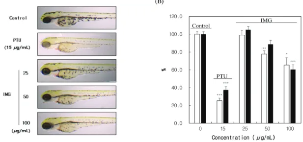
The number of melanin pigmentations on the zebrafish embryos treated with IMG (25, 50, 100 μg/mL) decreased in a dose-dependent manner by visual observation (Figure 2A).
The degree of inhibition of melanin pigmentation of IMG was
significantly weaker than that of PTU (sulfur-containing
tyrosinase inhibitor) used as a positive control group but
significantly higer than that of the untreated group. In order to
quantitatively confirm the amount of melanin, the total zebrafish
extract was used to determine the total content of melanin in
the zebrafish (Figure 2B). IMG decreased the total melanin
(A) (B)
Figure 2. Effects of IMG on melanin synthesis in zebrafish embryos. Synchronized embryos were treated with the indicated concentrations of PTU, IMG was dissolved in DMSO (0.1%) and then added to the embryo medium. (A) The change of phenotype on zebrafish was assessed using a microscope. (B) Total melanin content (white colored bar) and tyrosinase activity (black colored bar) were quantified using a spectro- photometer. Results shown are the mean of three independent experiments ± SD. *p < 0.05, **p
< 0.01, ***p < 0.001 versus control group.
(A) (B)
(C)
O
O
O O
HO
OH OH
OH
Figure 1. Effects of IMG on melanogenesis in B16-F10 melanoma. (A) Cell viability test was determined by MTT method. B16-F10 cells were cultured with 0-200 μg/mL of isomaltol glycoside for 24 h. (B) Melanin content in B16-F10 cells which were cultured with 0 – 200 μg/mL of IMG for 24 h was measured with triplicate experiments. Each value is expressed as the mean ± SD of triplicate determinations. *p < 0.05, **p < 0.01 versus control group. (C) Chemical structure of IMG.
content after 50 and 100 μg/mL treatment by about 20%
compared to the negative control and decreased the zebrafish tyrosinase activity by about 10%. These results were not concentration dependent. The content of melanin pigment was quantitatively measured in the IMG (50, 100 μg/mL)-treated group, which was about 20% lower than the untreated group, but no concentration dependence was observed. The tyrosinase activity in the zebrafish was reduced by about 10% in the IMG (50, 100 μg/mL) group and no concentration dependence was observed. The PTU (100 M) used as a positive control in this experiment reduced the melanin content by 78% and the tyrosinase activity by 64%. At this time, more than 95% of zebrafish survived PTU and IMG treatment (data not shown).
The zebrafish melanogenesis assay was examined three times each.
3.3. Mushroom Tyrosinase Activity of IMG
In cosmetics and pharmaceuticals, tyrosinase inhibitors are the most widely used antifouling agents[19]. Because tyrosinase is produced only in melanocytes, tyrosinase inhibitors are a very specific targeting agent without side effects on the
production of melanin in cells. Thus, the most common goal of whitening active agents is the direct discovery of inhibitors of tyrosinase catalysis[20].
IMG was treated in a mushroom tyrosinase assay system to determine whether the activity of tyrosinase was directly inhibited by IMG (Figure 3). In the untreated group (DMSO), when the tyrosinase activity was regarded as 100%, the kojic acid used as positive control showed a decrease in tyrosinase inhibitory activity in a concentration-dependent manner. However, the IMG treatment group seemed to slightly decrease the tyrosinase activity at the highest treatment concentration of 50 μg/mL, but the inhibition rate of tyrosinase inhibitory activity at that concentration did not deviate from the inhibition rate error range of the untreated group. Thus, it is believed that IMG does not directly inhibit the activity of mushroom tyrosinase. However, due to the structural differences between mushroom tyrosinase and mammlian tyrosinase, further experiments are needed to correlate the inhibitory activity of tyrosinase and IMG.
3.4. Effect of IMG on the Melanogenesis-related Signalling Pathway
MITF expression is one of the most important mechanisms of melanin production[21]. TRP-1 and TRP-2 are tyrosinase- related enzymes that play an important role in melanin synthesis, although their function is not fully understood. The tyrosinase family genes, TRP-1 and TRP-2, are tightly regulated by microphthalmia-associated transcription factor (MITF)[22].
Upregulation of MITF action promotes melanin production by activating tyrosinase and TRP-1, -2 expression, whereas down- regulation of MITF action inhibits the expression of related enzymes and inhibits melanin production. ERK and AKT, which are intracellular signalling enzymes, induce phosphor- ylation of serine-73 and serine-29 of MITF as up- stream regulators of MITF gene and induce down-regulation of MITF action. At this time, phosphorylated p-ERK and p-AKT interfere with the phosphorylation of MITF. Phosphorylated p-MITF does not act on the production of tyrosinase and tyrosinase- related enzymes. In other words, upregulation of p-ERK and p-AKT activity results in degradation of MITF and inhibition of tyrosinase and TRPs expression, thereby reducing melanin
Figure 3. Inhibitory effects on mushroom tyrosinase. Thetyrosinase inhibitory activity was expressed as the absorbance reduction rate of the sample solution and non-added sample solution. Results shown are the mean of three independent experiments ± SD. *p < 0.05 versus non-added sample.
production in melanocytes[23-25]
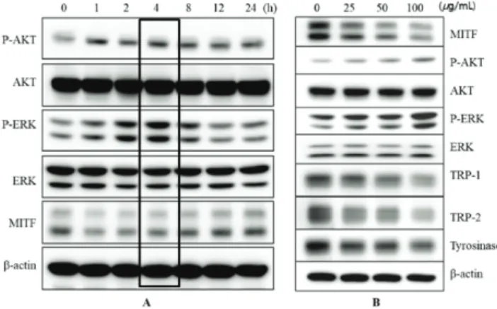
We investigated the effect of IMG on tyrosinase upstream enzymes in order to elucidate the mechanism of melanogenesis inhibition in melanoma. Figure 4A shows the expression pattern of AKTs, ERKs and MITF when treated with 100 μg/mL of IMG in B16-F10 cells. The expression of p-AKT and p-ERK increased with the elapsed time (0, 1, 2, 4, 8, 12, 24 h). These results were most prominent at 4 h after IMG treatment. Figure 4B shows the expression levels of AKTs, ERKs and MITF in cells treated with IMG (25, 50, 100 μg/mL) after 4 h.
Expressions of p-AKT and p-ERK increase and that of MITF decreases in a concentration-dependent manner. In particular, p-ERK clearly shows that the expression level increases with the IMG concentration. The expression of tyrosinase, TRP-1 and TRP-2 in B16-F10 cells was significantly decreased in those cells treated with IMG (25, 50, 100 μg/mL). As a result, increased concentrations of p-ERK and p-AKT predict that it promotes phosphorylation of MITF and interferes with the production of tyrosinase, TRP-1 and -2. Figure 4B shows that IMG increases the concentration of p-ERK and p-AKT and thereby decreases the concentration of MITF. The decrease in the concentration of MITF in B16-F10 cells by IMG is mediated by tyrosinase and TRP-1, -2 in a concentration-dependent manner. In conclusion, IMG phosp- horylates MITF by promoting ERK and AKT phosphorylation upstream of MITF and inhibits the production of downstream enzymes (tyrosinase, TRP-1, -2) that convert tyrosine to dopamine.
3.5. Contents of IMG in Red Ginseng Extract and Roots Various columns and solvents were investigated to enhance the resolution and sensitivity for isomaltol glycoside detection in P. ginseng samples. IMG is an extreme hydrophilic material that is not easily and ideally isolated in ODS columns. Therefore, for the separation of isomaltol glycoside, an NH
2column (NUCLEDUR 100-5 NH
2-RP, Machrey-Nagel) with acetonitrile (70 - 100%, in water) at a flow rate of 1.3 mL/min during 50 min was used in this experiment. The
Figure 4. Effects of IMG on melanogenesis factors. (A) The timedependant (0, 1, 2, 4, 8, 12, 24 h) expression pattern of AKTs, ERKs and MITF when treated with of isomaltol glycoside (100 μ g/mL) in B16-F10 cells. (B) The dose dependent (0, 25, 50, 100 μg/mL) expression pattern of of MITF, AKTs, ERKs, TRPs and tyrosinase when with IMG (4 h) in B16-F10 cells.
Figure 5. Chromatography of IMG (peak at 21-minute retention time) from Red ginseng samples by HPLC. For the separation of IMG, an NH2 column (NUCLEDUR 100-5 NH2-RP, Machrey-Nagel) with acetonitrile (70 - 100%, in water) at a flow rate of 1.3 mL/min during 50 min (UV detection at 285 nm) was used in this experiment. (A): Red ginseng root, (B): Red ginseng extract.