INTRODUCTION
A double outlet right ventricle (DORV) includes a broad spectrum of anatomic variants and associated malformations, and the optimal management remains controversial. The pre- ferred surgical approach for a DORV is an intraventricular tunnel with or without extracardiac procedures such as an arterial switch operation or a right ventricle-to-pulmonary artery conduit formation (Rastelli procedure). Regardless of its preoperative morphology and the type of operation, ob- structive subaortic lesions after biventricular repair of a DO- RV may develop, even in the presence of a sufficient left ven- tricular outflow tract and a non-restrictive ventricular septal defect (VSD). This report presents our 10-yr experience with the surgical management of left ventricular outflow tract ob- struction (LVOTO) after biventricular repair of a DORV.
MATERIALS AND METHODS
Between 1996 and 2006, 15 patients underwent reopera- tion for subaortic stenosis after biventricular repair of a DORV at Seoul National University Children’s Hospital and Sejong General Hospital. Two of the patients underwent biventricu- lar repair elsewhere. The other patients underwent biventricu- lar repair between 1985 and 2000, and 295 patients under- went biventricular repair in the same period. Therefore, the incidence of subaortic stenosis after biventricular repair of a DORV was 4.4% (13/295).
The diagnosis of a DORV and subaortic stenosis after biven- tricular repair of a DORV was based on a combination of pre- operative echocardiographic and angiographic findings as well as surgical inspection. A diagnosis of DORV was made with the application of the ‘50% rule’ that one great artery origi- nate entirely and the other more than 50% from the right ven- tricle (1, 2). The term ‘non-committed VSD’ was used to des-
374
Chang Young Kim1, Woong-Han Kim2, Jae Gun Kwak3, Woo-Sung Jang2, Chang-Ha Lee3, Dong Jin Kim2, Cheong Lim4, and Woo Ik Chang1
Department of Thoracic and Cardiovascular Surgery1, Ilsan Paik Hospital, College of Medicine, Inje University, Goyang; Department of Thoracic and Cardiovascular Surgery2, Seoul National University Children’s Hospital, Seoul; Department of Thoracic and Cardiovascular Surgery3, Sejong General Hospital, Buchon; Department of Thoracic and Cardiovascular Surgery4, Seoul National University Bundang Hospital, Seoul National University College of Medicine, Seongnam, Korea
Address for Correspondence Woong-Han Kim, M.D.
Department of Thoracic and Cardiovascular Surgery, Seoul National University College of Medicine, Seoul National University Children’s Hospital, 101 Daehak-ro, Jongno-gu, Seoul 110-774, Korea Tel : +82.2-2072-3637, Fax : +82.2-3672-3637 E-mail : [email protected]
Surgical Management of Left Ventricular Outflow Tract Obstruction after Biventricular Repair of Double Outlet Right Ventricle
Regardless of the preoperative morphology and the type of operation, left ventricu- lar outflow tract obstruction (LVOTO) after biventricular repair of double outlet right ventricle (DORV) may develop. This report presents our 10-yr experience with sur- gical management of LVOTO after biventricular repair of DORV. Between 1996 and 2006, 15 patients underwent reoperation for subaortic stenosis after biventricular repair of DORV. The mean age at biventricular repair was 23.3±18.3 months (1.1- 64.2). Biventricular repairs included tunnel constructions from the left ventricle to the aorta in 14 cases and an arterial switch operation in one. The mean left ventri- cle-to-aorta peak pressure gradient was 54.0±37.7 mmHg (15-140) after a mean follow-up of 9.5±6.3 yr. We performed extended septoplasty in nine patients and fibromuscular resection in six. There were no early or late mortality. There was one heart block and one aortic valve injury after an extended septoplasty, and two and one after a fibromuscular resection. No patient required reoperation for recurrent subaortic stenosis. The mean pressure gradient was 11.2±11.4 mmHg (0-34) after a mean follow-up of 5.6±2.7 yr. Extended septoplasty is a safe and effective method for the treatment of subaortic stenosis, especially in cases with a long-tunnel shaped LVOTO.
Key Words : DORV; Aortic Stenosis, Subvalvular
Received : 15 January 2009 Accepted : 14 May 2009
ⓒ 2010 The Korean Academy of Medical Sciences.
This is an Open Access article distributed under the terms of the Creative Commons Attribution Non-Commercial License (http://creativecommons.org/licenses/by-nc/3.0) which permits unrestricted non-commercial use, distribution, and reproduction in any medium, provided the original work is properly cited.
cribe a VSD which lies at a distance from both the aortic and pulmonary annulus greater than the aortic diameter (3, 4).
The term ‘DORV-TGA type’ (or Taussig-Bing anomaly) was a DORV with a subpulmonary VSD and without pulmonary stenosis, originating the aorta entirely and more than 50%
of the pulmonary artery from the right ventricle (5, 6).
Based on the surgical techniques used for subaortic steno- sis repair, we divided the patients into two subgroups; the Extended Septoplasty (ES) group (n=9) and the Fibro-Mus- cular Resection (FMR) group (n=6). Preoperative conven- tional angiography was performed in 13 patients (8 in the ES group, 5 in the FMR group). But left-sided angiography was not performed in one of them, in the FMR group, because of floating tissue in the left ventricular outflow tract (LVOT).
Therefore, there were 12 angiography-proven left ventricle- to-aorta peak pressure gradients. Magnetic resonance angiog- raphy or echocardiography were performed in the other 2 patients. In the ES group, the main cause of a reoperation was LVOTO with a peak pressure gradient of more than 50 mmHg except for one patient, who had a peak pressure gra- dient 15 mmHg and required a reoperation for right ventri- cle-to-pulmonary artery conduit stenosis. By contrast, in the FMR group, the main cause of reoperation was not LVOTO, but right ventricle-to-pulmonary artery conduit stenosis (n=
2), severe pulmonary regurgitation combined with a right ventricular outflow tract (RVOT) aneurysm (n=1), a neoaor- ta stenosis after an arterial switch operation (n=1), an ascend- ing aortic aneurysm (n=1) and pacemaker failure (n=1). All patients in the FMR group had a peak pressure gradient less than 30 mmHg. Therefore, the ES group had a significant- ly higher peak pressure gradient than the FMR group (70.8
±35.5 mmHg, n=8 and 20.5±4.9, n=4 respectively, P=
0.048). All but one patient in the ES group had a long-tun- nel shaped LVOTO. On the other hand, all but one in the FMR group had a localized LVOTO.
We reviewed the clinical records including operative reports, pre- and postoperative echocardiographic and angiographic studies retrospectively. Seoul National University Hospital Institutional Review Board (study approval number H-0603- 093-170) approved this study and individual consent for the study was waived due to its retrospective medical record review design.
Patients characteristics
Of the 15 patients, nine were boys and six were girls. Over- all, the mean age at the time of biventricular repair of the DORV was 23.3±18.3 months (ranged from 1.1 to 64.2 months, median age 23.1). The ES group was younger than the FMR group at the time of biventricular repair of the DO- RV; this difference was not significant (21.6±19.2 months and 25.9±18.2 respectively, P=0.864). The initial diagnosis of a DORV was the VSD type (DORV with subaortic VSD) in 3 patients, the Fallot type (DORV with subaortic or dou-
bly committed VSD and pulmonary outflow stenosis) in 8, the non-committed VSD type in 3 and TGA type in 1 patient.
Five patients in the ES group and four in the FMR group had an initial subaortic stenosis including a restrictive VSD or subaortic conal or septal hypertrophic lesions. One in the ES group had an anomalous tricuspid chordae attachment to the conal septum. The patient with the Taussig-Bing anoma- ly had a single coronary artery pattern from the right coronary sinus. One in the FMR group had an atrioventricular septal defect (AVSD). Two in the FMR group underwent modified Blalock-Taussig shunt and no patient underwent pulmonary artery banding before the biventricular repair.
Initial biventricular repair of the DORV
Fourteen patients underwent a tunnel construction from the left ventricle to the aorta and one patient with the Taus- sig-Bing anomaly in the FMR group underwent an arterial switch operation with a patch committing the left ventricle to the pulmonary artery (neo-aorta). Four patients in the ES group and two in the FMR group required anterior or antero- caudal VSD enlargement, resection of the subaortic conal or septal hypertrophy or both for the treatment of initial subaor- tic stenosis. Concomitant procedures were as follows: tricus- pid valve chordae transfer who had an anomalous tricuspid chordae attachment to the conal septum (n=1), mitral annu- loplasty (n=1) in the ES group and AVSD repair (n=1), tri- cuspid valvuloplasty (n=1) in the FMR group.
No patient had a LV-to-Aorta peak pressure gradient (DP) more than 20 mmHg by echocardiography at hospital dis- charge. Three patients required a permanent pacemaker im- plantation due to heart block in the FMR group. There were two reoperations after the biventricular repair: repair of a resid- ual VSD in the ES group (n=1), and mitral valve replacement in a patient with AVSD in the FMR group (n=1).
Surgical techniques
Fibromuscular resection was usually performed through transarterial approach and transatrial when needed. It depend- ed on surgeon’s preference whether extended septoplsty was performed. Extended septoplasty was first described at 1990 in patients with subaortic stenosis after VSD repair (7). We performed an extended septoplasty as follows (Figs. 1, 2). A longitudinal incision was made at the previous patch, and extended toward the apex, beyond the patch, into the apical interventricular septum. It was extended into the conal sep- tum avoiding direct injury to the aortic valve. The new patch was then trimmed along the extended incision, and inserted to secure a sufficient pathway in the LVOT.
Statistical analysis
Statistical analysis was performed using SPSS 12.0K soft-
ware (SPSS, Inc, Chicago, IL, USA). Continuous variables were compared using the Mann-Whitney U test and discrete vari- ables were analyzed using Fisher’s exact test. All data are ex- pressed as mean±standard deviation with ranges. The P values less than 0.05 were considered significant.
RESULTS
There were no early or late deaths and there was complete follow-up for all patients. Only one patient in ES group had a postoperative pressure gradient >20 mmHg (from 70 mmHg
by angiography to 25 by echocardiography) at hospital dis- charge. The overall time for patient follow-up ranged from 16.5 months to 10.9 yr with a mean follow-up of 5.6±2.7 yr. The overall recent peak pressure gradient was 11.2±11.4 mmHg (13.9±11.2 mmHg in ES group, and 7.2±11.3 in FMR group). Three patients had a newly developed peak pressure gradients >20 mmHg by recent echocardiographic follow-up (25 mmHg and 34 in ES group, 25 in FMR group respectively).
According to the intraoperative findings, all patients in the FMR group had LVOTO mainly caused by subarterial conal muscle hypertrophy with a non-restrictive VSD inlet. And eight patients had subarterial muscle hypertrophy or residu- al conal septum, who underwent simple fibromuscular resec- tion coincidentally, and 4 had a restirictive VSD inlet in the ES group.
The concomitant procedures were as follows: aortic valvu- loplasty (n=3:2 planned and 1 unexpected), tricuspid annu- loplasty (n=1), pulmonary valve replacement (n=3), right ventricular outflow tract widening (n=2) and main pulmo- nary artery angioplasty (n=1) in the ES group, and unexpect- ed aortic valvuloplasty (n=1), tricuspid valvuloplasty (n=1), pulmonary valve replacement (n=1), and a left and right pul- monary artery angioplasty (n=1) in FMR group.
The aortic valve was injured in one patient in each group during the procedures associated with the relief of subaortic stenosis. Primary repairs were done in these patients, and addi- tional pericardial patch repair of perforated site was perform- ed at 17 months after the operation for LVOTO in one pati- ent of FMR group. This patient showed moderate aortic regur- gitation by recent echocardiography. The other patient show- ed mild aortic regurgitation at 56 months postoperative echo- cardiography follow-up. Heart block, which required the im- plantation of a permanent pacemaker, developed in one pati- ent in the ES group and two in FMR group. No patient re-
Fig. 1. Illustrations showing the extended septoplasty. (A) A right ventriculotomy (dotted curve) is made in the right ventricular outflow tract.
(B) A longitudinal septal incision is made at the previous patch, and extended toward the apex, beyond the previous patch, into the inter- ventricular septum, and toward the aortic valve, into the conal septum avoiding direct injury to the aortic valve. (C) The new patch is then trimmed along the extended septal incision, and inserted to secure a redundant pathway in the left ventricular outflow tract.
A B C
aorta
aorta aorta
Fig. 2. Photograph, taken after the extended septal incision and some interrupted sutures for anchoring a new septal patch, show- ing the previous patch (arrow) and the septal incision (asterisk).
*
quired reoperation for recurrence of a LVOTO.
Table 1 includes preoperative, biventricular repair, reoper- ative and follow-up data which compared between the two subgroups.
DISCUSSION
Subaortic stenosis can occur after surgical repair of several congenital heart defects without an initial LVOTO such as coarctation of the aorta, atrioventricular septal defect, DORV, simple VSD, transposition of the great arteries with a VSD and pulmonary stenosis, an interrupted aortic arch, and tetral- ogy of Fallot (8, 9). In the case of a DORV, especially with a non-committed ventricular septal defect, new surgical tech- niques have been reported to resolve the problem of ventric- ular outflow tract stenosis (4, 10). Nevertheless, an intraven- tricular repair may form a long and akinetic area in the LVOT.
Subaortic stenosis, such as a restrictive ventricular septal defect and a subaortic conal or septal hypertrophy, which may develop after pulmonary artery banding, may be present before biventricular repair of the DORV. Subaortic stenosis is an inde- pendent risk factor for LVOTO after biventricular repair of a DORV (11) and it is important to relieve the subaortic steno- sis completely during the biventricular repair by means of VSD enlargement and resection of the conal septum (12). How- ever, subaortic stenosis after biventricular repair of a DORV
may develop in 3.5-5.5% of cases, even in the presence of a sufficient LVOT and a non-restrictive VSD (11, 13, 14).
Many possible factors can explain the development of a LVOTO after biventricular repair of a DORV. A subaortic
*calculated by means of echocardiography; �measured by angiography.
ES, extended septoplasty; FMR, fibro-muscular resection; DORV, double outlet right ventricle; VSD, ventricular septal defect; TB, Taussig-Bing; nc-VSD, non-committed ventricular septal defect; PS, pulmonary stenosis; ASO, arterial switch operation; DP, left ventricle-to-aorta peak pressure gradient; RV- PA, right ventricle-to-pulmonary artery; LVOTO, left ventricular outflow tract obstruction; NYHA Fc, New York Heart Association functional classification.
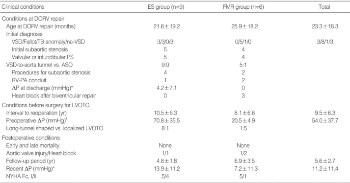
Clinical conditions ES group (n=9) FMR group (n=6) Total
Conditions at DORV repair
Age at DORV repair (months) 21.6±19.2 25.9±18.2 23.3±18.3
Initial diagnosis
VSD/Fallot/TB anomaly/nc-VSD 3/3/0/3 0/5/1/0 3/8/1/3
Initial subaortic stenosis 5 4
Valvular or infundibular PS 5 4
VSD-to-aorta tunnel vs. ASO 9:0 5:1
Procedures for subaortic stenosis 4 2
RV-PA conduit 1 2
DP at discharge (mmHg)* 4.2±7.1 0
Heart block after biventricular repair 0 3
Conditions before surgery for LVOTO
Interval to reoperation (yr) 10.5±6.3 8.1±6.6 9.5±6.3
Preoperative DP (mmHg)� 70.8±35.5 20.5±4.9 54.0±37.7
Long-tunnel shaped vs. localized LVOTO 8:1 1:5
Postoperative conditions
Early and late mortality None None
Aortic valve injury/Heart block 1/1 1/2
Follow-up period (yr) 4.8±1.8 6.9±3.5 5.6±2.7
Recent DP (mmHg)* 13.9±11.2 7.2±11.3 11.2±11.4
NYHA Fc, I/II 5/4 5/1
Table 1. Summary of data between the two groups
Fig. 3. Angiogram showing the long tunnel-shaped left ventricular outflow tract obstruction after biventricular repair of DORV.
septal or conal muscle hypertrophy or fibrous tissue deposits caused by turbulent flow and the sinuous shape of the tun- nelization (11); the decrease in the effective VSD size caused by the baffle itself or fibrous tissue around the VSD or the baffle (15); the diminution of the VSD orifice (11, 15). In addi- tion, growth of the heart without concomitant increases in size of the VSD and baffle (11) and retraction or kinking of the baffle patch (15). Rychik and coworkers suggested ‘geo- metric changes’ (16); the left ventricle undergoes geometric changes after a Rastelli operation or intraventricular repair for a DORV and subaortic obstruction may develop in pati- ents with the greatest degree of ventricular contraction and VSD diminution early after biventricular repair.
Subaortic stenosis includes a broad spectrum of pathology from a subaortic membrane to a tunnel-like obstruction, and surgical procedures are chosen based on the anatomical type of subaortic stenosis. The surgical management of the LVOTO remains a surgical challenge in view of the goal of preserva- tion of the aortic valve and the conduction system, ensuring complete relief of the LVOTO, and avoiding the recurrence of the LVOTO. In this study, all but one patient in the ES group had a long-tunnel shaped LVOTO (Fig. 3). On the other hand, all but one in the FMR group had a localized discrete LVOTO. Since the peak pressure gradient did not always corre- late with the severity and morphologic features of the LVOTO, it is not always an indication for a certain type of surgery. Kalfa and coworkers (9) reported five recurrent LVOTOs after 33 surgical procedures for secondary (acquired) subaortic steno- sis, after surgery for congenital heart anomalies. Belli and col- leagues (11) reported that none of the six patients who under- went extended septoplasty required further reoperation, but four of nine patients who underwent fibromuscular resection or septal patch enlargement required further reoperation, and they suggested (3) a ‘two stage management’: the biventric- ular repair and reoperation for progressive subaortic stenosis in patients with a DORV non-committed VSD type. Consid- ering the recurrence of LVOTO, extended septoplasty may be considered the preferred technique especially in cases with a long-tunnel shaped LVOTO, even if the left ventricle-to- aorta peak pressure gradient is not very high.
In this study, the fibromuscular resection group had a lower peak pressure gradient than the extended septoplasty group, because the fibromuscular resection group required closer observation so that early detection and surgery for the other causes could be performed before the LVOTO progressed.
Concerning the surgical relief of the LVOTO, the fibro- muscular resection and extended septoplasty are alternatives to consider on a case-by-case basis. In this study, the extend- ed septoplasty reduced the peak pressure gradient effective- ly from 70.8±35.5 mmHg to 13.9±11.2 especially in the patients with a long-tunnel shaped LVOTO. There was no difference in the early and late mortality, current functional status and morbidity, directly associated with the procedures, such as reoperation for recurrence and direct injury to the aor-
tic valve or conduction systems. The fibromuscular resection is useful in patients with a mild or discrete subaortic steno- sis especially combined with other sophisticated surgical prob- lems because it is relatively simple, less time-consuming and the operator can focus on other complex procedures. In this study, surgical results for the fibromuscular resection were satisfactory considering the effective reduction of subaortic stenosis, no death, easily combined with other sophisticated procedures and no recurrence of the LVOTO.
There were some limitations in this study. At first, the over- all follow-up period after surgery for the relief of subaortic stenosis (5.6±2.7 yr) is somewhat shorter than the overall interval from biventricular repair to reoperation (9.5±6.3 yr). Therefore, a further follow-up is needed, especially in three patients with newly developed peak pressure gradient
>20 mmHg. In addition, the main reason for reoperation was LVOTO with a high peak pressure gradient in the extended septoplasty group, however, there were other causes than a LVOTO in the fibromuscular resection group. Considering this baseline difference, it is difficult to compare the outcomes of the two groups directly. Finally, this study was not a con- trolled and randomized trial but a retrospective medical record review study. Because the initial biventricular repairs were performed between 1985 and 2000, there were some con- founding factors, such as technical improvements in the biven- tricular repair and understanding of LVOTO developmental mechanisms.
In conclusions, for surgical relief of subaortic stenosis after biventricular repairs of a DORV, fibromuscular resection and extended septoplasty are alternative options for surgery. Fibro- muscular resection shows satisfactory results in cases with mild, discrete subaortic stenosis especially when combined with other surgical problems, and extended septoplasty is a safe and effective method and ensures complete relief of subaortic stenosis. The extended septoplasty is an excellent choice for surgery, especially in cases with a long-tunnel shaped LVOTO.
ACKNOWLEDGMENT
The authors are grateful to the Medical Research Collabo- rating Center of Seoul National University Hospital for pro- viding a statistical review of this study.
REFERENCES
1. Stark J. Double-Outlet Ventricles. In: Stark J, de Leval M, editors, Surgery for Congenital Heart Defects, 2nd ed. Philadelphia: WB Saunders 1994; 437-46.
2. Stellin G, Ho SY, Anderson RH, Zuberbuhler JR, Siewers RD. The surgical anatomy of double-outlet right ventricle with concordant atrioventricular connection and non-committed ventricular septal defect. J Thorac Cardiovasc Surg 1991; 102: 849-55.
3. Belli E, Serraf A, Lacour-Gayet F, Hubler M, Zoghby J, Houyel L, Planche C. Double-outlet right ventricle with non-committed ventric- ular septal defect. Eur J Cardiothorac Surg 1999; 15: 747-52.
4. Lacour-Gayet F, Haun C, Ntalakoura K, Belli E, Houyel L, Marcsek P, Wagner F, Weil J. Biventricular repair of double outlet right ven- tricle with non-committed ventricular septal defect (VSD) by VSD rerouting to the pulmonary artery and arterial switch. Eur J Cardio- thorac Surg 2002; 21: 1042-8.
5. Stellin G, Zuberbuhler JR, Anderson RH, Siewers RD. The surgical anatomy of the Taussig-Bing malformation. J Thorac Cardiovasc Surg 1987; 93: 560-9.
6. Wetter J, Belli E, Sinzobahamvya N, Blaschzok HC, Brecher AM, Urban AE. Transposition of the great arteries associated with ven- tricular septal defect: surgical results and long-term outcome. Eur J Cardiothorac Surg 2001; 20: 816-23.
7. DeLeon SY, Ilbawi MN, Arcilla RA, Thilenius OG, Quinones JA, Duffy EC, Sulayman RF. Transatrial relief of diffuse subaortic steno- sis after ventricular septal defect closure. Ann Thorac Surg 1990; 49:
429-34.
8. Jahangiri M, Nicholson IA, del Nido PJ, Mayer JE, Jonas RA. Sur- gical management of complex and tunnel-like subaortic stenosis. Eur J Cardiothorac Surg 2000; 17: 637-42.
9. Kalfa D, Ghez O, Kreitmann B, Metras D. Secondary subaortic steno- sis in heart defects without any initial subaortic obstruction: a mul- tifactorial postoperative event. Eur J Cardiothorac Surg 2007; 32:
582-7.
10. Barbero-Marcial M, Tanamati C, Atik E, Ebaid M. Intraventricular repair of double-outlet right ventricle with noncommitted ventricu- lar septal defect: advantages of multiple patches. J Thorac Cardio- vasc Surg 1999; 118: 1056-67.
11. Belli E, Serraf A, Lacour-Gayet F, Inamo J, Houyel L, Planche C.
Surgical treatment of subaortic stenosis after biventricular repair of double-outlet right ventricle. J Thorac Cardiovasc Surg 1996; 112:
1570-80.
12. Lacour-Gayet F. Biventricular repair of double outlet right ventricle with noncommitted ventricular septal defect. Semin Thorac Cardio- vasc Surg Pediatr Card Surg Annu 2002; 5: 163-72.
13. Luber JM, Castaneda AR, Lang P, Norwood WI. Repair of double- outlet right ventricle: early and late results. Circulation 1983; 68 (3 pt 2): II144-7.
14. Kirklin JW, Pacifico AD, Blackstone EH, Kirklin JK, Bargeron LM Jr. Current risks and protocols for operations for double-outlet right ventricle. Derivation from an 18 year experience. J Thorac Cardio- vasc Surg 1986; 92: 913-30.
15. Rocchini AP, Rosenthal A, Castaneda AR, Keane JF, Jeresaty R.
Subaortic obstruction after the use of an intracardiac baffle to tun- nel the left ventricle to the aorta. Circulation 1976; 54: 957-60.
16. Rychik J, Jacobs ML, Norwood WI. Early changes in ventricular geometry and ventricular septal defect size following Rastelli oper- ation or intraventricular baffle repair for conotruncal anomaly. A cause for development of subaortic stenosis. Circulation 1994; 90 (5 Pt 2): II13-9.