• 검색 결과가 없습니다.

Antifungal Effect of Phenyllactic Acid Produced by Lactobacillus casei Isolated from Button Mushroom

N/A
N/A
Protected

Academic year: 2021

Share "Antifungal Effect of Phenyllactic Acid Produced by Lactobacillus casei Isolated from Button Mushroom"

Copied!
6
0
0

로드 중.... (전체 텍스트 보기)

전체 글

(1)

162 This is an Open-Access article distributed under the terms of the Creative Commons Attribution Non-Commercial License (http://

creativecommons.org/licenses/by-nc/3.0) which permits unrestricted non-commercial use, distribution, and reproduction in any medium, provided the original work is properly cited.

J. Mushrooms 2016 December, 14(4): 162-167 http://dx.doi.org/10.14480/JM.2016.14.4. 162 Print ISSN 1738-0294, Online ISSN 2288-8853

© The Korean Society of Mushroom Science

*Corresponding author E-mail : [email protected]

Tel : +82-42-821-6733, Fax : +82-42-823-9241 Received December 6, 2016

Revised December 16, 2016 Accepted December 21, 2016

Antifungal Effect of Phenyllactic Acid Produced by Lactobacillus casei Isolated from Button Mushroom

Jeoung Ah Yoo

1

, Chan-Jung Lee

2

, Yong-Gyun Kim

3

, Byung-Eui Lee

4

, and Min-Ho Yoon

1,

*

1

Department of Bio-Environmental Chemistry, College of Agriculture and Life Sciences, Chungnam National University, Daejeon 34134, Korea

2

Mushroom Research Division, National Institute of Horticultural & Herbal Science, RDA, Eumseong, Chungbuk 27712, Korea

3

Crop Research Division, Chungcheongnam-do Agricultural Research & Extension Services, Yesan 32418, Korea

4

Department of Chemistry, College of Natural Sciences, Soonchunghyang University, Asan 31538, Korea

ABSTRACT: Lactic acid bacteria (LAB) producing phenyllactic acid (PLA), which is known as antimicrobial compound, was

isolated from button mushroom bed and the isolated LAB was identified to Lactobacillus casei by 16 rRNA gene sequence analysis. Cell-free supernatant (CFS) from L. casei was assessed for both the capability to produce the antimicrobial compound PLA and the antifungal activity against three fungal pathogens ( Rhizoctonia solani, Botrytis cinerea, and Collectotricum aculatum).

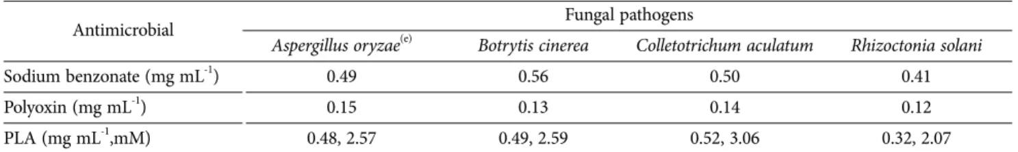
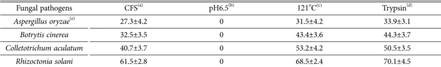
PLA concentration was investigated to be 3.23 mM in CFS when L. casei was grown in MRS broth containing 5 mM phenylpyruvic acid as precursor for 16 h. Antifungal activity demonstrated that all fungal pathogens were sensitive to 5% CFS (v/v) of L. casei with average growth inhibitions ranging from 34.58% to 65.15% (p < 0.005), in which R. solani was the most sensitive to 65.15% and followed by C. aculatum, and B. cinerea. The minimum inhibitory concentration (MIC) for commercial PLA was also investigated to show the same trend in the range of 0.35 mg mL-1 (2.11 mM) to 0.7 mg mL-1 (4.21 mM) at pH 4.0. The inhibition ability of CFS against the pathogens were not affected by the heating or protease treatment. However, pH modification in CFS to 6.5 resulted in an extreme reduction in their antifungal activity. These results may indicate that antifungal activities in CFS was caused by acidic compounds like PLA or organic acids rather than protein or peptide molecules.

KEYWORDS: Phenyllactic acid, Lactobacillus casei, Antifungal effect, Fermentation

Introduction

3-Phenyllactic acid (PLA), which is known as antimicrobial compound can be synthesized through the reduction of phenylpyruvic acid (PPA) by lactate dehydrogenase of lactic acid bacteria (LAB) and can be produced by many LAB (Lavermicocca et al., 2000).

The PLA was characterized having a broad inhibitory activity against yeast and bacteria as well as fungi

(Dieuleveux and Gueguen, 1998; Dieuleveux et al.,

1998-). Valerio et al. (2004) reported that the organic

acids such as PLA could be produced by a wide range

of LAB species but their production is strain dependent,

and presumed that the behavior of the antifungal activity

was positively related to the PLA content in the LAB

culture filtrate. Although the production of PLA varied

greatly based on the strains and species, it was known

that PLA can be produced by a wide range of LAB

species, such as Lactobacillus, Enterococcus, Weissella,

and Leuconostoc (Valerio et al., 2004). PLA was found

as metabolic by-products in LAB strains through the

amino acids degradation of phenylalanine and tyrosine,

respectively, by which these amino acids were

transaminated to 2-keto-carboxylic acids such as

phenylpyruvic acid (PPA) and 4-hydroxyphenypyruvic

acid (HPPA) (Yvon et al., 1997), and then the ketoacids

further reduced to the 2-hydroxy carboxylic acids (PLA)

(Li et al., 2007; Mu et al., 2010; Vermeulen et al.,

2006). The supplement of the above amino acids and

ketoacids in the initial culture broth for LAB

(2)

fermentation could effectively increase the corresponding 2-hydroxy acids (PLA) production, and the supplement of ketoacids was more significant than that of the original amino acids to improve the corresponding 2- hydroxy acids biosynthesis (Li et al., 2007; Mu et al., 2010).

Furthermore, PLA is an antimicrobial compound with a wide activity spectrum against some yeast such as Candida pulcherrima and Rhodotorula mucilaginosa (Schwenninger et al., 2008) and molds including some mycotoxigenic species such as Aspergillus ochraceus, and Penicillium citrinum (Valerio et al., 2004). In addition, PLA has been found to inhibit a range of Gram-positive and Gram-negative bacteria such as Staphylococcus aureus, Listeria monocytogenes, Enterococcus faecalis, and Klebsiella oxycota (Lavermicocca et al., 2000; Valerio et al., 2004). Recently, several LAB have been screened for their antifungal potential and their ability to produce PLA (Gerez et al., 2013; Ryan et al., 2011; Yoo et al, 2016), but additional studies are required in this field due to the wide diversity of both LAB and food spoilage molds. Therefore, the aim of this work was to isolate the novel LAB strains from button mushroom bed and to assess the antifungal activity of the isolated LAB against three fungal pathogens (R. solani, B. cinerea, and C. aculatum) and then to study their capability to produce the antimicrobial compound 3-phenyllactic acid and its relationship with the antifungal activity of the LAB.

Materials and methods

Microorganisms and culture media

Lactic acid bacteria (LAB) producing phenyllactic acid (PLA) were isolated from button mushroom bed in Buyeo-Gun, Chungchugnam-do province using deMan–

Rogosa–Sharpe (MRS, Difco, Detroit, USA) broth containing 5 mM phenylpyruvic acid (PPA, Sigma Co, USA). The isolated LAB was grown in MRS broth at 37

o

C for 24 h for preparation of the seed culture. A 2

% (v/v) inoculum of the seed was aseptically added to 5 mL of MRS for fermentation experiment. In order to evaluate the effect of PPA as precursor on PLA production, PPA were supplemented in the initial MRS broth with various concentrations. The fermentation was carried out in a 10 mL test tube at 37

o

C for 36 h without shaking. R. solani, B. cinerea, and C.

aculatumas fungal pathogens and A. oryzae as non-

pathogenic control were offered from Chungcheongnam- Do Agricultural Research & Extension Services and cultivated with potato dextrose broth (Difco, USA).

Identification of LAB

The partial sequencing of 16S rRNA for the LAB strain was done with the help of DNA sequencing service, SOLGENT, Daejeon, South Korea using universal primers, 27F (5’-AGAGTTTGATCCTGGCTCAG -3’) and 1492R (5’-GGTTACCTTGTTACGACTT -3’). The online program BLAST was used in identifying the related sequences with known taxonomic information available at the databank of NCBI (http://www.ncbi.nlm.nih.gov/BLAST).

A Phylogenetic tree was constructed using CLUSTAL X program (Thompson et al., 1997), which involved sequence alignment by neighbor joining method (Saitou and Nei, 1987) and maximum parsimony using the MEGA4 program (Kumar et al., 2001). Grouping of sequences was based on confidence values obtained by bootstrap analysis of 1,000 replicates. Gaps were edited in the BioEdit program and evolutionary distances were calculated using Kimura two parameter model.

Reference sequences were retrieved from GenBank under the accession numbers indicated in the trees.

Preparation of cell-free supernatants (CFS)

LAB colonies were precultured for 18 h at 37

o

C in 10 mL of MRS broth. An aliquot (200 µL) of the preculture was then inoculated into fresh sterile MRS broth (20 mL) and allowed to grow at 37

o

C without shaking for 72 h.

Cell free supernatants (CFS) were recovered by centrifugation (7200×g, 10 min), and the CFS filtered with 0.45-µm Millipore membrane were used for identifying PLA and screening their antifungal activity.

Identification of PLA

PLA in CFS was identified by HPLC as described by Valerio et al. (2004) and Gerez et al. (2010) with minor modifications. CFS were adjusted to pH 2.0 with 10 M formic acid and extracted three times with 20 mL of ethyl acetate. The extracts were dried using Na

2

SO

4

and concentrated in a rotary evaporator (Büchi model R210/

215, Flawil, Switzerland). The dried residues were

reconstituted with 5 mL of 2.5 mM H

3

PO

4

and the PLA

component filtered with 0.45 µm pore-size membrane

was analyzed in an HPLC system (Agilent, 1260

infinity, USA) fitted with an CAPCELL PAK C18

column (4.6×250 mm, 5 μm, Shiseido Co, Japan) at

(3)

30

o

C, using (A) 0.5% H

3

PO

4

(v/v) and (B) 0.5% H

3

PO

4

- CH

3

CN (V/V) as the mobile phases and a UV-visible detector set at 210 nm. Linear gradient elution was used with solvent A and solvent B at 1 mL min

-1

and A/B ratios of 80:20, 80:20, 0:100 and 0:100 with run times of 0, 12, 13, and 15 min, respectively (Li et al., 2007;

Mu et al., 2010). Commercial PLA was used as reference (retention time = 11.66 min).

Determination of antifungal activity

Antifungal activity was tested with cell-free supernatants of LAB by modification method of Wang et al. (2012). Briefly, CFS was mixed with the sterilized PDA (pH 4) to achieve a final concentration of 5-10%

for R. solani, B. cinerea, C. aculatum (v/v) and the mixture was poured into Petri dishes. Resulting media was centrally inoculated with 5 µL of the previously prepared fungal pathogen spore suspensions and incubated at 25°C. The sterile MRS broth instead of CFS in the same proportions was also used for control plates. During 8-days incubation period to grow the pathogens onto the plate, the area of mycelial growth in both treated and control plates were determined from the mean perpendicular diameter measurements assuming a circular growth. The percentage of growth inhibition (I) was calculated as I = 100(A

C

-A

T

)/A

C

, where A

T

are the area of mycelial growth in both treated and control plates, respectively. CFS were also subjected to heating (121°C, 15 min) treatment to evaluate the possible antifungal compounds present in each CFS besides PLA and the remaining antifungal activity of treated CFS was further assessed as described above.

Statistical analysis

Three independent replicates of each experiment were performed, and their results were expressed as mean values ± standard deviation. The data were subjected to analysis of variance (ANOVA) using SAS package (SAS, 1999). The Duncan’s Multiple Range Test (DMRT) was applied to test the significance of treatment means at P ≤ 0.05.

Results and Discussion

Isolation and identification of PLA producing LAB A PLA producing strain was selected by assessing the mycelial growth inhibition of R. solani on MRA media with culture filtrates of lactic acid bacteria isolated from

button mushroom bed and by finally confirming PLA in the culture filtrate through HPLC analysis. The selected strain showed a marked inhibition for mycelial growth of fungal pathogen and the perpendicular diameter of a circular growth was similar to commercial PLA used as control after 7 days of incubation (Fig. 1). According to 16S rRNA sequence analysis, the strain was identified as Lactobacillus casei. Comparison of the 16S rRNA sequence among available strains of Lactobacillus species showed high homology (> 99%) to L. casei ATCC393

T

. Neighbor-joining method was employed to construct the phylogenetic tree which illustrates the relationships of 16S r RNA sequence of strain and other Lactobacillus species (Fig 2).

Fig. 1. Growth of fungal pathogens in PDA after 10 days at 25

o

C in presence of CFS from Lactobacillus casei fermentation in MRS broth : (A) Rhizoctonia solani (PDA+500 mgL

-1

PLA), (B) Rhizoctonia solani (PDA+5 % CFS), (C) Colletotrichum aculatum (PDA+5 % CFS), (D) Botrytis cinerea (PDA+5 % CFS).

Fig. 2. Phylogenetic tree based on 16S rRNA gene sequences,

showing the position of Lactobacillus casei strain with respect

to related species. The scale bar indicates 0.005 substitutions

per nucleotide position and accession numbers are given in

parenthesis.

(4)

Cell growth

L. casei fermentation was performed at 37°C with MRS broth without PPA to assess PLA production and pH change. Growth and PLA production curves for L. casei are shown in Fig. 3. Cell growth reached to stationary phase after 12 h and PLA production progressively increased thereafter by the end of this stage. The highest production was reached to 0.58 mM after 36 h, in which pH was changed to lower the initial pH 6.5 to pH 4.0, indicating that the acidification of the media was directly related to PLA production by LAB. A similar results were described by Vermeulen et al. (2006) who reported PLA formation of L. sanfranciscensis DSM20451 continues to process by stationary phase cells. Valerio et al. (2004) also reported the production of PLA in L. plantarum ITM21B was found in synthetic media with a maximum production of 0.165 mM after 72 h. Recently, the growth conditions on LAB fermentations has become an actual topic in recent studies to achieve a maximum production of a given metabolite with antifungal properties such as PLA (Mu et al., 2012; Rodríguez et al., 2012).

Inhibitory effect of CFS against fungal pathogens Antifungal activity of CFS from L. casei demonstrated

that all fungal pathogens were sensitive to 5% CFS (v/

v) with average growth inhibitions ranging from 34.58%

to 65.15% (p < 0.005), in which R. solani was the most sensitive to 65.15% and followed by C. aculatum and B.

cinerea, (Table 1). The apparent results were also confirmed in further MIC experiments with commercial PLA and chemical antimicrobial agents. The fungal pathogens showed almost the similar sensitivities to pure PLA by following in the order R. solani, B. cinerea, C.

aculatum, and A. oryzae with MIC values in the range of 0.32 mg mL

-1

(2.07 mM) to 0.52 mg mL

-1

(3.06 mM) at pH 4.0 (Table 2). The obtained MIC values are compared to the result by Ryan et al. (2011) reporting an MIC for PLA of 15 mM against Aspergillus fumigatus J9. However, sodium benzoate and sodium propionate used as antifungal agents appeared to be more effective than PLA for the fungal pathogens.

Although PLA showed the least inhibition effect on mold growth, it can be well worth enough to natural and safe food preservatives (Prema et al., 2010; Ryan et al., 2011). Furthermore, CFS from L. casei was further subjected to heating (121

o

C, 20 min) and pH modification (6.5) in order to assess the characteristics of the antifungal compounds besides PLA. The inhibition characteristics of CFS against pathogens were not affected by the heating or protease treatment. However, pH modification in CFS to 6.5 caused an extreme reduction in their antifungal activity (Table 2). These results may indicate that antifungal activities in CFS are very likely to be lost by acidic compounds neutralizing pH of the media rather than proteins or peptides molecules being able to suffer by thermal denaturation.

It is known that the antimicrobial activity of the organic acids on molds and bacteria is pH-dependent and especially a maximum inhibition activity can be maintained at low pH values favoring the undissociated state of the acid molecule (Schillinger and Villareal, 2010). Similar results were obtained by Wang et al.

Fig. 3. Cell growth, pH variation and PLA production by Lactobacillus casei in MRA broth. All the fermentations were developed at 37

o

C for 72 h in MRA broth without the precursors.

Table 1. Minimal inhibitory concentrations of commercial phenyllactic acid (PLA) in the studied fungal pathogens

Antimicrobial Fungal pathogens

Aspergillus oryzae

(e)

Botrytis cinerea Colletotrichum aculatum Rhizoctonia solani

Sodium benzonate (mg mL

-1

) 0.49 0.56 0.50 0.41

Polyoxin (mg mL

-1

) 0.15 0.13 0.14 0.12

PLA (mg mL

-1

,mM) 0.48, 2.57 0.49, 2.59 0.52, 3.06 0.32, 2.07

*Minimal inhibitory concentrations were determined at pH 4.0 and 25

o

C in PDA.

(e): non-pathogenic fungi as control.

(5)

(2012) reported on the antifungal activity of CFS from L. plantarum IMAU10014 against B. cinerea, Glomerella cingulate, Phytophthora drechsleri, Penicillium citrinum, P. digitatum, and Fusarium oxysporum. Furthermore, growth inhibition properties in CFS also can be caused by synergism with other acidic compounds from LAB metabolism such as 4-hydroxyphenyllactic acid (HPLA) and organic acids (Hladíková et al., 2012; Schillinger and Villareal, 2010). In this study even though authors have not identified the existence of HPLA in CFS, conclusively, the inhibition effect was due to acidic metabolites in fermentation broth and PLA in CFS was concerned in the inhibition activity against fungal pathogens.

Conclusions

A lactic acid bacteria (LAB) producing PLA, which is known as antimicrobial compound was isolated from button mushroom bed and the isolated LAB was identified to L. casei by 16 rRNA gene sequence analysis. Cell-free supernatant (CFS) from L. casei was confirmed to possess both the capability to produce the antimicrobial compound PLA and the antifungal activity against three fungal pathogens (Rhizoctonia solani, Botrytis cinerea, and Collectotricum aculatum). L. casei showed 65% of conversion ratio by converting 3.23mM PLA from 5mM PPA of precursor for 16 h fermentation.Antifungal activity demonstrated that all fungal pathogens were sensitive to 5% CFS (v/v) of L.

casei with average growth inhibitions ranging from 27.32% to 69.05% (p < 0.005), in which R. solani was the most sensitive to 69.05%. The minimum inhibitory concentration (MIC) for commercial PLA also appeared the same trend with the result of growth inhibition in the range of 0.35 mg mL

-1

(2.11 mM) to 0.7 mg mL

-1

(4.21 mM) at pH 4.0. The inhibition ability of CFS

against the pathogens were not affected by the heating or protease treatment. However, pH modification in CFS to 6.5 caused an extreme reduction in their antifungal activity. These results may indicate that antifungal activities in CFS were caused by acidic compounds like PLA or organic acids rather than proteins or peptides molecules.

References

Dieuleveux V, Lemarinier S, Gueguen M. 1998.

Antimicrobial spectrum and target site of D-3-phenyllactic acid. Int J Food Microbiol. 40:177-183.

Dieuleveux V, Gueguen M. 1998. Antimicrobial effects of D-3-phenyllactic acid on Listeria monocytogenesin TSB- YE medium, milk, and cheese. J Food Prot. 61:1281- 1285.

Gerez CL, Torres MJ, Font de Valdez G, Rollán G. 2013.

Control of spoilage fungi by lactic acid bacteria. Biol Control. 64:231-237.

Hladikova Z, Smetankova J, Greif G, Greifkova M. 2012.

Antimicrobial activity of selected lactic acid cocci and production of organic acids. Acta Chim Slov. 5:80-85.

Kumar S, Tamura K, Jakobsen IB, Nei M. 2001. MEGA2:

molecular evolutionary genetics analysis software.

Bioinformatics. 17: 1244-1245.

Lavermicocca P, Valerio F, Evidente A, Lazzaroni S, Corsetti A, Gobbetti M. 2000. Purification and characterization of novel antifungal compounds from the sourdough Lactobacillus plantarum strain 21B. Appl Environ Microbiol 66:4084-4090.

Li X, Jiang B, Pan B. 2007. Biotransformation of phenylpyruvic acid to phenyllactic acid by growing and resting cells of a Lactobacillus sp. Biotechnol Lett 29:593-597.

Mu W, Yang Y, Jia J, Zhang T, Jiang B. 2010. Production of 4-hydroxyphenyllactic acid by Lactobacillus sp. SK007 fermentation. J Biosci Bioeng. 109:369-371.

Mu W, Yu S, Zhu L, Jiang B, Zhang T. 2012. Production of 3-phenyllactic acid and 4-hydroxyphenyllactic acid by Pediococcus acidilactici DSM 20284 fermentation. Eur Food Res Technol. 235:581-585.

Prema P, Smila D, Palavesam A, Immanuel G. 2010.

Table 2. Percentage of growth inhibition (I) of selected fungi by CFS obtained from Lactobacillus casei fermentation with some modifications

Fungal pathogens CFS

(a)

pH6.5

(b)

121

o

C

(c)

Trypsin

(d)

Aspergillus oryzae

(e)

27.3±4.2 0 31.5±4.2 33.9±3.1

Botrytis cinerea 32.5±3.5 0 43.4±3.6 44.3±3.7

Colletotrichum aculatum 40.7±3.7 0 53.2±4.2 50.5±3.5

Rhizoctonia solani 61.5±2.8 0 68.5±2.4 70.1±4.5

(a) PDA treated with CFS, (b) PDA treated with pH-modified CFS (pH 6.5), (c) PDA with heat-treated CFS (121

o

C, 20 min), and (d) PDA

treated with CFS + Trypsin. (e): non-pathogenic fungi as control. All plates were prepared with a 5% CFS (v/v). The percentage of growth inhi-

bition (I) was calculated as I = 100 (A

C

– A

T

)/AC, where A

T

are the area of mycelial growth in both treated and control plates, respectively.

(6)

Production and characterization of an antifungal compound (3-phenyllactic acid) produced by Lactobacillus plantarum strain. Food Bioprocess Tech. 3:379-386.

Rodriguez N, Salgado JM, Cortes S, Dominguez J.M. 2012.

Antimicrobial activity of D-3-phenyllactic acid produced by fed-batch process against Salmonella enterica. Food Control. 25:274-284.

Ryan LAM, Zannini E, Dal Bello F, Pawlowska A. 2011.

Lactobacillus amylovorus DSM 19280 as a novel food- grade antifungal agent for bakery products. Int J Food Microbiol. 146:276-283.

Saitou N, Nei M. 1987. The neighbor-joining method: a new method for reconstructing phylogenetic trees. Mol Biol Evol. 4:406-425.

Schillinger U, Villareal JV. 2010. Inhibition of Penicillium nordicumin MRS medium by lactic acid bacteria isolated from foods. Food Control. 21:107-111.

Schwenninger SM, Lacroix C, Truttmann S, Jans C, Spördli C, Bigler L, Meile L. 2008. Characterization of low- molecular-weight antiyeast metabolites produced by a food protective Lactobacillus–Propionibacterium coculture.

J Food Prot. 71:2481–2487.

Thompson JD, Gibson1 TJ, Plewniak F, Jeanmougin F, Higgins DG. 1997. The CLUSTAL_X windows interface:

flexible strategies for multiple sequence alignment aided by quality analysis tools. Nucleic Acids Res.25: 4876-

4882.

Valerio F, Lavermicocca P, Pascale M, Visconti A. 2004.

Production of phenyllactic acid by lactic acid bacteria: an approach to the selection of strains contributing to food quality and preservation. FEMS Microbiol Lett. 233:289- 295.

Vermeulen N, Ganzle MG, Vogel RF. 2006. Influence of peptide supply and cosubstrates on phenylalanine metabolism of Lactobacillus sanfranciscensis DSM20451T and Lactobacillus plantarum TMW1.468. J Agri Food Chem. 54:3832-3839.

Wang H, Yan Y, Wang J, Zhang H, Qi W. 2012. Production and characterization of antifungal compounds produced by Lactobacillus plantarum IMAU0014. PLoS ONE.

7:e29452.

Yoo JA, Lim YM, Yoon MH. 2016. Production and antifungal effect of 3-phenyllactic acid (PLA) by lactic acid bacteria. J Appl Biol Chem. 59:173-178

Yvon M, Thirouin S, Rijnen L, Fromentier D, Gripon JC.

1997. An aminotransferase from Lactococcus lactis initiates conversion of amino acids to cheese flavor compounds. Appl. Environ. Microbiol. 63:414-419.

Zheng Z, Ma C, Gao C, Li F, Qin J, Zhang H, Wang K, Xu

P. 2011. Efficient conversion of phenylpyruvic acid to

phenyllactic acid by using whole cells of Bacillus

coagulans SDM. PLoS ONE. 6(4):e19030.

수치

Fig. 1.  Growth  of  fungal  pathogens  in  PDA  after  10  days  at 25 o C in presence of CFS from Lactobacillus casei fermentation in MRS broth : (A) Rhizoctonia solani (PDA+500 mgL -1 PLA), (B) Rhizoctonia solani (PDA+5 % CFS), (C) Colletotrichum aculat
Table 1. Minimal inhibitory concentrations of commercial phenyllactic acid (PLA) in the studied fungal pathogens
Table 2. Percentage of growth inhibition (I) of selected fungi by CFS obtained from Lactobacillus casei fermentation with some modifications

참조

관련 문서

Anti-fungal effect of isolated bactrial strains was tested in vitro by incubating in potato dextrose agar with isolates of four fungal plant pathogens

In this study, a titanium surface was modified with acrylic acid (AA) using a plasma treatment and immobilized with bioactive arginine- glycine-aspartic acid (RGD) peptide,

Spoilage potential of psychrotrophic lactic acid bacteria (LAB) species: Leuconostoc gelidum subsp. gasicomitatum and Lactococcus piscium , on sweet bell

The purpose and necessity of this study was to investigate the effects of lactic acid fatigue and body oxidation through myofascial relaxation exercise using small tools

Probiotic properties of lactic acid bacteria isolated from mukeunji, a long-term ripened kimchi. Journal of Food

Comparison of genotypes using REP-PCR and Integron-IS26 PCR results among 78 IRAB strains isolated from 2 university hospitals.. The result of REP-PCR genotypes and

Effect of amino acid addition in culture media on the mycelial growth and g-aminobutyric acid (GABA) production of Sparassis latifolia..

The Effect of Hydrogen Peroxide on the Citric Acid Leaching of Heavy Metal from Soil.. 지도교수