INTRODUCTION
Treatment with statin, a 3-hydroxy-3-methylglutaryl-coenzyme reductase inhibitor, has been reported to reduce adverse clini- cal events in both primary and secondary prevention studies.1-6 Studies have also shown that intensive lipid-lowering therapy significantly reduces the risk of coronary events compared with moderate lipid-lowering therapy.7,8 Although long-term clinical outcomes have improved following statin therapy, previous an- giographic studies have shown only trivial changes in angio- graphic lumen dimension in statin-treated patients.9,10 Howev- er, several intravascular ultrasound (IVUS) studies have clearly
Early Effects of Intensive Lipid-Lowering Treatment on Plaque Characteristics Assessed by Virtual Histology Intravascular Ultrasound
Jung-Hee Lee1,2*, Dong-Ho Shin1,3*, Byeong-Keuk Kim1,3, Young-Guk Ko1,3, Donghoon Choi1,3, Yangsoo Jang1,3,4, and Myeong-Ki Hong1,3,4
1Division of Cardiology, Severance Cardiovascular Hospital, Yonsei University Health System, Seoul;
2Cardiovascular Division, Yeungnam University College of Medicine, Yeungnam University Medical Center, Daegu;
3Cardiovascular Research Institute, Yonsei University College of Medicine, Seoul;
4Severance Biomedical Science Institute, Yonsei University College of Medicine, Seoul, Korea.
Purpose: The effects of short-term intensive lipid-lowering treatment on coronary plaque composition have not yet been suffi- ciently evaluated. We investigated the influence of short-term intensive lipid-lowering treatment on quantitative and qualitative changes in plaque components of non-culprit lesions in patients with acute coronary syndrome.
Materials and Methods: This was a prospective, randomized, open-label, single-center trial. Seventy patients who underwent both baseline and three-month follow-up virtual histology intravascular ultrasound were randomly assigned to either an inten- sive lipid-lowering treatment group (ezetimibe/simvastatin 10/40 mg, n=34) or a control statin treatment group (pravastatin 20 mg, n=36). Using virtual histology intravascular ultrasound, plaque was characterized as fibrous, fibro-fatty, dense calcium, or necrotic core. Changes in plaque components during the three-month lipid-lowering treatment were compared between the two groups.
Results: Compared with the control statin treatment group, there was a significant reduction in low-density lipoprotein choles- terol in the intensive lipid-lowering treatment group (-20.4±17.1 mg/dL vs. -36.8±17.4 mg/dL, respectively; p<0.001). There were no statistically significant differences in baseline, three-month follow-up, or serial changes of gray-scale intravascular ultrasound parameters between the two groups. The absolute volume of fibro-fatty plaque was significantly reduced in the intensive lipid-low- ering treatment group compared with the control group (-1.5±3.4 mm3 vs. 0.8±4.7 mm3, respectively; p=0.024). A linear correlation was found between changes in low-density lipoprotein cholesterol levels and changes in the absolute volumes of fibro-fatty plaque (p<0.001, R2=0.209).
Conclusion: Modification of coronary plaque may be attainable after only three months of intensive lipid-lowering treatment.
Key Words: Coronary artery disease, coronary vessels, ultrasonography, cholesterol, anticholesteremic agents Yonsei Med J 2016 Sep;57(5):1087-1094
http://dx.doi.org/10.3349/ymj.2016.57.5.1087 pISSN: 0513-5796 · eISSN: 1976-2437
Received: November 11, 2015 Revised: February 23, 2016 Accepted: March 11, 2016
Corresponding author: Dr. Myeong-Ki Hong, Division of Cardiology, Severance Cardiovascular Hospital, Yonsei University College of Medicine, 50-1 Yonsei-ro, Seodaemun-gu, Seoul 03722, Korea.
Tel: 82-2-2228-8458, Fax: 82-2-393-2041, E-mail: [email protected]
*Jung-Hee Lee and Dong-Ho Shin contributed equally to this work.
•The authors have no financial conflicts of interest.
© Copyright: Yonsei University College of Medicine 2016
This is an Open Access article distributed under the terms of the Creative Com- mons Attribution Non-Commercial License (http://creativecommons.org/licenses/
by-nc/3.0) which permits unrestricted non-commercial use, distribution, and repro- duction in any medium, provided the original work is properly cited.
demonstrated the benefits of statin treatments, which were sig- nificantly associated with regression or no progression of coro- nary plaque.11-13
Statin treatments have been recommended for the stabili- zation of vulnerable plaque and improvements of long-term clinical outcomes in patients with acute coronary syndrome (ACS).14,15 Several studies have reported the long-term effects of statin treatments on coronary plaque composition.16-18 How- ever, studies evaluating the early effects of lipid-lowering treat- ment on coronary plaque composition are limited.19 In the pres- ent study, using virtual histology (VH)-IVUS, we evaluated and compared short-term (three months) quantitative and qualita- tive changes in plaque components in ACS patients who received either intensive lipid-lowering or low-dose statin treatment.
MATERIALS AND METHODS
Study design
This trial was a prospective, randomized, open-label, single- center trial to evaluate the early effects of intensive lipid-lower- ing treatment (ezetimibe/simvastatin 10/40 mg) on plaque characteristics in ACS patients compared with the effects of control statin treatment (pravastatin 20 mg) (ClinicalTrials.gov Identifier: NCT01857843). Patients with the clinical presenta- tion of ACS who underwent a percutaneous coronary interven- tion of culprit lesions were eligible for the participation in this study. Patients were at least 20 years old at the clinical presenta- tion of ACS, and had de novo lesions with diameter stenosis
<50% by visual estimation, which were located in non-culprit vessels; reference vessel diameter was >3.0 mm and the seg- ment length of 10–20 mm. Patient exclusion criteria were as fol- lows: 1) failed percutaneous coronary intervention of culprit le- sions; 2) is a candidate for coronary artery bypass graft surgery;
3) is in cardiogenic shock; 4) has a history of use of lipid-lower- ing agents before enrollment; 5) has significant hepatic dys- function (≥3 times the normal reference values); 6) has signifi- cant renal dysfunction (serum creatinine >2.0 mg/dL); 7) has significant leukopenia, thrombocytopenia, anemia, or known bleeding diathesis; 8) is pregnant or potentially childbearing;
and 9) has saphenous vein graft lesions. We initially estimated that 160 patients were required to undergo randomization. How- ever, because the enrollment of study patients was very slow, this study was prematurely terminated. The main reasons for slow enrollment were a small number of lipid-lowering treat- ment-naïve patients and the refusal to undergo a three-month follow-up angiography. Subsequently, a total of 70 patients were randomly allocated in a ratio of approximately 1:1 to ei- ther the intensive lipid-lowering treatment (ezetimibe 10 mg/
simvastatin 40 mg, n=34) or control statin treatment (pravas- tatin 20 mg, n=36). All patients were followed at out-patient clinics after the hospital discharge. This study was approved by the Institutional Review Board of our institute and written in-
formed consent was obtained from each patient.
IVUS examination and analysis
Baseline and three-month follow-up gray-scale and VH-IVUS examinations, in the region of interest segments of non-culprit lesions, were performed after an intracoronary administration of 0.2 mg nitroglycerin using a motorized transducer pullback system (0.5 mm/s). The 2.9-Fr IVUS imaging catheter (Eagle Eye, Volcano Corp, Rancho Cordova, CA, USA) with a 20-MHz phased-array transducer was used. Conventional gray-scale quantitative IVUS analyses were performed according to the criteria of the clinical expert consensus document on IVUS to include the external elastic membrane (EEM), lumen, plaque, and media (P&M; P&M=EEM minus lumen) volumes.20 Quan- titative and qualitative volumetric VH-IVUS analyses were per- formed along a 10-mm segment (centered on the segment with minimal lumen area) with the use of an off-line software pro- gram (QIVUS®, Medis Medical Imaging Systems, Leiden, the Netherlands) and a manual contour correction of both the lu- men and EEM interface. VH-IVUS analysis classified color-cod- ed tissue as dark-green (fibrous), yellow-green (fibro-fatty), white (dense calcium), or red (necrotic core).21-23 VH-IVUS analyses were reported in absolute amounts and as a percentage (rela- tive amounts) of plaque volume. All IVUS images were ana- lyzed at the core laboratory (Cardiovascular Research Center, Seoul, Korea) by analysts who were blinded to the patient and treatment procedure information. Based on reproducible land- marks, such as calcium deposits or side branches, the same segments were identified and analyzed in the baseline and three-month follow-up IVUS examinations.
Statistical analyses
Statistical analyses were performed using SPSS (version 20.0.0, IBM, Armonk, NY, USA). Data are expressed as number (%) or mean±standard deviation. Comparisons were made using χ-square statistics, Fisher’s exact test, or Student’s t-tests (paired or unpaired, as appropriate). Pearson’s correlation analysis was performed to evaluate the correlation between the changes in low-density lipoprotein cholesterol (LDL-C) levels and changes in the absolute volume of plaque components. A p-value of
<0.05 was considered to be statistically significant.
RESULTS
Baseline clinical characteristics are summarized in Table 1. No significant differences were found in the baseline clinical char- acteristics between the two treatment groups. Baseline and three-month follow-up laboratory findings are shown in Table 2. Compared with the control statin treatment group, three- month follow-up total cholesterol and LDL-C levels were sig- nificantly lower in the intensive lipid-lowering treatment group.
The relative percentages of change in LDL-C from baseline to
three-month follow-up were significantly different between the control statin treatment and intensive lipid-lowering treatment (-20.4±17.1% vs. -36.8±17.4%, respectively; p<0.001) groups.
Gray-scale IVUS analysis showed no statistically significant changes of EEM, lumen, and P&M volume from baseline to the three-month follow-up in both groups. There were no signifi- Table 1. Baseline Clinical Characteristics*
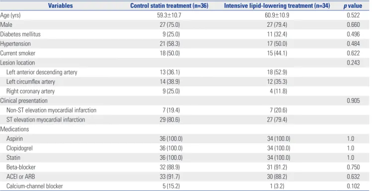
Variables Control statin treatment (n=36) Intensive lipid-lowering treatment (n=34) p value
Age (yrs) 59.3±10.7 60.9±10.9 0.522
Male 27 (75.0) 27 (79.4) 0.660
Diabetes mellitus 9 (25.0) 11 (32.4) 0.496
Hypertension 21 (58.3) 17 (50.0) 0.484
Current smoker 18 (50.0) 15 (44.1) 0.622
Lesion location 0.243
Left anterior descending artery 13 (36.1) 18 (52.9)
Left circumflex artery 14 (38.9) 12 (35.3)
Right coronary artery 9 (25.0) 4 (11.8)
Clinical presentation 0.905
Non-ST elevation myocardial infarction 7 (19.4) 7 (20.6)
ST elevation myocardial infarction 29 (80.6) 27 (79.4)
Medications
Aspirin 36 (100.0) 34 (100.0) 1.0
Clopidogrel 36 (100.0) 34 (100.0) 1.0
Statin 36 (100.0) 34 (100.0) 1.0
Beta-blocker 32 (88.9) 31 (91.2) 0.750
ACEI or ARB 33 (91.7) 30 (88.2) 0.632
Calcium-channel blocker 5 (15.2) 1 (3.2) 0.102
ACEI, angiotensin converting enzyme inhibitor; ARB, angiotensin receptor blocker.
*Values are n (%) or mean±SD.
Table 2. Laboratory Findings*
Variables Control statin treatment (n=36) Intensive lipid-lowering treatment (n=34) p value Total cholesterol (mg/dL)
Baseline 196.8±38.4 190.8±24.7 0.442
Three-month follow-up 153.3±38.5† 129.7±30.3† 0.006
∆Total cholesterol -43.6±37.3 -61.1±42.1 0.070
LDL cholesterol (mg/dL)
Baseline 119.1±29.9 111.4±22.0 0.230
Three-month follow-up 92.2±21.8† 68.1±15.3† <0.001
∆LDL cholesterol -26.9±20.4 -43.4±24.1 0.003
HDL cholesterol (mg/dL)
Baseline 39.4±6.3 36.0±8.9 0.072
Three-month follow-up 40.7±8.8 37.8±8.7 0.173
∆HDL cholesterol 1.3±8.8 1.8±7.5 0.793
Triglyceride (mg/dL)
Baseline 136.8±80.9 120.6±90.4 0.435
Three-month follow-up 131.1±64.9 127.6±76.3 0.836
∆Triglycerides -1.8±94.4 7.1±56.9 0.637
hsCRP (mg/L)
Baseline 7.6±17.0 5.2±8.3 0.474
Three-month follow-up 2.1±2.5 3.8±7.2 0.201
∆hsCRP -5.5±17.4 -1.3±10.9 0.250
LDL, low-density lipoprotein; HDL, high-density lipoprotein; hsCRP, high-sensitive C-reactive protein.
∆ indicates changes from baseline to three-month follow-up.
*Values are mean±SD, †p<0.05 for comparison between baseline and three-month follow-up levels.
Table 3. Intravascular Ultrasound Analysis between Intensive Lipid-Lowering Treatment and Control Statin Treatment*
Baseline Three-month follow-up p value Change in three months p value† Gray-scale intravascular ultrasound analysis
External elastic membrane volume (mm3) 0.730
All patients (n=70) 154.1±57.9 150.9±63.6 0.758 -3.2±23.8
Control statin treatment (n=36) 163.5±60.9 161.3±71.2 0.886 -2.2±30.2
Intensive lipid-lowering treatment (n=34) 144.1±53.6 140.0±53.2 0.750 -4.2±14.6
Lumen volume (mm3) 0.598
All patients 71.6±32.2 70.2±34.1 0.793 -1.4±16.3
Control statin treatment 77.0±32.8 74.6±36.0 0.762 -2.4±20.3
Intensive lipid-lowering treatment 65.9±31.1 65.5±31.9 0.957 -0.4±10.8
Absolute total plaque volume (mm3) 0.231
All patients 82.5±32.3 80.8±35.9 0.773 -1.7±13.7
Control statin treatment 86.5±34.7 86.7±41.9 0.981 0.3±17.0
Intensive lipid-lowering treatment 78.3±29.5 74.5±27.4 0.596 -3.7±9.0
Virtual histology intravascular ultrasound analysis
Absolute volume of fibro-fatty plaque (mm3) 0.024
All patients 5.6±3.9 5.2±4.8 0.575 -0.3±4.3
Control statin treatment 6.0±3.8 6.7±5.8 0.568 0.8±4.7
Intensive lipid-lowering treatment 5.2±4.0 3.7±2.6 0.063 -1.5±3.4
Absolute volume of fibrous plaque (mm3) 0.229
All patients 30.6±14.6 29.3±15.7 0.600 -1.3±9.5
Control statin treatment 31.2±14.4 31.2±17.7 0.999 -0.1±11.6
Intensive lipid-lowering treatment 30.1±14.9 27.3±13.1 0.420 -2.8±6.5
Absolute volume of necrotic core (mm3) 0.415
All patients 12.3±8.9 12.9±11.4 0.704 0.6±5.6
Control statin treatment 13.6±10.0 13.7±13.7 0.977 0.1±6.4
Intensive lipid-lowering treatment 10.9±7.5 12.2±8.5 0.516 1.2±4.6
Absolute volume of dense calcium (mm3) 0.746
All patients 5.1±6.8 5.1±8.0 0.982 0.0±4.0
Control statin treatment 6.5±8.4 6.4±10.2 0.960 -0.2±5.4
Intensive lipid-lowering treatment 3.7±4.4 3.7±4.6 0.957 0.1±1.8
Percentage of fibro-fatty plaque volume (%) 0.235
All patients 10.9±6.3 10.3±8.1 0.625 -0.5±7.4
Control statin treatment 11.4±7.6 11.8±10.2 0.865 0.5±8.9
Intensive lipid-lowering treatment 10.4±4.8 8.8±4.6 0.161 -1.6±5.3
Percentage of fibrous plaque volume (%) 0.200
All patients 59.4±11.0 59.7±15.6 0.896 0.3±13.1
Control statin treatment 57.4±11.5 59.6±19.3 0.545 2.3±16.6
Intensive lipid-lowering treatment 61.6±10.2 59.8±10.7 0.481 -1.7±7.7
Percentage of necrotic core volume (%) 0.208
All patients 20.6±8.2 21.4±9.6 0.584 0.8±8.8
Control statin treatment 20.7±8.3 20.2±10.4 0.832 -0.5±9.1
Intensive lipid-lowering treatment 20.4±8.2 22.7±8.7 0.285 2.1±8.3
Percentage of dense calcium volume (%) 0.866
All patients 8.3±7.5 7.8±7.5 0.529 -0.5±6.4
Control statin treatment 9.3±9.1 8.8±9.0 0.806 -0.5±8.3
Intensive lipid-lowering treatment 7.3±5.1 6.7±5.3 0.677 -0.3±3.8
*Values are mean±SD, †p value for comparison of changes in intravascular ultrasound variables from baseline to three-month follow-up between intensive lipid- lowering treatment and control statin treatment.
cant differences found for serial changes of EEM, lumen, and P&M volume between the two groups (Table 3).
The findings from VH-IVUS analysis are shown in Table 3.
While there appeared to be a tendency of reduced absolute fi- bro-fatty plaque volume in the intensive lipid-lowering treat- ment group (from 5.2±4.0 mm3 at baseline to 3.7±2.6 mm3 at the three-month follow-up, p=0.063), there were no significant changes in absolute fibro-fatty plaque volume in the control statin treatment group. The reduction of absolute fibro-fatty plaque volume from baseline to the three-month follow-up was greater in the intensive lipid-lowering treatment group com- pared with the control statin treatment group (-1.5±3.4 mm3 vs.
0.8±4.7 mm3, p=0.024) (Table 3, Fig. 1). However, there were no statistically significant changes in fibrous, necrotic core, and dense calcium volume from baseline to the three-month fol- low-up between the two groups.
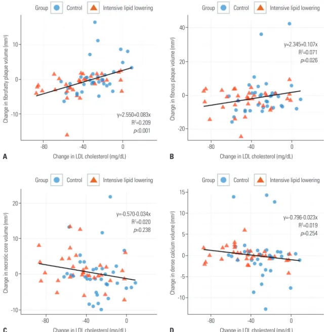
A significant linear correlation was found between the changes in LDL-C from baseline to the three-month follow-up and that of absolute fibro-fatty plaque volume (p<0.001, R2=0.209) and fi- brous plaque volume (p=0.026, R2=0.071) (Fig. 2). In the multi- variate analyses including statin groups, changes in LDL-C were still the independent predictor of changes in fibro-fatty plaque volume (p<0.001) while not for fibrous plaque volume (p=0.055). Treatment group itself was not an independent pre- dictor for changes in fibro-fatty (p=0.289) and fibrous plaque volume (p=0.652).
No major adverse cardiovascular events, such as cardiovas- cular mortality, myocardial infarction, or stroke occurred dur- ing the study period for patients in either of the two groups. As for the adverse effects of drugs, 3 episodes of myalgia and/or general weakness were reported, 2 in the control and 1 in the intensive lipid lowering group.
DISCUSSION
This randomized study showed that significant changes in cor- onary plaque components (i.e., reduction of absolute volume of fibro-fatty plaque) as well as decreases in LDL-C levels were observed early (at three-month follow-up) in ACS patients who were given an intensive lipid-lowering treatment. There was a significant linear correlation between the changes in LDL-C levels and changes in absolute volume of fibro-fatty plaque.
Statins have several beneficial properties beyond their lipid- lowering effect, including atherosclerotic plaque stabilization, oxidative stress reduction, enhancement of endothelial func- tion, a decrease in vascular inflammation, and improvements of vascular healing after stent implantation.24,25 Previous gray- scale IVUS studies showed that statin treatment was associated with regression or no progression of coronary artery atheroscle- rotic plaque.11-13 Ezetimibe is a member of a class of non-statin agents that inhibit the absorption of cholesterol from the intes- tine by blocking the Niemann-Pick-like 1 receptor and reduce the absorption of both dietary and biliary cholesterol by 54% to 65%.26,27 The combination treatment of ezetimibe and statin in- hibits both the cholesterol synthesis and intestinal cholesterol absorption, resulting in approximately 18% greater reduction in LDL-C levels than the treatment with statin alone.28,29 A recent randomized study showed that an additional decrease in LDL- C levels after the addition of ezetimibe to statin therapy was as- sociated with a reduction of cardiovascular events compared with statin mono-therapy in stabilized ACS patients.30
Several VH-IVUS studies evaluated the effect of statin treat- ment on coronary plaque components with respect to different types or dosages of statins and the duration of statin treatment.16-19 In a recent study, 24-month maximally-intensive statin treat- ment in 36 rosuvastatin (40 mg)- and 35 atorvastatin (80 mg)- treated patients resulted in a coronary atheroma regression and a reduction in fibro-fatty components.18 In an another study,
Fig. 1. Changes in absolute volume of fibro-fatty plaque. (A) The absolute volume of fibro-fatty plaque changed from 6.0±3.8 mm3 at baseline to 6.7±5.8 mm3 at three-month follow-up after control statin treatment (pravastatin 20 mg). (B) The absolute volume of fibro-fatty plaque decreased from 5.2±4.0 mm3 at baseline to 3.7±2.6 mm3 at three-month follow-up after intensive lipid-lowering treatment (ezetimibe/simvastatin, 10/40 mg).
A B
Fibrofatty plaque volume
(mm
3) Fibrofatty plaque volume
(mm
3) 20
15
10
5
0
20
15
10
5
0
Baseline Baseline
Control Intensive lipid lowering
Follow-up Follow-up
12-month treatment with fluvastatin (60 mg/day, n=40) result- ed in a significant regression of plaque volume and significant reduction of fibro-fatty volume compared with a control group (n=40).16 The results from a randomized study that evaluated six-months of statin treatment showed that there were higher percentages of plaque volume regression and lower percentag- es of necrotic core expansion in higher-dose atorvastatin (40 mg)-treated (n=20) than in lower-dose atorvastatin (10 mg)- treated patients (n=20),17 and a randomized study involving only two to three weeks of statin treatment showed significant plaque regression and reduction of fibro-fatty components in pitavastatin (2 mg)-treated (n=80), but not in atorvastatin (10 mg)-treated patients (n=80).19
In the present study, three-month intensive lipid-lowering
treatment resulted in a significant reduction in fibro-fatty plaque volume, which is in accordance with previous studies.16,18,19 Plaque regression was not significantly different between the control statin and intensive lipid-lowering treatment groups (0.3±17.0 mm3 vs. -3.7±9.0 mm3, respectively; p=0.231). For the evaluation of early effects of lipid-lowering treatment on coro- nary plaque components, a statin versus statin comparison was performed in the above-mentioned previous study,19 while the present study compared statin mono-therapy with the addition of ezetimibe to statin therapy.
Although this study is not without some limitations, it dem- onstrates the early positive effects of intensive lipid-lowering treatment. Small sample size, due to the single study site, may have a potential for selection bias. Also, most of the VH-IVUS
Fig. 2. Correlation between change in low-density lipoprotein (LDL)-cholesterol and change in absolute volume of each plaque component. (A) Fibro-fatty plaque, (B) fibrous plaque, (C) necrotic core, and (D) dense calcium. Blue circles denote control group, and red triangles are for intensive lipid-lowering group.
A
C
B
D
Change in fibrofatty plaque volume (mm3)Change in necrotic core volume (mm3) Change in fibrous plaque volume (mm3)Change in dense calcium volume (mm3)
10
0
-10
20
10
0
-10
40
20
0
-20
15 10 5 0 -5 -10 -80 -40 0
-80 -40 0
-80 -40 0
-80 -40 0 Change in LDL cholesterol (mg/dL)
Group Control Intensive lipid lowering
Group Control Intensive lipid lowering
Group Control Intensive lipid lowering
Group Control Intensive lipid lowering
Change in LDL cholesterol (mg/dL)
Change in LDL cholesterol (mg/dL)
Change in LDL cholesterol (mg/dL) y=2.550+0.083x
R2=0.209 p<0.001
y=-0.570-0.034x R2=0.020 p=0.238
y=2.345+0.107x R2=0.071 p=0.026
y=-0.796-0.023x R2=0.019 p=0.254
parameters studied were not found to be different at the levels of statistical significance between the two groups, with the ex- ception of the absolute volume of fibro-fatty plaque. Consider- ing that the purpose of the present study was to evaluate the early effects of intensive lipid-lowering treatment on changes in coronary plaque components, these results suggest that the ab- solute volume of fibro-fatty plaque may be the most sensitive parameter affected by the intensive lipid-lowering treatment.
Furthermore, changes in the absolute volume of fibro-fatty plaque may be a potential early indicator of efficacy in intensive lipid-lowering treatment regimens.
In conclusion, the most significant effects of intensive lipid- lowering treatment, such as the addition of ezetimibe to statin therapy, on coronary plaque modification may appear early dur- ing treatment. Therefore, it may be necessary to consider early aggressive LDL-C control by intensive lipid-lowering for the initiation of rapid and effective plaque modification in ACS pa- tients.
ACKNOWLEDGEMENTS
This study was supported by a grant from the Korea Healthcare Technology Research & Development Project, Ministry for Health, Welfare & Family Affairs, Republic of Korea (Nos.
A085136 and A102064), the Mid-career Researcher Program through NRF grant funded by the MEST, Republic of Korea (No.
2015R1A2A2A01002731) and the Cardiovascular Research Cen- ter, Seoul, Korea.
REFERENCES
1. Randomised trial of cholesterol lowering in 4444 patients with cor- onary heart disease: the Scandinavian Simvastatin Survival Study (4S). Lancet 1994;344:1383-9.
2. Prevention of cardiovascular events and death with pravastatin in patients with coronary heart disease and a broad range of initial cholesterol levels. The Long-Term Intervention with Pravastatin in Ischaemic Disease (LIPID) Study Group. N Engl J Med 1998;339:
1349-57.
3. Heart Protection Study Collaborative Group. MRC/BHF Heart Protection Study of cholesterol lowering with simvastatin in 20,536 high-risk individuals: a randomised placebo-controlled trial. Lan- cet 2002;360:7-22.
4. Pitt B, Waters D, Brown WV, van Boven AJ, Schwartz L, Title LM, et al. Aggressive lipid-lowering therapy compared with angioplasty in stable coronary artery disease. Atorvastatin versus Revascular- ization Treatment Investigators. N Engl J Med 1999;341:70-6.
5. Sacks FM, Pfeffer MA, Moye LA, Rouleau JL, Rutherford JD, Cole TG, et al. The effect of pravastatin on coronary events after myo- cardial infarction in patients with average cholesterol levels. Cho- lesterol and Recurrent Events Trial investigators. N Engl J Med 1996;335:1001-9.
6. Schwartz GG, Olsson AG, Ezekowitz MD, Ganz P, Oliver MF, Wa- ters D, et al. Effects of atorvastatin on early recurrent ischemic events in acute coronary syndromes: the MIRACL study: a ran- domized controlled trial. JAMA 2001;285:1711-8.
7. Cannon CP, Braunwald E, McCabe CH, Rader DJ, Rouleau JL,
Belder R, et al. Intensive versus moderate lipid lowering with statins after acute coronary syndromes. N Engl J Med 2004;350:1495-504.
8. LaRosa JC, Grundy SM, Waters DD, Shear C, Barter P, Fruchart JC, et al. Intensive lipid lowering with atorvastatin in patients with stable coronary disease. N Engl J Med 2005;352:1425-35.
9. Effect of simvastatin on coronary atheroma: the Multicentre Anti- Atheroma Study (MAAS). Lancet 1994;344:633-8.
10. Jukema JW, Bruschke AV, van Boven AJ, Reiber JH, Bal ET, Zwind- erman AH, et al. Effects of lipid lowering by pravastatin on pro- gression and regression of coronary artery disease in symptomatic men with normal to moderately elevated serum cholesterol levels.
The Regression Growth Evaluation Statin Study (REGRESS). Cir- culation 1995;91:2528-40.
11. Nissen SE, Tuzcu EM, Schoenhagen P, Brown BG, Ganz P, Vogel RA, et al. Effect of intensive compared with moderate lipid-lower- ing therapy on progression of coronary atherosclerosis: a random- ized controlled trial. JAMA 2004;291:1071-80.
12. Nissen SE, Nicholls SJ, Sipahi I, Libby P, Raichlen JS, Ballantyne CM, et al. Effect of very high-intensity statin therapy on regression of coronary atherosclerosis: the ASTEROID trial. JAMA 2006;295:
1556-65.
13. Okazaki S, Yokoyama T, Miyauchi K, Shimada K, Kurata T, Sato H, et al. Early statin treatment in patients with acute coronary syn- drome: demonstration of the beneficial effect on atherosclerotic lesions by serial volumetric intravascular ultrasound analysis dur- ing half a year after coronary event: the ESTABLISH Study. Circu- lation 2004;110:1061-8.
14. Virmani R, Burke AP, Farb A, Kolodgie FD. Pathology of the vul- nerable plaque. J Am Coll Cardiol 2006;47(8 Suppl):C13-8.
15. Libby P, Schoenbeck U, Mach F, Selwyn AP, Ganz P. Current con- cepts in cardiovascular pathology: the role of LDL cholesterol in plaque rupture and stabilization. Am J Med 1998;104:14S-8S.
16. Nasu K, Tsuchikane E, Katoh O, Tanaka N, Kimura M, Ehara M, et al. Effect of fluvastatin on progression of coronary atherosclerotic plaque evaluated by virtual histology intravascular ultrasound.
JACC Cardiovasc Interv 2009;2:689-96.
17. Lee SW, Hau WK, Kong SL, Chan KK, Chan PH, Lam SC, et al. Vir- tual histology findings and effects of varying doses of atorvastatin on coronary plaque volume and composition in statin-naive pa- tients: the VENUS study. Circ J 2012;76:2662-72.
18. Puri R, Libby P, Nissen SE, Wolski K, Ballantyne CM, Barter PJ, et al. Long-term effects of maximally intensive statin therapy on changes in coronary atheroma composition: insights from SAT- URN. Eur Heart J Cardiovasc Imaging 2014;15:380-8.
19. Toi T, Taguchi I, Yoneda S, Kageyama M, Kikuchi A, Tokura M, et al. Early effect of lipid-lowering therapy with pitavastatin on regres- sion of coronary atherosclerotic plaque. Comparison with atorv- astatin. Circ J 2009;73:1466-72.
20. Mintz GS, Nissen SE, Anderson WD, Bailey SR, Erbel R, Fitzgerald PJ, et al. American College of Cardiology Clinical Expert Consen- sus Document on Standards for Acquisition, Measurement and Reporting of Intravascular Ultrasound Studies (IVUS). A report of the American College of Cardiology Task Force on Clinical Expert Consensus Documents. J Am Coll Cardiol 2001;37:1478-92.
21. Nair A, Kuban BD, Tuzcu EM, Schoenhagen P, Nissen SE, Vince DG. Coronary plaque classification with intravascular ultrasound radiofrequency data analysis. Circulation 2002;106:2200-6.
22. Rodriguez-Granillo GA, García-García HM, Mc Fadden EP, Val- gimigli M, Aoki J, de Feyter P, et al. In vivo intravascular ultrasound- derived thin-cap fibroatheroma detection using ultrasound ra- diofrequency data analysis. J Am Coll Cardiol 2005;46:2038-42.
23. Hong MK, Park DW, Lee CW, Lee SW, Kim YH, Kang DH, et al. Ef- fects of statin treatments on coronary plaques assessed by volu-
metric virtual histology intravascular ultrasound analysis. JACC Cardiovasc Interv 2009;2:679-88.
24. Moreno PR, Fuster V. The year in atherothrombosis. J Am Coll Car- diol 2004;44:2099-110.
25. Suh Y, Kim BK, Shin DH, Kim JS, Ko YG, Choi D, et al. Impact of statin treatment on strut coverage after drug-eluting stent im- plantation. Yonsei Med J 2015;56:45-52.
26. Sudhop T, Lütjohann D, Kodal A, Igel M, Tribble DL, Shah S, et al.
Inhibition of intestinal cholesterol absorption by ezetimibe in hu- mans. Circulation 2002;106:1943-8.
27. Davis HR Jr, Zhu LJ, Hoos LM, Tetzloff G, Maguire M, Liu J, et al.
Niemann-Pick C1 Like 1 (NPC1L1) is the intestinal phytosterol and cholesterol transporter and a key modulator of whole-body cho-
lesterol homeostasis. J Biol Chem 2004;279:33586-92.
28. Ballantyne CM, Blazing MA, King TR, Brady WE, Palmisano J. Ef- ficacy and safety of ezetimibe co-administered with simvastatin compared with atorvastatin in adults with hypercholesterolemia.
Am J Cardiol 2004;93:1487-94.
29. Ballantyne CM, Abate N, Yuan Z, King TR, Palmisano J. Dose-com- parison study of the combination of ezetimibe and simvastatin (Vytorin) versus atorvastatin in patients with hypercholesterol- emia: the Vytorin Versus Atorvastatin (VYVA) study. Am Heart J 2005;149:464-73.
30. Cannon CP, Blazing MA, Giugliano RP, McCagg A, White JA, Ther- oux P, et al. Ezetimibe added to statin therapy after acute coronary syndromes. N Engl J Med 2015;372:2387-97.