Renal ischemia-reperfusion (IR) causes remote liver damage
5
0
0
전체 글
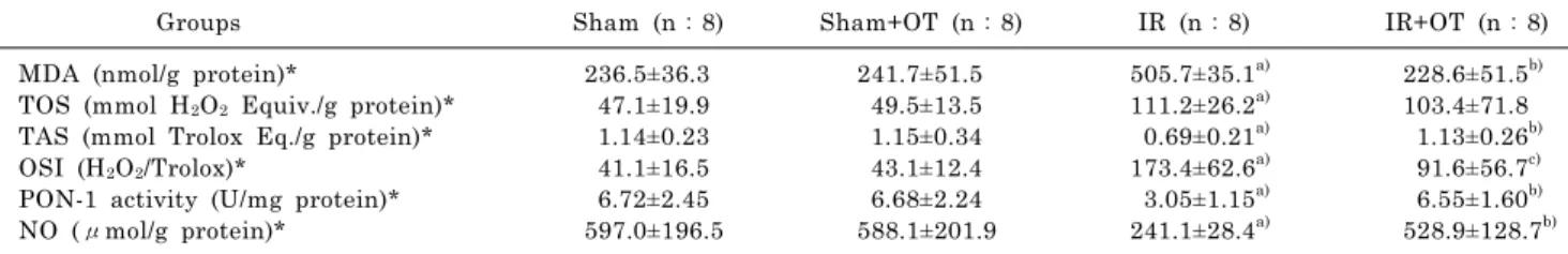
(2) 170. A Tas Hekimoglu, et al. was initiated after obtaining approval from Dicle University Local Committee on Animal Research Ethics. We meticulously complied with the principles for the “Protection of Animal Rights” specified by the National Institutes of Health (NIH) during the entire course of the study. Experimental method and procedure The rats were put under anaesthesia with ketamine (75 mg/kg i.p.) and xylazine (8 mg/kg i.p.). Body temperature o was preserved in every part of surgery at 37±1 C. All rats were submitted to surgical exposure of the left and right renal pedicles via midline incision. To create renal ischemia, both renal pedicles were blocked for 45 min with vascular clamps. After 45 min of blockage, the clamps were taken away, and kidneys were examined to endure reperfusion for 24 hours. The rats were randomly divided into four different groups (n=8). The groups were as follows: (1) Sham operated group; (2) Sham operated+OT group (3) Renal IR group; (4) Renal IR+OT group. OT (500μg/kg) (Sigma, St. Louis, MO, USA) was administered subcutaneously 12 and 24 hours before and immediately after ischemia. By the termination of experimental procedure, the rats were sacrificed. The excised liver tissue specimens for biochemical assay were weighed, and instantly stored o at −70 C. Part of liver was fixed in 10% neutral formalin for histological examination. Biochemical analyses The liver tissues were washed with 1.15% ice-cold KCl, minced, then homogenized in five volumes (w/v) of the same solution. Measurements were performed on the supernatant of the homogenate that was prepared at 14.000 rpm o for 30 min at +4 C. The protein density of the tissue was determined by the method of Lowry [12]. Lipid peroxidation level in the liver was represented as malondialdehyde (MDA). It was determined by the method of Ohkawa et al. [13]. Consistent with this method, samples were mixed with sodium dodecyl sulfate, acetic acid and 2-thiobarbituric acid. After vortexing, tissue samples were incubated for 1 o h in 95 C and butanol-pyridine 15:1 (v/v) was added. The mixture was shaken for 10 min and then centrifuged. Butanol-pyridine layer was measured fluorometrically (UV-1205 Shimadzu) at 552 nm. Data are shown as nmol/g protein. Nitric oxide (NO) levels were assessed with Griess’ method [14]. Paraoxonase (PON-1) activity was evaluated spectrophotometrically by modified Eckerson [15] method. The total antioxidant status (TAS) of supernatant fractions was evaluated by using a novel automated and colorimetric measurement method developed by Erel [16]. Hydroxyl radicals, the most potent biological radicals, are produced in this method. In the assay, the ferrous ion solution present in reagent 1 is mixed with hydrogen peroxide, which is present in reagent 2. The subsequently produced radicals, such as brown-colored dianisidine radical cations produced by the hydroxyl radicals, are also potent radicals. Using this method, the antioxidative effect of the sample is measured against the potent free radical reactions initiated by the produced hydroxyl radicals. The assay has excellent precision values lower than 3%. The assay is calibrated with Trolox (Vitamin E), and the TAS results are expressed as nmol Trolox equivalent/mg protein. The total oxidant status (TOS) of supernatant fractions was measured by using a novel automated and colorimetric measure-. ment method [17]. Oxidants present in the sample oxidize the ferrous ion-o-dianisidine complex to ferric ion. The oxidation reaction is increased by glycerol molecules, which are abundantly present in the reaction medium. The ferric ion makes a colored complex with xylenol orange in an acidic medium. The color intensity, which can be measured spectrophotometrically, is related to the total amount of oxidant molecules present in the sample. The assay is calibrated with hydrogen peroxide, and the results are expressed in terms of nmol H2O2 equivalent/mg protein [18]. The TOS/ TAS ratio was considered to be the oxidative stress index (OSI). The units of liver tissue TOS and TAS were μmole H2O2 Equiv./gram protein and mmole H2O2 Equiv./gram protein, respectively [19]. Histopathological assessment Liver injury was analyzed under light microscopy. All tissue specimens were taken from the central part of the right lobe of the liver and were fixed in 10% neutral buffered formalin solutions, refined for implanting in paraffin by routine protocols, and 4μm thick sections were then cut by microtome. The sections were stained with HematoxylinEosin by using a routine protocol and examined with a photomicroscope (Eclipse 50i, Nikon). Congestion, hepatocyte vacuolization, sinusoidal dilatation, central vein dilatation, Kupffer cell activation and periportal inflammation were scored as 0 (no change), 1 (minimal change), 2 (moderate change), and 3 (severe change) and were assessed in a blinded manner. Statistical analysis Statistical analysis was performed by using GraphPad Prisma V3 program (GraphPad Software, Inc. San Diego, USA). Data were shown as means±standard deviation, and Kruskal-Wallis test was used for analysis. In the event of significant results, the Mann-Whitney U test was employed for comparisons of differences between the two independent groups. Histopathological results were expressed as median values and analyzed by Kruskal-Wallis and Mann-Whitney U test. A p value <0.05 was considered statistically significant.. RESULTS Biochemical results The MDA, TOS, TAS, OSI and NO levels and PON-1 enzyme activities in the liver tissues are presented in Table 1. MDA, TOS and OSI levels were notably higher, and TAS, NO and PON-1 levels were notably lower in the IR as compared to the sham operated and sham+OT groups (p<0.01). However, TAS, NO and PON-1 levels were notably increased in oxytocin-treated group compared to the IR group (p<0.01). MDA levels were notably decreased in IR+OT group compared to the IR group (p<0.05). Histopathological results The histopathological findings are shown in Table 2. Light microscopic evaluation revealed that IR group resulted in widespread hepatic injury with marked congestion, hepatocyte vacuolization, sinusoidal dilatation,.
(3) 171. Oxytocin Ameliorates Remote Liver Injury. Table 1. Levels of MDA, TOS and TAS, OSI, PON-1 and NO in groups Groups. Sham (n:8). MDA (nmol/g protein)* TOS (mmol H 2O 2 Equiv./g protein)* TAS (mmol Trolox Eq./g protein)* OSI (H 2O 2/Trolox)* PON-1 activity (U/mg protein)* NO (μmol/g protein)*. Sham+OT (n:8). 236.5±36.3 47.1±19.9 1.14±0.23 41.1±16.5 6.72±2.45 597.0±196.5. IR (n:8). IR+OT (n:8) a). 241.7±51.5 49.5±13.5 1.15±0.34 43.1±12.4 6.68±2.24 588.1±201.9. 505.7±35.1 a) 111.2±26.2 a) 0.69±0.21 a) 173.4±62.6 a) 3.05±1.15 a) 241.1±28.4. b). 228.6±51.5 103.4±71.8 b) 1.13±0.26 c) 91.6±56.7 b) 6.55±1.60 b) 528.9±128.7. Results are presented as means±standard deviation. *p<0.01 for Kruskal Wallis test. a)p<0.01 as compared to the sham operated and sham+OT groups, b)p<0.01 as compared to the IR group. c)p<0.05 as compared to the IR group. IR, ischemia-reperfusion; OT, oxytocin; MDA, malondialdehyde; TOS, total oxidant status; TAS, total antioxidant status; OSI, oxidative stress index; PON-1, paraoxonase activity; NO, nitric oxide.. Table 2. Histopathological findings in the different study groups Sham Congestion Hepatocyte vacuolization Sinusoidal dilatation Central vein dilatation Kupffer cell activation Periportal inflammation. Sham+OT. 0 0 0 0 0 0. 0 0 0 0 0 0. IR. IR+OT. p. 1 2 3 2 2 2. 0 0.5 1 0.5 0.5 1. p<0.05 p<0.05 p<0.05 p<0.05 p<0.05 p<0.05. Results are presented as median values IR group was compared with the other groups. IR, ischemia-reperfusion; OT, oxytocin. 0: no change, 1: minimal change, 2: moderate change, 3: severe change.. central vein dilatation, Kupffer cell activation and periportal inflammation (Fig. 1C). Histopathological alterations that were observed in IR group were significantly improved in IR+OT group (Fig. 1D). There were minimal changes in sham (Fig. 1A) and sham+OT (Fig. 1B) groups.. DISCUSSION Acute renal failure appears in almost 5% of hospitalized patients and up to 30% of patients in intensive care units [20,21]. Acute renal ischemic damage persists to be linked with a high mortality rate and arises in many clinical situations, such as urinary tract surgery, septic shock cases, organ transplantation and trauma [22]. Numerous studies have shown that renal injury affects additional organs including the liver [1,5,22]. In this work, we have investigated the potential effect of OT on liver injury triggered by renal I/R. To that end, we measured the levels of liver MDA, TOS, TAS, NO and PON-1 activity. IR of the tissue was determined to be connected with lipid peroxidation, which is an autocatalytic process causing oxidative demolition of the cellular membranes, and their breakdown can contribute to the generation of toxic, reactive products and cell death [5,23-25]. Lipid peroxidation, as a free radical-producing system, has been proposed to be tightly linked to IR-triggered tissue damage, and MDA is an important parameter of oxidative stress and a good pointer of lipid peroxidation [5,26,27]. In this research, in renal IR group, the level of MDA notably elevated in the liver tissue, while subcutaneously administered OT repressed MDA raising notably. This finding indicates that renal IR induced lipid peroxidation is likely to be improved with OT administration in liver tissue. In our study, we evaluated oxidative status as TOS and. TAS along with the measurement of OSI, a pointer of oxidative stress, which reveals the redox balance between oxidation and antioxidation [28]. Because of difficulties in measuring the levels of different oxidant molecules such as superoxide radical anion, hydrogen peroxide one by one, we measured TOS in serum as previously described by Erel [17]. Likewise, we appraised TAS, instead of measuring antioxidant molecules separately following the methods of Erel [29] and Cikrikcioglu et al. [30]. Lately, it has been widely documented that OSI may reveal the oxidative status more correctly than TOS or TAS level alone [31,32]. In the present study, the levels of TOS and OSI were notably augmented, and the level of TAS was notably reduced in IR group compared with sham operated group. On the other hand, in OT treated IR rats, TAS level was notably increased with respect to IR group. These results showed that renal IR leads to increased oxidative stress in liver tissue, and that the rising of oxidative stress was prevented by application of OT. PON-1 is an enzyme associated with high-density lipoprotein that is mainly secreted by the liver. Although its physiological function has not been completely clarified, it seems that PON-1 is responsible for hydrolyzing lipid peroxides, also playing a major role in the antioxidant system [33,34]. PON-1 protects liver against inflammation, liver disease and fibrosis [35,36]. In this research, we revealed decreased PON-1 activity in the liver tissues of IR rats compared to sham operated rats. OT treatment reversed the reduced activity of PON-1 in IR+OT group. OT treatment prevented the reduced activity of PON-1 in liver tissue. These findings demonstrate that a decrease in PON-1 activity is related to oxidative damage in the liver tissues caused by renal IR and OT treatment reverses the decline of PON-1 activity in these tissues. Augmented PON-1 activity through OT treatment.
(4) 172. A Tas Hekimoglu, et al. Fig. 1. Photomicrographs of Hematoxylin and Eosin stained sections from liver of rats. Minimal changes observed in sham (A) and sham+OT (B) groups. Congestion, hepatocyte vacuolization, sinusoidal dilatation, central vein dilatation, Kupffer cell activation and periportal inflammation are seen in IR group (C) sections. Histopathological alterations that were observed in IR group were significantly improved in IR+OT group (D). cv, central vein; v, vacuoles; kc, Kuppfer cells (inset); asterisk, inflammatory cells (inset); c, congestion; arrows, sinusoidal dilatation; IR, ischemia-reperfusion; OT, oxytocin.. in these tissues may be associated with antioxidant and anti-inflammatory effect of OT. NO has protective effects on cells during IR. NO has been demonstrated to inhibit oxidative stress, cytokine release, apoptosis, adhesion and aggregation of neutrophil leukocytes [24,37-39]. Several studies have shown a relationship between NO and IR injury [10,24,39,40]. A reduction of NO during IR is generally caused by endothelial dysfunction and reduction of endothelial nitric oxide synthase activity [41,42]. In this study, renal IR reduced the NO level, and OT treatment increased NO content in hepatic tissue. These findings showed that renal IR injury is associated with endothelial dysfunction and reduction of nitric oxide synthase activity. OT, which is revealed to be protective in IR induced remote liver injury, may exhibit its effect by increasing NO synthase activity and releasing NO from vascular endothelium. In our research, we also investigated the histopathological consequences of exogenously administered OT in liver tissue. In IR group, intense hepatocyte vacuolization, evident sinusoidal dilatation, central vein dilation, Kupffer. cell activation and periportal inflammation were observed. These alterations pointed out a severe damage in liver paranchyme, and the impairment of liver tissues in IR group was notably more severe than the IR+OT group. These data show that renal ischemia provokes destructive alterations in liver histology, oxidative stress and induces a diminution in hepatic antioxidant capacity. OT ameliorates remote liver injury triggered by renal ischemia-reperfusion, and this preservation is related with suppression of inflammation and regulation of oxidant-antioxidant status.. REFERENCES 1. Kadkhodaee M, Golab F, Zahmatkesh M, Ghaznavi R, Hedayati M, Arab HA, Ostad SN, Soleimani M. Effects of different periods of renal ischemia on liver as a remote organ. World J Gastroenterol. 2009;15:1113-1118. 2. Park SW, Chen SW, Kim M, Brown KM, Kolls JK, D’Agati VD, Lee HT. Cytokines induce small intestine and liver injury after.
(5) Oxytocin Ameliorates Remote Liver Injury. renal ischemia or nephrectomy. Lab Invest. 2011;91:63-84. 3. Serteser M, Koken T, Kahraman A, Yilmaz K, Akbulut G, Dilek ON. Changes in hepatic TNF-alpha levels, antioxidant status, and oxidation products after renal ischemia/reperfusion injury in mice. J Surg Res. 2002;107:234-240. 4. Miyazawa S, Watanabe H, Miyaji C, Hotta O, Abo T. Leukocyte accumulation and changes in extra-renal organs during renal ischemia reperfusion in mice. J Lab Clin Med. 2002;139:269278. 5. Kaçmaz A, User EY, Sehirli AO, Tilki M, Ozkan S, Sener G. Protective effect of melatonin against ischemia/reperfusion-induced oxidative remote organ injury in the rat. Surg Today. 2005;35:744-750. 6. Petersson M, Wiberg U, Lundeberg T, Uvnäs-Moberg K. Oxytocin decreases carrageenan induced inflammation in rats. Peptides. 2001;22:1479-1484. 7. Barberis C, Mouillac B, Durroux T. Structural bases of vasopressin/oxytocin receptor function. J Endocrinol. 1998;156: 223-229. 8. Liberzon I, Young EA. Effects of stress and glucocorticoids on CNS oxytocin receptor binding. Psychoneuroendocrinology. 1997;22:411-422. 9. Petersson M, Lundeberg T, Sohlström A, Wiberg U, UvnäsMoberg K. Oxytocin increases the survival of musculocutaneous flaps. Naunyn Schmiedebergs Arch Pharmacol. 1998; 357:701-704. 10. Tuğtepe H, Sener G, Biyikli NK, Yüksel M, Cetinel S, Gedik N, Yeğen BC. The protective effect of oxytocin on renal ischemia/reperfusion injury in rats. Regul Pept. 2007;140:101-108. 11. Düşünceli F, Işeri SO, Ercan F, Gedik N, Yeğen C, Yeğen BC. Oxytocin alleviates hepatic ischemia-reperfusion injury in rats. Peptides. 2008;29:1216-1222. 12. Lowry OH, Rosebrough NJ, Farr AL, Randall RJ. Protein measurement with the Folin phenol reagent. J Biol Chem. 1951;193:265-275. 13. Ohkawa H, Ohishi N, Yagi K. Assay for lipid peroxides in animal tissues by thiobarbituric acid reaction. Anal Biochem. 1979;95:351-358. 14. Cortas NK, Wakid NW. Determination of inorganic nitrate in serum and urine by a kinetic cadmium-reduction method. Clin Chem. 1990;36:1440-1443. 15. Eckerson HW, Wyte CM, La Du BN. The human serum paraoxonase/arylesterase polymorphism. Am J Hum Genet. 1983;35:1126-1138. 16. Erel O. A novel automated method to measure total antioxidant response against potent free radical reactions. Clin Biochem. 2004;37:112-119. 17. Erel O. A new automated colorimetric method for measuring total oxidant status. Clin Biochem. 2005;38:1103-1111. 18. Ozturk E, Balat O, Acılmıs YG, Ozcan C, Pence S, Erel Ö. Measurement of the placental total antioxidant status in preeclamptic women using a novel automated method. J Obstet Gynaecol Res. 2011;37:337-342. 19. Aycicek A, Erel O, Kocyigit A. Increased oxidative stress in infants exposed to passive smoking. Eur J Pediatr. 2005;164: 775-778. 20. Star RA. Treatment of acute renal failure. Kidney Int. 1998; 54:1817-1831. 21. Gu J, Chen J, Xia P, Tao G, Zhao H, Ma D. Dexmedetomidine attenuates remote lung injury induced by renal ischemia-reperfusion in mice. Acta Anaesthesiol Scand. 2011;55:1272-1278. 22. Awad AS, Kamel R, Sherief MA. Effect of thymoquinone on hepatorenal dysfunction and alteration of CYP3A1 and spermidine/spermine N-1-acetyl-transferase gene expression induced by renal ischaemia-reperfusion in rats. J Pharm Pharmacol. 2011;63:1037-1042. 23. Eschwège P, Paradis V, Conti M, Holstege A, Richet F, Detève. 24. 25. 26. 27. 28. 29. 30.. 31. 32.. 33. 34.. 35.. 36.. 37. 38. 39. 40. 41. 42.. 173. J, Ménager P, Legrand A, Jardin A, Bedossa P, Benoit G. In situ detection of lipid peroxidation by-products as markers of renal ischemia injuries in rat kidneys. J Urol. 1999;162:553557. Işeri SO, Gedik IE, Erzik C, Uslu B, Arbak S, Gedik N, Yeğen BC. Oxytocin ameliorates skin damage and oxidant gastric injury in rats with thermal trauma. Burns. 2008;34:361-369. Vaghasiya JD, Sheth NR, Bhalodia YS, Jivani NP. Exaggerated liver injury induced by renal ischemia reperfusion in diabetes: effect of exenatide. Saudi J Gastroenterol. 2010;16:174-180. Jang HS, Han JH, Jeong JY, Sohn UD. Protective Effect of ECQ on Rat Reflux Esophagitis Model. Korean J Physiol Pharmacol. 2012;16:455-462. Jung J, Nam Y, Sohn UD. Inhibitory Effects of ECQ on Indomethacin-Induced Gastric Damage in Rats. Korean J Physiol Pharmacol. 2012;16:399-404. Davies GR, Simmonds NJ, Stevens TR, Grandison A, Blake DR, Rampton DS. Mucosal reactive oxygen metabolite production in duodenal ulcer disease. Gut. 1992;33:1467-1472. Erel O. A novel automated direct measurement method for total antioxidant capacity using a new generation, more stable ABTS radical cation. Clin Biochem. 2004;37:277-285. Cikrikcioglu MA, Hursitoglu M, Erkal H, Kınas BE, Sztajzel J, Cakirca M, Arslan AG, Erek A, Halac G, Tukek T. Oxidative stress and autonomic nervous system functions in restless legs syndrome. Eur J Clin Invest. 2011;41:734-742. Harma M, Harma M, Erel O. Increased oxidative stress in patients with hydatidiform mole. Swiss Med Wkly. 2003;133: 563-566. Akkoc H, Kelle I, Tunik S, Bahceci S, Sencar L, Ayaz E, Nergiz Y, Erdinc L, Erdinc M. Protective effect of ethyl pyruvate on liver injury in streptozotocin-induced diabetic rats. Acta Gastroenterol Belg. 2012;75:336-341. Aviram M, Rosenblat M. Paraoxonases 1, 2, and 3, oxidative stress, and macrophage foam cell formation during atherosclerosis development. Free Radic Biol Med. 2004;37:1304-1316. Yıldırım S, Kısa F, Karadeniz A, Yıldırım A, Karakoc A, Can I, Kara A, Simsek N. Effects of pomegranate seed extract on liver paraoxonase and bcl-xL activities in rats treated with cisplatin. J Med Plants Res. 2012;6:2317-2323. Marsillach J, Bertran N, Camps J, Ferré N, Riu F, Tous M, Coll B, Alonso-Villaverde C, Joven J. The role of circulating monocyte chemoattractant protein-1 as a marker of hepatic inflammation in patients with chronic liver disease. Clin Biochem. 2005;38:1138-1140. Hashemi M, Bahari A, Hashemzehi N, Moazeni-Roodi A, Shafieipour S, Bakhshipour A, Ghavami S. Serum paraoxonase and arylesterase activities in Iranian patients with nonalcoholic fatty liver disease. Pathophysiology. 2012;738:1-5. Siriussawakul A, Zaky A, Lang JD. Role of nitric oxide in hepatic ischemia-reperfusion injury. World J Gastroenterol. 2010;16:6079-6086. Phillips L, Toledo AH, Lopez-Neblina F, Anaya-Prado R, Toledo-Pereyra LH. Nitric oxide mechanism of protection in ischemia and reperfusion injury. J Invest Surg. 2009;22:46-55. Morsy MA. Protective effect of lisinopril on hepatic ischemia/ reperfusion injury in rats. Indian J Pharmacol. 2011;43:652655. Bae EH, Kim SW. Alteration of nitric oxide synthase and guanylyl cyclase activity in rats with ischemia/reperfusion renal injury. Korean J Physiol Pharmacol. 2006;10:337-341. Köken T, Inal M. The effect of nitric oxide on ischemia-reperfusion injury in rat liver. Clin Chim Acta. 1999;288:55-62. Uhlmann D, Uhlmann S, Spiegel HU. Endothelin/nitric oxide balance influences hepatic ischemia-reperfusion injury. J Cardiovasc Pharmacol. 2000;36(5 Suppl 1):S212-214..
(6)
수치

관련 문서
[r]
This paper proposes the improved real time object detection and distance measurement system using IR-UWB radar.. Firstly, the IR-UWB signal is applied for
제조된 유리 시편의 IR 영역에서 Transmission, OH absorption, IR edge를 비 교 분석하기 위하여 Fourier Transform Infrared Spectrometer(Jasco FTIR-4200)를 이용하여
Relaxation effects of active tension by propofol on spontaneous activity(S.A) and oxytocin(OT) induced contractions of rat uterine myometrium..
Effects of Nitroglycerin(Ni) on Spontaneous Activity(S.A) and Oxytocin (OT) Induced Contractions in the Uterine Smooth Muscle. Data are expressed as mean
(net expected) Status, start construction Start or commercial operation. FNPP 1 for Pevek
Fresh samples were immediately tested for antioxidant activity, and antioxidant activity was compared according to gestational age and measured the difference
Free radical의 변화는 free radical의 변화와 밀접한 관계가 있는 항산화 효소인 SOD, catalase와 free radical에 의한 세포 손상 시에 증가되는
