http://dx.doi.org/10.5625/lar.2013.29.3.168
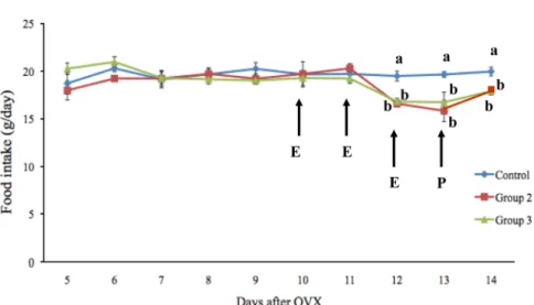
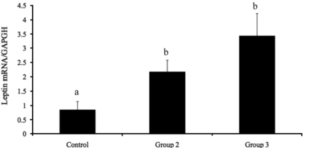
Effects of estrogen on food intake, serum leptin levels and leptin mRNA expression in adipose tissue of female rats
Wirasak Fungfuang
1,5, Misao Terada
2, Noriyuki Komatsu
3, Changjong Moon
4, Toru R. Saito
1*
1
Behavioral Neuroscience Laboratory, Graduate School of Veterinary Medicine, Nippon Veterinary and Life Science University, Musashino, Tokyo, Japan
2
Department of Laboratory Animal Science, Dokkyo University School of Medicine, Mibu, Tochigi, Japan
3
Civil International Corporation, Ueno, Taito, Japan
4
Department of Veterinary Anatomy, College of Veterinary Medicine, Chonnam National University, Gwangju, Korea
5