The Association of Heart Rate Variability with Parkinsonian Motor Symptom Duration
Dorji Harnod,
1Shu-Hui Wen,
2Shin-Yuan Chen,
3and Tomor Harnod
31Surgical Intensive Care Unit, Shin Kong Wu Ho-Su Memorial Hospital, Taipei;
2Department of Public Health, College of Medicine, Tzu Chi University, Hualien;
3Department of Neurosurgery, Buddhist Tzu Chi General Hospital, College of Medicine, Tzu Chi University, Hualien, Taiwan.
Received: September 30, 2013 Revised: February 5, 2014 Accepted: February 20, 2014
Corresponding author: Dr. Tomor Harnod, Department of Neurosurgery,
Buddhist Tzu Chi General Hospital, College of Medicine, Tzu Chi University, 707, Sec. 3, Chung-Yang Road, Hualien, Taiwan.
Tel: 886 3 856 1825 ext. 2228, Fax: 886 3 857 7161 E-mail: [email protected]
∙ The authors have no financial conflicts of interest.
© Copyright:
Yonsei University College of Medicine 2014 This is an Open Access article distributed under the terms of the Creative Commons Attribution Non- Commercial License (http://creativecommons.org/
licenses/by-nc/3.0) which permits unrestricted non- commercial use, distribution, and reproduction in any medium, provided the original work is properly cited.
Purpose: Impaired cardiovascular autonomic regulation is a non-motor symptom of Parkinson’s disease (PD) and may increase long-term morbidity. This study ap- plied frequency-domain analysis of heart rate variability (HRV) to understand the progression of sympathetic and parasympathetic cardiac regulation in patients with PD. Materials and Methods: In this cross-sectional study, 21 male and 11 female Taiwanese patients with advanced PD and 32 healthy gender- and age-matched subjects were enrolled. To minimize artifacts due to subject motion, daytime elec- trocardiograms for 5 minutes were recorded in awake patients during levodopa-on periods and controls. Using fast Fourier transformation, heart rate variables were quantified into a high-frequency power component [0.15--0.45 Hz, considered to reflect vagal (parasympathetic) regulation], low-frequency power component (0.04--0.15 Hz, reflecting mixed sympathetic and parasympathetic regulation), and low-frequency power in normalized units (reflecting sympathetic regulation). The significance of between-group differences was analyzed using the paired t-test.
Pearson correlation analysis and stepwise regression analysis were applied to as- sess the correlation of patient age, PD duration, and disease severity (represented by the Unified Parkinson’s Disease Rating Scale) with each heart rate variables.
Results: Impaired HRV is significantly correlated with the duration of PD, but not with disease severity and patient age. Meanwhile, parasympathetic heart rate vari- able is more likely than sympathetic heart rate variable to be affected by PD. Con- clusion: PD is more likely to affect cardiac parasympathetic regulation than sym- pathetic regulation by time and the heart rate variables have the association with Parkinsonian motor symptom duration.
Key Words: Autonomic, heart rate variability, Parkinson’s disease
INTRODUCTION
Cardiovascular autonomic regulation has been reported to be impaired in Parkin-
son’s disease (PD) and may increase the long-term morbidity of patients with this
disease.
1-3Moreover, the deterioration of functional performance in Parkinsonian pa-
tients with impaired autonomic function may be more rapid, and these patients prob-
ing movements, the time required to walk a distance of 7 meters, tremorography, cognitive performance (the Mini- Mental State Examination score), and brain magnetic reso- nance imaging images.
For ruling out the autonomic deterioration from other med- ical issues, none of the enrolled patients had evidence of ar- rhythmia, ischemic heart disease, heart failure, diabetes mel- litus, multiple system atrophy, pure autonomic failure, PD with dementia as well as Parkinsonism with other brain dis- eases, such as traumatic brain injury or stroke.
9Patients who were taking propranolol or atenolol were also excluded be- cause of the sympatholytic effects of such medications.
Thirty-two age- and gender-matched healthy subjects were enrolled as the control group. The study protocol was ap- proved by the Institutional Review Board of the Buddhist Tzu Chi General Hospital. All of the subjects gave their written informed consent at enrollment.
Heart rate recording and frequency-domain analysis of HRV
Since many muscle tremors would be recorded in a Parkin- sonian patient during a long-term heart rate recording, espe- cially in the levodopa-off period (without levodopa or do- pamine agonist, etc. for at least 12 hours), daytime electro- cardiograms (ECG) for 5 min were recorded in awake patients during levodopa-on periods (with levodopa use).
Each subject lay quietly in a comfortable head-up 45-de- gree position during the heart rate recording. Lead I ECG signals were recorded using an analog-to-digital converter ably require higher dosage of levodopa supplementation.
3Heart rate change is primarily determined by cardiac auto- nomic regulation. Heart rate variability (HRV) is defined by irregularities in the interval between normal sinus beats.
4,5Frequency-domain analysis of HRV is a sophisticated and non-invasive tool for studying sympathetic and parasympa- thetic regulation of heart rate. The standard procedures and interpretation of HRV analysis were first reported in 1996.
6We have applied a modification of these procedures to in- vestigate cardiac autonomic dysregulation in children with epilepsy.
7In this case-control study on a cohort of patients with advanced PD, we used the same technology to investi- gate the changes of HRV in adult Parkinsonian patients.
MATERIALS AND METHODS
Study population
We enrolled 32 Taiwanese patients with PD (21 male and 11 female; mean age: 62.2 years, range: 44--79 years), who planned to be treated by subthalamic deep brain stimulation at the Buddhist Hualien Tzu Chi General Hospital, Taiwan (Table 1). All patients met the clinical criteria for PD that at least two of the cardinal symptoms are present. The core as- sessment program including an acute levodopa test to mea- sure the effects of levodopa on PD was used in all patients.
8The following was assessed: Unified Parkinson’s Disease Rating Scale (UPDRS) score, behavior from videotaped clips, Hoehn and Yahr (H-Y) stage, timing of rapid alternat-
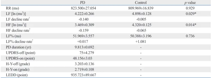
Table 1. Clinical Features and Heart Rate Variables of Age- and Sex-Matched PD and Control Groups
PD Control p value
RR (ms) 823.500±27.054 809.969±16.839 0.929
LF [ln (ms2)] 4.222±0.266 4.898±0.128 0.029*
LF decline rate† -0.140 -0.005
HF [ln (ms2)] 3.469±0.309 4.320±0.125 0.014*
HF decline rate† -0.159 -0.065
LF% (nu) 51.969±3.557 50.388±3.196 0.736
LF% decline rate† +0.017 +1.081
PD duration (yr) 9.813±0.692 -
UPDRS-off (point) 75±4.279 -
UPDRS-on (point) 48.156±3.03 -
H-Y-off (grade) 3.203±0.136 -
H-Y-on (grade) 2.719±0.108 -
LEDD (point) 935.723±89.667 -
RR, interval between two neighboring R waves; LF, low frequency power; HF, high frequency power; LF%, LF/(HF+LF) in normalized units; PD, Parkinson’s disease; UPDRS-off, Unified Parkinson’s Disease Rating Scale in levodopa-off period; UPDRS-on, Unified Parkinson’s Disease Rating Scale in levodopa-on period; H-Y-off, Hoehn and Yahr stage in levodopa-off period; H-Y-on, Hoehn and Yahr stage in levodopa-on period; LEDD, levodopa equivalent daily dose.
*p<0.05.
†The estimated change of value/year of duration.
HF values between PD and control groups were significant.
The PD group had significantly lower LF [4.222±0.266 ln (ms
2) vs. 4.898±0.128 ln (ms
2), p=0.028] and lower HF [3.469±0.309 ln (ms
2) vs. 4.320±0.125 ln (ms
2), p=0.029]
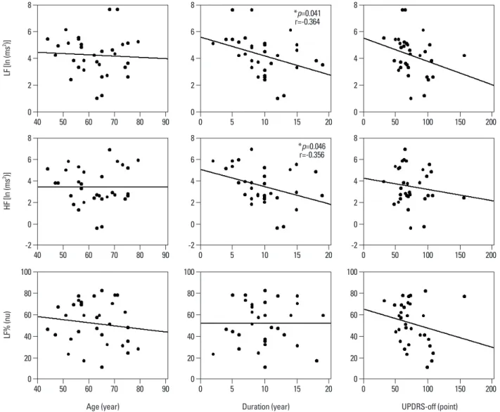
when compared to the age- and gender-matched control group. In the patient group, Pearson correlation analysis re- vealed that the rate of LF decline significantly correlated with PD duration (r=-0.364, p=0.041) but not with disease severity (UPDRS-off) and patient age (Fig. 1, Table 2).
Similarly, the results indicated that lower HF was correlated with longer PD duration (r=-0.356, p=0.046). After adjust- ment for possible confounders, a stepwise regression using age, gender, PD duration, and UPDRS-off score still found significant correlation of heart rate variables such as LF and HF with long PD duration (Table 3). Furthermore, the re- sults indicated that the duration of PD explained 13.3% and 12.7% of the variance in LF and HF measures, respectively.
No variables were significantly correlated with the LF%.
Meanwhile, increase in UPDRS-off was significantly corre- lated with PD duration with a slope of 2.578 points per year.
DISCUSSION
Cardiovascular autonomic dysregulation in PD has been at- tributed to either central or peripheral autonomic regulatory impairment,
1-3,11-14involving the hypothalamus, insular cor- tex, locus coeruleus, dorsal motor nucleus of the vagus, in- termediolateral nucleus of the thoracic cord, sympathetic ganglia, and sacral parasympathetic nuclei. Thus, PD af- fects both sympathetic and parasympathetic divisions, each of which can be evaluated by frequency-domain analysis of HRV.
4-7In this study, although the sympathetic indicator of autonomic cardiac regulation, LF%, was not significantly changed, the total and parasympathetic indicators (LF and HF) decreased significantly and correlated with disease du- ration. Some previous studies reported that levodopa re- placement in Parkinsonian patients could decrease central sympathetic outflow, an unusual effect caused by the cen- tral D2 agonist action of levodopa.
15,16Based on it, some au- thors recommended monoamine oxidase B inhibitors to avoid the effect of levodopa on sympathetic regulation.
17We thought that the discordant changes in autonomic sympa- thetic and parasympathetic divisions in our results might stem from the effects of levodopa treatment and leave the cardiac parasympathetic dysregulation being more sensitive in the HRV study.
with a sampling rate of 512 Hz. Frequency-domain analysis was performed using a nonparametric method of fast Fouri- er transformation (FFT). The direct current component was deleted and a Hamming window was used to attenuate the leakage effect. For each time segment (288 s; 2048 data points), our algorithm estimated the power spectrum densi- ty on the basis of FFT. The resulting power spectrum was corrected for attenuation resulting from the sampling pro- cess and the use of a Hamming window.
10The power spec- trum was subsequently quantified into standard frequency- domain measurements as defined by the Task Force of the European Society of Cardiology and the North American Society of Pacing and Electrophysiology. The frequency- domain measurements included R-R intervals (the intervals between two neighboring R waves, RR) and heart rate vari- ables: high-frequency power (0.15--0.45 Hz, HF), low-fre- quency power (0.04--0.15 Hz, LF), and LF% [LF/(HF+LF) in normalized units]. The HF and LF data were logarithmi- cally transformed to correct for any skew in the distribu- tion. The LF reflected contributions from mixed sympathet- ic and parasympathetic divisions. The HF was considered to reflect vagal (parasympathetic) regulation and the LF%
was considered to mirror sympathetic regulation.
6,7,10Statistical analysis
All measures are presented as the mean and standard error (SE) of the mean. Differences in clinical features and heart rate variables between the matched PD and control groups were analyzed by the paired t-test. Pearson correlation coef- ficient (r) was used to measure correlations of patient age, PD duration, and disease severity (represented by UPDRS score in levodopa-off period, UPDRS-off) with heart rate variables including LF, HF, and LF%. Stepwise regression analysis was conducted to identify those factors associated with heart rate variables. All analyses were performed us- ing SPSS (now called PASW) version 17.0 (SPSS Inc., Chicago, IL, USA) and statistical assessments were evalu- ated at the 0.05 level of significance.
RESULTS
Table 1 demonstrated that the PD group had 9.813±0.692
years (mean±SE) in the duration of disease. The UPDRS
scores and H-Y stages of the PD group were: 75±4.279 in
UPDRS-off, 48.156±3.03 in UPDRS-on, 3.203±0.136 in
H-Y-off, and 2.719±0.108 in H-Y-on. Differences in LF and
cular autonomic regulation. In this study with focus on pure PD itself, we excluded the patients with cardiac arrhythmia, ischemic heart disease, heart failure, diabetes mellitus, mul- PD is not a rare degenerative disorder in aged population.
However, aged people usually suffer from multiple degener- ative diseases and many of those might affect the cardiovas-
Table 2. Correlations of LF, HF, and LF% with Patient Age, PD Duration and UPDRS-Off Score
Age PD duration UPDRS-off
LF
Pearson correlation -0.059 -0.364 -0.279
p value 0.749 0.041* 0.122
HF
Pearson correlation 0.004 -0.356 -0.142
p value 0.981 0.046* 0.437
LF%
Pearson correlation -0.133 0.003 -0.213
p value 0.468 0.986 0.241
LF, low frequency power; HF, high frequency power; LF%, LF/(HF+LF) in normalized units; PD, Parkinson’s disease; UPDRS-off, Unified Parkinson’s Disease Rating Scale in levodopa-off period.
*p<0.05.
Fig. 1. The correlations of patient age, PD duration, and UPDRS-off score with heart rate variables. Pearson correlation analysis showed that the rates of LF and HF decline were significantly correlated with PD duration. LF, low frequency power; HF, high frequency power; LF%, LF/(HF+LF) in normalized units;
UPDRS-off, Unified Parkinson’s Disease Rating Scale in levodopa-off period; PD, Parkinson’s disease. *p<0.05.
0
-2
0
0
-2
0
0
-2
0 2
2
0
40
20
2
2
0
40
20
2
2
0
40
20 4
4
60
4
4
60
4
4
60 6
6
80
6
6
80
6
6
80 8
8
100
8
8
100
8
8
100 LF [ln (ms2 )]HF [ln (ms2 )]LF% (nu)
40
40
40
Age (year) Duration (year) UPDRS-off (point)
0
0
0
0
0
0 50
50
50
5
5
5
50
50
50 60
60
60 70
70
70
10
10
10
100
100
100 80
80
80
15
15
15
150
150
150 90
90
90
20
20
20
200
200
200
*p=0.041 r=-0.364
*p=0.046 r=-0.356
tor affecting the results of cardiac autonomic regulation in the patients of this study; third, to minimize the artifacts from muscle tremor, we decided that the better experimen- tal design was to combine short-term heart rate recordings with HRV spectral analysis during the levodopa-on peri- od.
20,21However, all heart rate variables were recorded and analyzed during daytime to avoid major circadian effects.
22Obtaining recordings and heart rate variables in gender- and age-matched subjects in the same position can minimize in- ter-individual anthropometric and posture confounding ef- fects. Therefore, we believe that the results of this case-con- trol study show that PD duration is the main factor affecting changes in HRV. In the future, we are planning to study pa- tients either in the early or late stages of their disease to elu- cidate more about the development of autonomic dysregu- lation in patients with PD.
In conclusion, PD is more likely to affect cardiac para- sympathetic regulation than sympathetic regulation by time, and the heart rate variables have the association with Par- kinsonian motor symptom duration.
REFERENCES
1. Jost WH. Autonomic dysfunctions in idiopathic Parkinson’s dis- ease. J Neurol 2003;250 Suppl 1:I28-30.
2. Oka H, Mochio S, Onouchi K, Morita M, Yoshioka M, Inoue K.
Cardiovascular dysautonomia in de novo Parkinson’s disease. J Neurol Sci 2006;241:59-65.
3. Lucetti C, Gambaccini G, Del Dotto P, Ceravolo R, Logi C, Rossi G, et al. Long-term clinical evaluation in patients with Parkinson’s disease and early autonomic involvement. Parkinsonism Relat Disord 2006;12:279-83.
4. Vanderlei LC, Pastre CM, Hoshi RA, Carvalho TD, Godoy MF.
Basic notions of heart rate variability and its clinical applicability.
Rev Bras Cir Cardiovasc 2009;24:205-17.
5. Stein PK, Bosner MS, Kleiger RE, Conger BM. Heart rate vari- ability: a measure of cardiac autonomic tone. Am Heart J 1994;
127:1376-81.
6. Heart rate variability. Standards of measurement, physiological in- terpretation, and clinical use. Task Force of the European Society of Cardiology and the North American Society of Pacing and
tiple system atrophy, pure autonomic failure, PD with de- mentia as well as Parkinsonism with other brain diseases, such as traumatic brain injury or stroke, at the enrollment. All of them were ever reported to affect the autonomic regulation and might interfere the study results. Although we had only 32 patients left in the study group, the statistically significant results made us believe that the study number was enough to make the unquestionable conclusions in the association between the heart rate variables and PD duration. These re- sults are important because cardiovascular autonomic dys- function is a relatively under-recognized problem of PD and is related to the management in disease progression. It may be associated with the patients’ life quality, and life span, and then may increase care-givers’ burdens.
The progressive loss of cardiac autonomic regulation, both in sympathetic and parasympathetic divisions from middle age to old age is correlated with the aging itself.
10,18In the present study, the estimated annual rate of LF (-0.140 vs. -0.005) and HF (-0.159 vs. -0.065) decline was faster in the PD patients aged 44 to 79 years (Table 1). Meanwhile, the UPDRS-off increased at an estimated rate of 2.578 points per year (in agreement with a report from western coun- tries),
19and UPDRS-off significantly correlates with dis- ease duration. Since there is no standard way to grade the function of the autonomic nervous system using the UP- DRS, applying heart rate variables as progressing biomark- ers in PD may be suggested.
There were 32 patients with mean age of 62.2 years and mean disease duration of 9.8 years enrolled in this study.
Their mean H-Y stage was 3.2 and mean UPDRS was 75 during the levodopa-off period because all enrolled patients at our hospital were receiving subthalamic deep brain stimu- lation for advanced PD. The major limitations of this study were; first, no subjects with early stage PD were enrolled because the cost of deep brain stimulation is still relatively high and cannot routinely be given in early stage of the dis- ease; second, the use of a combination of anti-Parkinsonian medications for advanced PD could be a confounding fac- Table 3. Relationships between LF, HF, and PD Duration
Variable Factor Regression
coefficient 95% confidence interval r square p value
LF Constant 5.597 4.188 7.007 <0.001
Duration -0.14 -0.274 -0.006 0.133 0.041*
HF Constant 5.03 3.388 6.672 <0.001
Duration -0.159 -0.315 -0.003 0.127 0.046*
LF, low frequency power; HF, high frequency power; PD, Parkinson’s disease.
*p<0.05.
neider J, et al. Effects of subthalamic nucleus stimulation and le- vodopa on the autonomic nervous system in Parkinson’s disease. J Neurol Neurosurg Psychiatry 2007;78:742-5.
16. Bouhaddi M, Vuillier F, Fortrat JO, Cappelle S, Henriet MT, Rumbach L, et al. Impaired cardiovascular autonomic control in newly and long-term-treated patients with Parkinson’s disease: in- volvement of L-dopa therapy. Auton Neurosci 2004;116:30-8.
17. Löhle M, Reichmann H. Controversies in neurology: why mono- amine oxidase B inhibitors could be a good choice for the initial treatment of Parkinson’s disease. BMC Neurol 2011;11:112.
18. Byrne EA, Fleg JL, Vaitkevicius PV, Wright J, Porges SW. Role of aerobic capacity and body mass index in the age-associated de- cline in heart rate variability. J Appl Physiol (1985) 1996;81:743- 50.
19. Jankovic J, Kapadia AS. Functional decline in Parkinson disease.
Arch Neurol 2001;58:1611-5.
20. Kallio M, Suominen K, Bianchi AM, Mäkikallio T, Haapaniemi T, Astafiev S, et al. Comparison of heart rate variability analysis methods in patients with Parkinson’s disease. Med Biol Eng Com- put 2002;40:408-14.
21. Lucreziotti S, Gavazzi A, Scelsi L, Inserra C, Klersy C, Campana C, et al. Five-minute recording of heart rate variability in severe chronic heart failure: correlates with right ventricular function and prognostic implications. Am Heart J 2000;139:1088-95.
22. Nakagawa M, Iwao T, Ishida S, Yonemochi H, Fujino T, Saikawa T, et al. Circadian rhythm of the signal averaged electrocardio- gram and its relation to heart rate variability in healthy subjects.
Heart 1998;79:493-6.
Electrophysiology. Eur Heart J 1996;17:354-81.
7. Harnod T, Yang CC, Hsin YL, Shieh KR, Wang PJ, Kuo TB.
Heart rate variability in children with refractory generalized epi- lepsy. Seizure 2008;17:297-301.
8. Langston JW, Widner H, Goetz CG, Brooks D, Fahn S, Freeman T, et al. Core assessment program for intracerebral transplanta- tions (CAPIT). Mov Disord 1992;7:2-13.
9. Takase B, Kurita A, Noritake M, Uehata A, Maruyama T, Nagayo- shi H, et al. Heart rate variability in patients with diabetes mellitus, ischemic heart disease, and congestive heart failure. J Electrocar- diol 1992;25:79-88.
10. Kuo TB, Lin T, Yang CC, Li CL, Chen CF, Chou P. Effect of ag- ing on gender differences in neural control of heart rate. Am J Physiol 1999;277(6 Pt 2):H2233-9.
11. Benarroch EE, Schmeichel AM, Parisi JE. Involvement of the ventrolateral medulla in parkinsonism with autonomic failure.
Neurology 2000;54:963-8.
12. Papapetropoulos S, Mash DC. Insular pathology in Parkinson’s disease patients with orthostatic hypotension. Parkinsonism Relat Disord 2007;13:308-11.
13. Wakabayashi K, Takahashi H. Neuropathology of autonomic ner- vous system in Parkinson’s disease. Eur Neurol 1997;38 Suppl 2:2-7.
14. Isaias IU, Marotta G, Pezzoli G, Sabri O, Schwarz J, Crenna P, et al. Enhanced catecholamine transporter binding in the locus coe- ruleus of patients with early Parkinson disease. BMC Neurol 2011;11:88.
15. Ludwig J, Remien P, Guballa C, Binder A, Binder S, Schattsch-