A Simple and Efficient Multiplex PCR Assay for the
Identification of Mycobacterium Genus and Mycobacterium tuberculosis Complex to the Species Level
Yeun Kim,1 Yeonim Choi,1 Bo-Young Jeon,1 Hyunwoo Jin,1,2 Sang-Nae Cho,3 and Hyeyoung Lee1
1Department of Biomedical Laboratory Science, College of Health Sciences, Yonsei University, Wonju;
2Department of Clinical Laboratory Science, College of Health Sciences, Catholic University of Pusan, Busan;
3Department of Microbiology, Yonsei University College of Medicine, Seoul, Korea.
Received: September 19, 2012 Revised: October 25, 2012 Accepted: October 29, 2012
Corresponding author: Dr. Hyeyoung Lee, Department of Biomedical Laboratory Science, College of Health Sciences, Yonsei University, 1 Yonseidae-gil, Wonju 220-710, Korea.
Tel: 82-33-760-2740, Fax: 82-33-760-2561 E-mail: [email protected]
∙ The authors have no financial conflicts of interest.
© Copyright:
Yonsei University College of Medicine 2013 This is an Open Access article distributed under the terms of the Creative Commons Attribution Non- Commercial License (http://creativecommons.org/
licenses/by-nc/3.0) which permits unrestricted non- commercial use, distribution, and reproduction in any medium, provided the original work is properly cited.
Purpose: The Mycobacterium tuberculosis complex comprises M. tuberculosis, M. bovis, M. bovis bacillus Calmette-Guérin (BCG) and M. africanum, and causes tuberculosis in humans and animals. Identification of Mycobacterium spp. and M.
tuberculosis complex to the species level is important for practical use in microbi- ological laboratories, in addition to optimal treatment and public health. Materials and Methods: A novel multiplex PCR assay targeting a conserved rpoB sequence in Mycobacteria spp., as well as regions of difference (RD) 1 and RD8, was devel- oped and evaluated using 37 reference strains and 178 clinical isolates. Results:
All mycobacterial strains produced a 518-bp product (rpoB), while other bacteria produced no product. Virulent M. tuberculosis complex strains, M. tuberculosis, M. bovis and M. africanum, produced a 254-bp product (RD1), while M. bovis BCG, M. microti and nontuberculous mycobacteria produced no RD1 region product. Additionally, M. tuberculosis and M. africanum produced a 150-bp prod- uct (RD8), while M. bovis and M. bovis BCG produced a 360-bp product (deleted form of RD8). M. microti and nontuberculous mycobacteria produced no RD8 re- gion product. This assay identified all Mycobacterium spp. and all M. tuberculosis complex strains to the species level. Conclusion: The multiplex PCR assay of the present study could be implemented as a routine test in microbiology laboratories, and may contribute to more effective treatment and surveillance of tuberculosis stemming from the M. tuberculosis complex.
Key Words: M. tuberculosis complex, multiplex PCR, rpoB, RD1, RD8
INTRODUCTION
Tuberculosis is a global health problem, with approximately 8.8 million new cases and 1.4 million deaths reported in 2010.1 The disease is caused by members of the Mycobacterium tuberculosis (M. tuberculosis) complex, a group of closely related species and subspecies, which include M. tuberculosis, M. africanum, M. bovis, M.
bovis bacillus Calmette-Guérin (BCG), M. microti, M. canettii, M. pinnipedii, and
berculosis and M. bovis, but is absent in M. bovis BCG, al- lowing for M. tuberculosis and M. bovis to be differentiated from M. bovis BCG.11,12 Additionally, a novel RD8 is pres- ent in M. tuberculosis and M. africanum that is deleted in M. bovis and M. bovis BCG and absent in M. microti and nontuberculous mycobacteria.14
In this study, we developed a rapid and efficient multi- plex PCR assay that enables not only the identification of the genus Mycobacterium using the rpoB sequence con- served in all mycobacteria, but also the identification of the M. tuberculosis complex to the species level, using RD1 and RD8 sequences. The assay was shown to be simple and efficient in identifying Mycobacterium genus and distin- guishing the M. tuberculosis complex to the species level.
MATERIALS AND METHODS
Bacterial strains
The bacterial strains used in this study are listed in Table 1.
Seventy-six clinical M. tuberculosis isolates from humans and 52 clinical M. bovis isolates from cattle were provided kindly from Yonsei University College of Medicine, Seoul, Korea. Twenty-seven clinical M. avium isolates and 23 clini- cal M. intracellulare isolates from humans were provided by the Korea Institute of Tuberculosis, Osong, Korea.
Culture media and DNA extraction
Bacterial culture and chromosomal DNA isolation were performed as previously described.13 Briefly, bacteria were cultured on Middlebrook 7H9 media (Difco, Detroit, MI, USA) supplemented with 10% Middlebrook OADC En- richment media (BBL, Sparks, MD, USA) at 37°C for 2 to 4 weeks. Bacterial cells were collected by centrifugation at 10000×g, resuspended in distilled water, and boiled for 10 min. Cell debris was sedimented by centrifugation for 2 min at 10000×g, and the resultant DNA concentration was measured using a spectrophotometer (260 nm, Amersham Pharmacia Biotech Inc., Piscataway, NJ, USA). DNA was stored at -20°C prior to use.
PCR primer design
The primers used in this study are listed in Table 2. The ge- nome sequence of M. tuberculosis H37Rv was imported into the Vector NTI® Software (Life technologies, Grand Is- land, NY, USA), and rpoB, RD1 and RD8 were created ac- cording to the genome address. Primers were selected by M. mungi.2 M. tuberculosis is the most common cause of
tuberculosis in humans. M. bovis causes tuberculosis in hu- mans and animals, and is responsible for 10 to 15% of new human tuberculosis cases in developing countries.3 M. bo- vis BCG, a live attenuated strain of M. bovis, is a widely used anti-tuberculosis vaccine, and is used for the treatment of bladder cancer.4 However, M. bovis BCG is known to lead to complications, such as osteomyelitis, abscesses, lymphadenitis, and dissemination of M. bovis BCG, most severely in human immunodeficiency virus-infected neo- nates and children. In M. bovis BCG-treated patients, com- plications such as pneumonitis, hepatitis and noncaseating granulomas have been reported.5
Identification of the M. tuberculosis complex to the spe- cies level is important for public health, epidemiology and treatment.6 In the laboratory, tuberculosis is diagnosed by acid-fast bacilli (Ziehl-Neelsen) staining and culturing of mycobacteria, followed by identification of the cultured mycobacteria. For more rapid identification, several poly- merase chain reaction (PCR)-based methods have been de- veloped, and PCR assay that targets M. tuberculosis-specific sequences, including IS6110 and the MPT64 gene, has been widely applied.7 However, while this assay can differentiate the M. tuberculosis complex from nontuberculous myco- bacteria, it cannot identify the M. tuberculosis complex to the species level.7 For rapid and efficient identification of the M. tuberculosis complex, several PCR-based methods have been developed.8-10 However, these are rarely used as a routine test in diagnostic laboratories as they require two rounds of PCR reaction and electrophoresis to distinguish the M. tuberculosis complex from other mycobacteria and to identify M. tuberculosis complex to the species level.11,12 Moreover, identification of nontuberculous mycobacteria to the species level is of greater importance due to the in- creased incidence of infection with nontuberculous myco- bacteria. Thus, more rapid and efficient methods of identifi- cation are needed.
A well-validated probe for the Mycobacterium genus tar- gets the conserved sequence of the rpoB gene, which en- codes the β subunit of RNA polymerase, and not only facil- itates identification of all Mycobacterium spp., but also distinguishes Mycobacterium spp. at the species level by restriction enzyme analysis.13 In a comparative genomics study of M. tuberculosis H37Rv, 16 regions of the genome (regions of difference, RD) were shown to be deleted in M.
bovis and M. bovis BCG.10 One of them, RD1, is present in virulent M. tuberculosis complex strains, including M. tu-
1.5 units of Taq DNA polymerase (Bioneer, Daejeon, Ko- rea), and 20 ng of genomic DNA as the template. PCR am- plification was performed in an automated thermal cycler (PTC-100 Programmable Thermal Controller, MJ Research, Inc., Waltham, MA, USA). The cycling parameters were 94°C for 5 min, followed by 35 cycles of denaturation at 94°C for 30 s, annealing at 68°C for 30 s and a final exten- sion at 72°C for 7 min. Sterile distilled water was used as a negative control. Analysis of PCR products was performed by electrophoresis in ethidium bromide (0.5 µg/mL)-stained 1.7 % (w/v) agarose gels.
RESULTS
Validation of the PCR method
PCR conditions were optimized with four sets of primer pairs using M. tuberculosis H37Rv, M. bovis AN5 and M.
bovis BCG Pasteur as representative strains (Figs. 1 and 2), and confirmed using all bacterial strains by gel electropho- resis. The PCR products for each target sequence from each set of primer pairs were also confirmed. All reference my- cobacterial strains consistently yielded a single 518-bp product with primers rpoB-F and rpoB-R, but other bacteri- al strains yielded no products, indicating that all mycobac- terial strains contained the specific rpoB gene (Table 3). M.
tuberculosis H37Rv, M. africanum and M. bovis AN5 con- sistently yielded a single 254-bp product with RD1-F and RD1-R primers, but other bacterial strains, including all M.
bovis BCG strains and M. microti, yielded no product with RD1-F and RD1-R primers, indicating the RD1 sequence is present in virulent M. tuberculosis complex strains. M. tu- berculosis and M. africanum type strains consistently yield- ed a single 150-bp product with RD8-F and RD8-T-R prim- ers, but yielded no product with RD8-F and RD8-T-R primers, indicating the presence of the RD8 sequence. Con- versely, M. bovis and all M. bovis BCG strains consistently yielded a single 360-bp product with RD8-F and RD8-B-R primers, but yielded no product with RD8-F and RD8-T-R primers, indicating a deletion of the RD8 sequence in M.
bovis, including M. bovis BCG (Fig. 1, Table 3). However, other bacteria, including nontuberculous mycobacteria and M. microti, yielded no product with either RD8-F and RD8- T-R or RD8-F and RD8-B-R primers.
Multiplex PCR on reference strains and clinical isolates PCR products using the four sets of primer pairs were per- analysis of the appropriate genomic regions using Primer 3
software (http://frodo.wi.mit.edu/) (Fig. 1, Table 2).
PCR amplification
The amplification reaction mixture consisted of 10 mM Tri- HCl, 50 mM KCl, 1.5 mM MgCl2, 200 µM of each primer, Table 1. Bacterial Strains Used in This Study
Species (no. of strains) Reference no./Source
M. tuberculosis H37Rv ATCC27294
M. tuberculosis Erdman ATCC35801
M. tuberculosis (76) YUMC
M. bovis AN5 ATCC35726
M. bovis (52) YUMC
M. bovis BCG Pasteur YUMC
M. bovis BCG Danish YUMC
M. bovis BCG Tokyo YUMC
M. bovis BCG Tice YUMC
M. abscessus ATCC19977
M. africanum ATCC25420
M. avium ATCC25291
M. avium ATCC35719
M. avium (27) KIT
M. celatum ATCC51130
M. celatum ATCC51131
M. chelonae ATCC19237
M. fortuitum ATCC49403
M. fortuitum ATCC49404
M. gilvum ATCC43909
M. gordonae ATCC14470
M. intracellulare ATCC13950
M. intracellulare (23) KIT
M. kansasii ATCC12478
M. marinum ATCC927
M. microtii ATCC19422
M. mucogenicum ATCC49649
M. peregrinum ATCC14467
M. senegalense ATCC35796
M. septicum ATCC700731
M. smegmatis ATCC19420
M. szulgai ATCC 35799
M. terrae ATCC15755
M. ulcerans ATCC19423
Enterococcus faecalis ATCC29212
E. faecium ATCC35667
Escherichia coli ATCC25922
Haemophilus infulenza ATCC49247
Micrococcus lutens ATCC49732
Pseudomonas aeroginosa ATCC27853
Staphylococcus aureus ATCC29213
YUMC, Yonsei University College of Medicine, Seoul, Korea; KIT, Korea Institute of Tuberculosis, Osong, Korea; BCG, bacillus Calmette-Guérin.
results identical to M. tuberculosis (Fig. 3). However, multi- plex PCR of M. microti yielded only a 518-bp product, simi- lar to nontuberculous mycobacteria.
Multiplex PCR on M. bovis AN5 and 52 clinical M. bovis isolates yielded 518-, 360- and 254-bp products, indicating the presence of rpoB and RD1, as well as the deletion of RD8 (Fig. 5). All four M. bovis BCG strains yielded 518- and 360-bp products, indicating the presence of rpoB, the formed on 30 reference strains and 178 clinical isolates
(Figs. 3, 4 and 5). Multiplex PCR of M. tuberculosis H37Rv and M. tuberculosis Erdman yielded three 518-, 254- and 150-bp products, indicating the presence of rpoB, RD1 and RD8, respectively (Fig. 4). Multiplex PCR of 76 clinical M.
tuberculosis isolates provided results identical to the M. tu- berculosis reference strain (Fig. 4). Multiplex PCR of M. af- ricanum, a member of the M. tuberculosis complex, yielded
Fig. 2. Multiplex PCR for M. tuberculosis, M. bovis and M. bovis bacillus Calmette-Guérin (BCG) strains. PCR products were separated by electrophoresis and visualized. Lanes M: molecular size marker (100-bp ladder); 1: M. bovis BCG Pasteur; 2: M. bovis BCG Tice; 3: M. bovis BCG Tokyo; 4: M. bovis AN5; 5: M.
tuberculosis H37Rv; 6: negative control. Expected sizes of PCR products are shown. RD, regions of difference; PCR, polymerase chain reaction; ESAT-6, early secreted antigenic target 6 kDa protein.
500 300 200 100
rpoB (518 bp) RD8 absent (518 bp) ESAT-6 (254 bp) RD8 present (150 bp) bp M 1 2 3 4 5 6
Fig. 1. Schematic diagram showing regions flanking the direct repeat region in M. tuberculosis H37Rv, M. bovis and M. bovis bacillus Calmette-Guérin (BCG). This region is specifically deleted in M. bovis and M. bovis BCG, indicated by dotted lines. Primer positions are indicated by small arrows. RD, regions of difference.
M. tuberculosis &
M. africanum
Region specifically deleted in M. bovis and M. bovis BCG
M. bovis & BCG
Rv3616c ephA Rv3618 esxV esxW PPE65 PE32 IpqG hpt
Rv3616c lpqG hpt
(150 bp)
(360 bp)
(-6.3 Kb) RD8
RD8
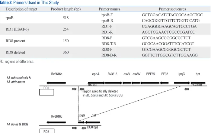
Table 2. Primers Used in This Study
Description of target Product length (bp) Primer names Primer sequences
rpoB 518 rpoB-F GCTGGACATCTACCGCAAGCTGC
rpoB-R CAGCGGGTTGTTCTGGTCCATG
RD1 (ESAT-6) 254 RD1-F CGAGGGGAAGCAGTCCCTGA
RD1-R AGGTCGAACTCGCCCGATCC
RD8 present 150 RD8-F GTCGAAGCGGGGCGCTCT
RD8-T-R GCGCAACGGATTTCCATCGT
RD8 deleted 360 RD8-F GTCGAAGCGGGGCGCTCT
RD8-B-R GGTTCTTGGCGTCTTGGAAGG
RD, regions of difference.
DISCUSSION
We developed a simple and effective multiplex PCR assay that enabled the differentiation of Mycobacteria spp. from other bacteria and identified the M. tuberculosis complex to the species level. Our analysis using reference strains and clinical isolates suggested that the multiplex PCR assay is specific for M. tuberculosis, M. bovis and M. bovis BCG, as well as nontuberculosis mycobacteria. The conserved rpoB sequence of the mycobacteria was used to differentiate the genus Mycobacterium from other bacteria, resulting in data specific for all Mycobacteria spp. tested.13 Moreover, the fragment length polymorphism of the 518-bp PCR prod- ucts targeting rpoB were effective for species identification (data not shown). This sequence offers advantages over oth- er target sequences, including IS6110 and 16S RNA. IS6110 can be used for the detection of M. tuberculosis complex, but cannot differentiate members of the M. tuberculosis complex.7 16S RNA has been used for the identification of Mycobacteria spp., but is unable to provide identification of absence of RD1 and the deletion of RD8. Multiplex PCR of
23 nontuberculous mycobacteria reference strains and 50 clinical nontuberculous mycobacteria isolates yielded only a 518-bp product, indicating the absence of RD1 and RD8.
Multiplex PCR of other non-mycobacterial bacteria yielded no positive results. The data were consistently reproducible, and complete concordance between independent research- ers was confirmed.
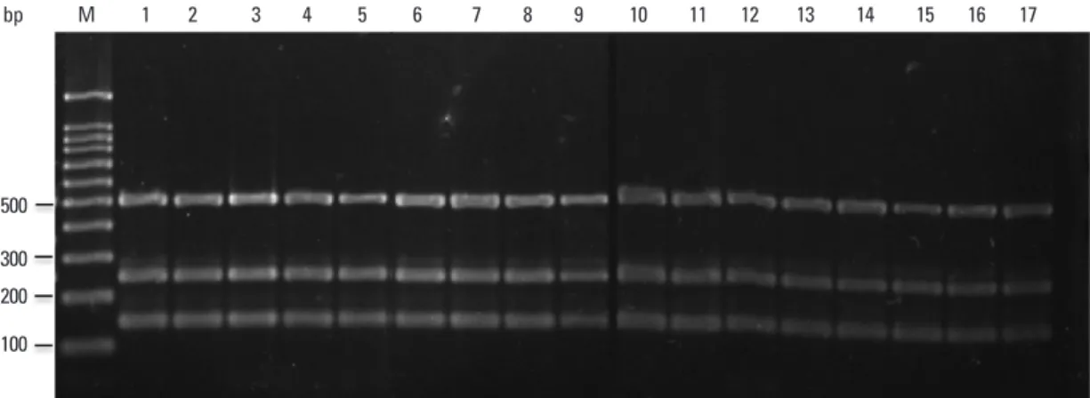
Fig. 3. Multiplex PCR for the Mycobacterium species. PCR products were separated by electrophoresis and visualized. Lanes M: molecular size marker (100- bp ladder); 1: M. avium; 2: M. africanum; 3: M. abscesses; 4: M. celatum; 5: M. chelonae; 6: M. fortuitum; 7: M. gordonae; 8: M. intracellulare; 9: M. kansasii;
10: M. marinum; 11: M. microtii; 12: M. phlei; 13: M. septicum; 14: M. szulgai; 15: M. terrae; 16: M. ulcerans; PCR, polymerase chain reaction.
Fig. 4. Multiplex PCR for clinical M. tuberculosis isolates. PCR products were separated by electrophoresis and visualized. Lanes M: molecular size marker (100-bp ladder); 1-15: M. tuberculosis isolates; 16: M. tuberculosis H37Rv; 17: M. tuberculosis Erdman; PCR, polymerase chain reaction.
500 300 200 100
bp M 1 2 3 4 5 6 7 8 9 10 11 12 13 14 15 16
500 300 200 100
bp M 1 2 3 4 5 6 7 8 9 10 11 12 13 14 15 16 17
Table 3. Signature Patterns of Positive and Negative Multi- plex PCR Used to Discriminate Mycobacterial Strains
Organism
PCR result for target
rpoB RD1 RD8
present RD8 deleted
M. tuberculosis + + + -
M. bovis + + - +
M. bovis BCG + - - +
M. africanum + + + -
M. microti + - - -
NTM + - - -
Other bacteria - - - -
RD, regions of difference; BCG, bacillus Calmette-Guérin; PCR, polymerase chain reaction; NTM, non-tuberculous Mycobacteria.
not used routinely in clinical microbiology laboratories as they are time consuming and costly. Recently, multiplex PCR assays have been proposed as a diagnostic method due to a single reaction and multiple targets.7,11,12 The present as- say is the first multiplex PCR assay shown to identify the Mycobacterium genus and distinguish the M. tuberculosis complex to the species level by targeting the rpoB sequence, as well as RD1 and RD8 sequences, respectively.
This assay can be used as a routine diagnostic test in mi- crobiology laboratories and may contribute to determination of an optimal treatment regimen, improve the surveillance of tuberculous disease due to members of the M. tuberculo- sis complex, and aid in the identification of the M. tubercu- losis complex and nontuberculous mycobacteria.
ACKNOWLEDGEMENTS
This study was supported by a grant of the Korea Health- care Technology R&D Project, Ministry of Health & Wel- fare, Republic of Korea (A010024, HYL) and by the Yon- sei University Research Fund of 2000 (in part).
REFERENCES
1. Raviglione M, Marais B, Floyd K, Lönnroth K, Getahun H, Mi- gliori GB, et al. Scaling up interventions to achieve global tuber- culosis control: progress and new developments. Lancet 2012;379:
1902-13.
2. Ernst JD, Trevejo-Nuñez G, Banaiee N. Genomics and the evolu- tion, pathogenesis, and diagnosis of tuberculosis. J Clin Invest 2007;117:1738-45.
3. de la Rua-Domenech R. Human Mycobacterium bovis infection in the United Kingdom: incidence, risks, control measures and re- view of the zoonotic aspects of bovine tuberculosis. Tuberculosis
M. tuberculosis complex to the species level, due to their high similarity.15 The present assay was based on the detec- tion of RD1 and RD8 in order to distinguish members of the M. tuberculosis complex. Using this assay, RD1 was detected in all M. tuberculosis complex strains, including M. tuberculosis, M. bovis and M. africanum, but not M. bo- vis BCG or M. microti, in accordance with previous re- ports.11,12 RD8 was detected in M. tuberculosis and M. afri- canum, but a deleted form of RD8 was detected in M. bovis and M. bovis BCG. Interestingly, this sequence was absent in M. microti even though M. microti belongs to the M. tu- berculosis complex. This result was in agreement with pre- vious reports that demonstrated the presence of RD8 in M.
tuberculosis, but not M. microti.16 In our study, the M. africa- num reference strain (ATCC 25420) contained RD8; howev- er, investigation using other M. africanum strains would be necessary since Vasconcellos, et al.17 reported RD8 to be present in the West African-1 lineage, but absent from the West African-2 lineage. Our assay using RD8 possessed advantages over other RDs, including RD4 and RD9, since RD8 can distinguish mycobacteria to three groups, M. tu- berculosis and M. africanum, M. bovis and M. bovis BCG, and M. microti and nontuberculous mycobacteria, by deter- mining the presence of RD8, the deletion of RD8 or the ab- sence of RD8, respectively. Conversely, RD4 and RD9 can distinguish between M. tuberculosis and M. bovis, includ- ing M. bovis BCG, but not between M. africanum and M.
microtii.11 Multiplex PCR assays that target RD8 combined with RD1 effectively distinguish the M. tuberculosis com- plex to the species level.
Several methods of identifying the M. tuberculosis com- plex currently exist, including high-performance liquid chro- matography,18 restriction fragment length polymorphism,7,8 and spoligotyping.19 However, these assays are generally
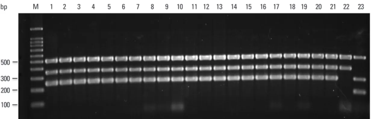
Fig. 5. Multiplex PCR for clinical M. bovis isolates. PCR products were separated by electrophoresis and visualized. Lanes M: molecular size marker (100-bp ladder); 1-20: M. bovis isolates; 21: M. bovis AN5; 22: M. bovis BCG Pasteur; 23: M. tuberculosis H37Rv. BCG, bacillus Calmette-Guérin; PCR, polymerase chain reaction.
500 300 200 100
bp M 1 2 3 4 5 6 7 8 9 10 11 12 13 14 15 16 17 18 19 20 21 22 23
Mycobacterium tuberculosis complex in clinical specimens. J Clin Microbiol 2011;49:2562-7.
12. Pinsky BA, Banaei N. Multiplex real-time PCR assay for rapid identification of Mycobacterium tuberculosis complex members to the species level. J Clin Microbiol 2008;46:2241-6.
13. Lee H, Park HJ, Cho SN, Bai GH, Kim SJ. Species identification of mycobacteria by PCR-restriction fragment length polymor- phism of the rpoB gene. J Clin Microbiol 2000;38:2966-71.
14. Gordon SV, Eiglmeier K, Garnier T, Brosch R, Parkhill J, Barrell B, et al. Genomics of Mycobacterium bovis. Tuberculosis (Edinb) 2001;81:157-63.
15. Dobner P, Feldmann K, Rifai M, Löscher T, Rinder H. Rapid identification of mycobacterial species by PCR amplification of hypervariable 16S rRNA gene promoter region. J Clin Microbiol 1996;34:866-9.
16. Frota CC, Hunt DM, Buxton RS, Rickman L, Hinds J, Kremer K, et al. Genome structure in the vole bacillus, Mycobacterium micro- ti, a member of the Mycobacterium tuberculosis complex with a low virulence for humans. Microbiology 2004;150(Pt 5):1519-27.
17. Vasconcellos SE, Huard RC, Niemann S, Kremer K, Santos AR, Suffys PN, et al. Distinct genotypic profiles of the two major clades of Mycobacterium africanum. BMC Infect Dis 2010;10:80.
18. Butler WR, Jost KC Jr, Kilburn JO. Identification of mycobacteria by high-performance liquid chromatography. J Clin Microbiol 1991;29:2468-72.
19. Streicher EM, Victor TC, van der Spuy G, Sola C, Rastogi N, van Helden PD, et al. Spoligotype signatures in the Mycobacterium tuberculosis complex. J Clin Microbiol 2007;45:237-40.
(Edinb) 2006;86:77-109.
4. Lamm DL, Stogdill VD, Stogdill BJ, Crispen RG. Complications of bacillus Calmette-Guerin immunotherapy in 1,278 patients with bladder cancer. J Urol 1986;135:272-4.
5. Kamphuis JT, Buiting AG, Miseré JF, van Berge Henegouwen DP, van Soolingen D, Rensma PL. BCG immunotherapy: be cautious of granulomas. Disseminated BCG infection and mycotic aneu- rysm as late complications of intravesical BCG instillations. Neth J Med 2001;58:71-5.
6. O’Reilly LM, Daborn CJ. The epidemiology of Mycobacterium bovis infections in animals and man: a review. Tuber Lung Dis 1995;76 Suppl 1:1-46.
7. Sankar S, Ramamurthy M, Nandagopal B, Sridharan G. An ap- praisal of PCR-based technology in the detection of Mycobacteri- um tuberculosis. Mol Diagn Ther 2011;15:1-11.
8. Frothingham R. Differentiation of strains in Mycobacterium tuber- culosis complex by DNA sequence polymorphisms, including rapid identification of M. bovis BCG. J Clin Microbiol 1995;33:840-4.
9. Kjeldsen MK, Bek D, Rasmussen EM, Priemé A, Thomsen VØ.
Line probe assay for differentiation within Mycobacterium tuber- culosis complex. Evaluation on clinical specimens and isolates in- cluding Mycobacterium pinnipedii. Scand J Infect Dis 2009;41:
635-41.
10. Parsons LM, Brosch R, Cole ST, Somoskövi A, Loder A, Bretzel G, et al. Rapid and simple approach for identification of Mycobac- terium tuberculosis complex isolates by PCR-based genomic dele- tion analysis. J Clin Microbiol 2002;40:2339-45.
11. Halse TA, Escuyer VE, Musser KA. Evaluation of a single-tube multiplex real-time PCR for differentiation of members of the