18
A few studies on patients with branch retinal vein occlusion (BRVO) have suggested that intravitreal corticosteroid can reduce macular edema and increase visual acuity.
1-3However, these studies did not evaluate the effectiveness of triamcinolone with respect to the timing of the intravitreal injection. In addition, follow-up periods were inadequate or patients who had undergone multiple injections of intravitreal triamcinolone or combined treatment with laser photo- coagulation were included. The purpose of this study was to directly compare the effect of an early versus a late intravitreal triamcinolone as a primary treatment for macular edema due to BRVO.
Patients and Methods
This study had been performed according to the Declaration
of Helsinki of the World Medical Association. This retrospective study included 20 eyes of 20 patients who received a single injection of intravitreal triamcinolone (4 mg/0.1 ml) as an only treatment for macular edema from BRVO between July 2004 and June 2005, and who had a post-injection follow-up time of >6 months. Of these 20 patients, 10 had a disease duration from onset to injection of
≤ 3 months and the other 10 had a duration of >3 months.
We designated these as the early treatment and late treatment groups, respectively. Best-corrected Snellen visual acuity (converted to logarithms of the minimal angle of resolution [logMAR]), intraocular pressure (IOP) by Goldmann applanation tonometry, and foveal thickness on optical coherence tomography (Stratus OCT
TM; Carl Zeiss Meditec Inc., Dublin, CA, USA) were measured at baseline and at 1, 3, and 6 months post-injection. The difference between baseline and postoperative measurements in logMAR visual acuity and foveal thickness were compared in each group, using the two-tailed Student's t test and the chi-square test.
Statistical significance was accepted at p<0.05.
The protocol was approved by the Institutional Review Board of Seoul National University Hospital, Seoul, Korea,
Received: November 15, 2006 Accepted: February 2, 2007 Reprint requests to Hum Chung, MD. Department of Ophthal- mology, Seoul National University College of Medicine, 28 Yeongun- dong, Chongno-gu, Seoul 110-744, Korea. Tel: 82-2-2072-2438, Fax:
82-2-741-3187, E-mail: [email protected]
Early versus Late Intravitreal Triamcinolone Acetonide for Macular Edema associated with Branch Retinal Vein
Occlusion
Joo Youn Oh, MD.
1,2, Je Hyun Seo, MD.
1,2, Jae Kyoun Ahn, MD.
1,2, Jang Won Heo, MD.
3, Hum Chung, MD.
1,2Department of Ophthalmology, Seoul National University College of Medicine
1, Seoul, Korea Seoul Artificial Eye Center, Seoul National University Hospital Clinical Research Institute
2, Seoul, Korea
Seoul Municipal Boramae Hospital
3, Seoul, Korea
Purpose: To compare the effect of early versus late intravitreal injection of triamcinolone in patients with macular edema due to branch retinal vein occlusion (BRVO).
Methods: Twenty eyes of 20 patients with macular edema from BRVO, including 10 with duration after onset of ≤3 months and 10 with duration of >3 months, were treated using a single intravitreal triamcinolone injection (4 mg/0.1 ml). Best-corrected visual acuity and foveal thickness by optical coherence tomography were measured 1, 3, and 6 months post-injection.
Results: In patients that received treatment after a disease duration of ≤3 months, visual acuity and foveal thickness significantly improved from baseline over 6 months of follow-up. However, in those with a duration of > 3 months, improvements in visual acuity and foveal thickness, though apparent at 1 month, were not maintained at 3 and 6 months post-triamcinolone.
Conclusions: Intravitreal triamcinolone is more effective in patients with BRVO who are treated earlier.
Korean Journal of Ophthalmology 21(1):18-20, 2007
Key Words: Branch retinal vein occlusion, Intravitreal triamcinolone, Macular edema
JY Oh, et al. INTRAVITREAL TRIAMCINOLONE FOR MACULAR EDEMA
19 and informed consent was obtained from the patients.
Results
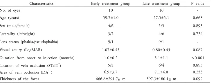
Patient demographics and characteristics of BRVO are shown in Table 1; no statistically significant differences were found between the two groups. In the early treatment group, mean visual acuity significantly improved from a baseline of 1.07±0.45 logMAR units to 0.74±0.51 (p=0.021) at 1 month, 0.63±0.43 (p=0.012) at 3 months, and 0.34±0.33 (p=0.005) at 6 months post-injection. In this group, mean foveal thickness was 666.8±291.7 μm at baseline, and significantly reduced to 233.8±77.2 μm (p<0.001) at 1 month, 351.1±180.8 μm (p=0.026) at 3 months, and 446.2±
266.6 μm (p=0.029) at 6 months. However, in the late
treatment group, though mean visual acuity significantly improved from baseline 0.80±0.45 to 0.47±0.37 (p=0.039) at 1 month, it did not further improve at 3 months (0.54±
0.34, p=0.080) or 6 months (0.60±0.53, p=0.226). Similarly, mean foveal thickness decreased significantly from baseline 597.3±180.1μm to 344.0±183.3 μm (p=0.030) at 1 month, but results did not significantly improve at 3 months (464.0
±246.8 μm, p=0.594) or 6 months (545.1±204.6 μm, p=0.952). Comparisons of mean visual acuity and foveal thickness in the two groups are shown in figures 1 and 2, respectively. One patient in the early treatment group and two in the late treatment group experienced an elevated IOP during follow-up, and one in the late treatment group required trabeculectomy.
Fig. 1. LogMAR visual acuity results in the early and late treatment groups. A significant sustained improvement was evident in the early treatment group throughout the subsequent 6-month follow-up, but in the late treatment group, the initial improvement at 1 month was not sustained at 3 and 6 months.
0 0.2 0.4 0.6 0.8 1 1.2 1.4 1.6
Baseline 1 3 6
Time after injection (months)
LogMAR visual acuity
Early treatment Late treatment
Table 1. Patient demographics and characteristics of the retinal vein occlusion in the early and late treatment groups Characteristics Early treatment group Late treatment group P value
No. of eyes 10 10 -
Age (years) 59.7±1.0 57.5±5.1 0.663
Sex (male/female) 4/6 5/5 0.893
Laterality (left/right) 3/7 4/6 0.734
Lens status (phakia/pseudophakia) 9/1 9/1 -
Visual acuity (LogMAR) 1.07±0.45 0.80±0.45 0.087
Duration from onset to injection (months) 1.0±0.2 5.1±1.1 <0.001
Location of vein occlusion (ST/IT
*) 5/5 6/4 0.893
Area of vein occlusion (DA
†) 6.9±3.7 7.1±4.0 0.253
Thickness of the fovea 666.8±291.7μm 597.3±180.1μm 0.092
*
ST : Superotemporal, IT : Inferotemporal.
†
DA : Disc area.
Fig. 2. Foveal thickness by optical coherence tomography in the early and late treatment groups. A significant improvement was observed in the early treatment group throughout the 6-month follow-up, but the improvement observed at 1month was not further sustained in the late treatment group.
0 200 400 600 800 1000 1200
Baseline 1 3 6
Time after injection (months)
Foveal thickness (um)
Early treatment Late treatment