Vascular Neurology 2010;2:26-30
ISSN 2092-6855
Introduction
Cerebral amyloid angiopathy (CAA) commonly presents with lobar intracerebral hemorrhage in the elderly. Other manifes- tations such as cerebellar hematomas, dementia, transient isch- emic attack, seizures and cerebral vasculitis have been described although they are unusual.
1,2Dementia can result from the re- current lobar hemorrhage or can occur due to a coexistent Al- zheimer’s disease (AD) in 80% of cases. CAA consists of depo- sition of amyloid in brain arterioles, capillaries, and leptomenin- geal vessels. In AD, CAA is due to the deposition of amyloid al- pha protein (Abeta) within the adventitia and media of leptomen- ingeal and brain parenchymal arteries.
3The most common form of CAA results from deposition of the amyloid-(A) peptide in the walls of cerebral vessels, gradually replacing the smooth muscle cell layer. The vast majority of patients diagnosed with AD also have CAA.
4A major consequence of CAA is fatal lobar cerebral hemorrhage, and it also appears to play a role in isch- emic brain lesions and leukoariaosis. The role of Apolipopro- tein E (ApoE) in the genetics and pathogenesis of AD has been well established. As with AD, the 4 allele of ApoE is a risk fac- tor for developing CAA, whereas the 2 allele is a risk factor for
developing hemorrhage associated with CAA. ApoE2 may play protecting against Ab deposition and the development of AD.
5-7Our case differs from the case recently published.
Case Report
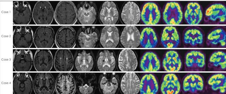
There were five patients, two female and three male, with age range from 56 years old to 69 years old and mean age of 61.3 years old. All patients underwent neuropsychological test and brain magnetic resonance imaging (MRI) and brain positron emission tomography (PET) (Fig. 1)(Table 1, 2 and 3).
Case 1
A 56-year-old man was admitted with an 3-week history of rapidly progressive change in personality, memory impairment and apathy. She had episodes of numbness spreading from Lt.
hand to arm and face, mild memory loss. There were no depres- sive symptoms. There was no past history of diabetes, hyperten- sion, renal failure, exposure to toxic chemicals, radiation, or drugs.
There was no family history. She was a non-smoker and drank little alcohol. Neurologic, ophthalmologic and general examina- tion, were otherwise normal. She scored 23/30 on her mini men-
Cerebral Amyloid Angiopathy Presenting as Alzheimer’s Disease: Four Case Reports
YoungSoon Yang,
1Hee-Jeong Seo,
1Yong Woo Noh,
1Yong Tae Kwak,
1Il-Woo Han,
1Choong-Soon Lee
21
Departments of Neurology,
2Psychiatry, Hyoja Geriatric Hospital, Yongin, Korea
Received February 26, 2010 Revised
April 2, 2010
Accepted April 13, 2010Correspondence Il-Woo Han, MD Department of Neurology, Hyoja Geriatric Hospital, 33 Sangha-dong, Giheung-gu, Yongin 446-512, Korea
Tel +82-31-288-0600 Fax +82-31-288-0539 E-mail [email protected]Cerebral amyloid angiopathy (CAA) is well known to present with lobar intracerebral hemor- rhage, dementia or transient neurological events. CAA is a microangiopathy that frequently co- occurs with Alzheimer’s disease (AD) and appears to increase with age. In AD, CAA is due to the deposition of amyloid alpha protein (Abeta) within the adventitia and media of leptomeningeal and brain parenchymal arteries. We present 4 cases with rapidly progressive dementia in whom magnetic resonance imaging brain showed multiple microbleed in the whole cerebral lobe, mainly parieto-occipital lobe. Brain positron emission tomography revealed symmetrical hypometabo- lism in temporal and parietal cortex as features of AD. Apolipoprotein E (ApoE) genotype re- vealed Apo e3/e3. The presence of one or two ApoE E4 alleles is considered to be a risk factor for AD. Evidence suggests that the ApoE4 isoform promotes fibril formation of the amyloid b protein (Ab). ApoE2, on the other hand, may play an opposite role, protecting against Ab deposition and the development of AD. The role of ApoE in the genetics and pathogenesis of AD has been well established. As with AD, the 4 allele of ApoE is a risk factor for developing CAA, whereas the 2 al- lele is a risk factor for developing hemorrhage associated with CAA. We found ApoE3 in 4 pa- tients, and we can’t prospect AD accompanied with CAA in ApoE genotype.
Vascular Neurology 2010;2:26-30
Key Wordsaa Cerebral amyloid angiopathy, Alzheimer’s disease, ApoE genotype.
tal status examination (MMSE). Routine blood exam, metabolic and endocrine work up, thyroid function, serum B
12and red cell folate, venereal disease research laboratory test (VDRL), rheu- matic arthritis (RA) factor, chest X-ray, electrocardiography (ECG), electroencephalography (EEG) were normal. MRI brain showed multiple microbleed subsequent Rt. basal ganglia hemorrhage with negative follow-up MRI. So this patient is diagnosed as pro- bable CAA according to boston criteria.
8Brain PET revealed symmetrical hypometabolism in temporal and parietal cortex as features of AD. ApoE genotype revealed Apo e3/e3.
Case 3
A 69-year-old woman was admitted with an 8-week history of rapidly progressive change in personality, memory impairment and apathy. She had increasing difficulty in getting dressed, cook- ing and coordinating household tasks and wandered aimlessly around the house. There were no depressive symptoms.
There was no past history of diabetes, hypertension, renal fail- ure, exposure to toxic chemicals, radiation, or drugs. There was no family history. She was a non-smoker and drank little alco- hol. Neurologic, ophthalmologic and general examination, were otherwise normal. She scored 25/30 on her MMSE. Routine blood exam, metabolic and endocrine work up, thyroid func- tion, serum B
12and red cell folate, VDRL, RA factor, chest X-ray, ECG, EEG were normal. MRI brain showed multiple micro- bleed in the whole cerebral lobe, mainly parieto-occipital lobe.
So this patient is diagnosed as probable CAA according to bos- ton criteria.
8Brain PET revealed symmetrical hypometabolism in temporal and parietal cortex as features of AD. ApoE geno- type revealed Apo e3/e3.
Discussion
CAA is characterized by cerebrovascular amyloid deposition.
Mild CAA is not associated with clinical manifestations, while severe CAA may cause cerebrovascular disorders such as lobar cerebral hemorrhage and leukoencephalopathy, and may pres- ent with dementia.
4,9CAA is found in the leptomeningeal and cortical vessels of the cerebral lobes. As for the distribution of CAA in the brain, the occipital lobe is most frequently and se- verely affected with CAA. CAA is also found in the cerebellum.
10In contrast, CAA is uncommon in the basal ganglia, thalamus, brainstem, and white matter.
4Development of CAA is not cor- related with the presence of common cerebrovascular risk fac- tors including hypertension, diabetes mellitus, and hyperlipid- emia, or with severity of atherosclerosis of the cerebral arteries.
10Progressive dementia is frequently found in patients with CAA.
Ta bl e 1. C lin ic al c ha rc te ris tic s of p at ie nt s Pa tie nt nu m be r A ge Se x Ye ar s
V as cu la r C lin ic al p re se nt at io n Ev id en ce fo r C A A (e du ca tio n) ris k fa ct or 1 56 M 12 N on e Ep iso de s o f n um bn es s s pr ea di ng f ro m L t. ha nd to a rm a nd fa ce , m ild m em or y lo ss
M ul tip le sm al l c or tic al h em or rh ag es o n M RI in cl ud in g p re vi ou s s ub se qu en t R t. fro nt op ar ie ta l w ith n eg at iv e a ng io gr am a nd C T 2 59 F
06 Hy pe rlip id em ia Ep iso de s o f p ar es th es ia s s pr ea di ng f ro m L t. sh ou ld er to h an d
M ul tip le sm al l c or tic al h em or rh ag es a nd P re vi ou s s ub se qu en t R t. pa rie to oc ci pi ta l h em or rh ag e w ith n eg at iv e an gi og ra m 3 69 F 16 N on e Ep iso de s o f v isu al h al lu ci na tio ns a nd m isp er ce pt io ns
M ul tip le sm al l c or tic al h em or rh ag es o n M RI , i nc lu di ng h em or rh ag e ne ar R t. ce nt ra l s ul cu s 4 61 M 16 N on e Pr of ou nd d em en tia , p ro gr es siv e ov er 2 y ea rs; M ul tip le lo ba r h em or rh ag es in cl ud in g su bs eq ue nt h em or rh ag es o f L p on s CAA
: cerebral amyloid angiopathy, MRI : magnetic resonance imaging, CT : computed tomography.
Table 2. Results of ApoE genotype
Case 1 Case 2 Case 3 Case 4
ApoE genotype ε3/ ε3 ε3/ ε3 ε3/ ε4 ε3/ ε3
ApoE: apolipoprotein E.
Pathomechanisms underlying the dementia are not uniform, including vascular dementia (VD) due to CAA, coexistence of AD, mixed dementia of VD and AD, and a vascular variant of AD.
11In AD patients, soluble Ab and ApoE levels in the cere- brospinal fluid have been reported to be significantly lower in AD patients with CAA than in those without CAA.
12A recently
described genetic risk factor for AD is the e4 allele of the ApoE gene. The ApoE e4 allele is found at a frequency of approximate- ly 0.4 in sporadic cases of late-onset AD, roughly three-fold its frequency in the general population. ApoE e4 is associated with an increased density of senile plaques in AD and a greater like- lihood of Ab deposition following head trauma, raising the pos- Table 3. Results of neuropsychological test
Case 1 Case 2 Case 3 Case 4
KMMSE
Orientation to time
05 05 05 05Orientation to place
04 04 04 04Registration
03 03 03 03Attention and calculation
02 00 03 03Recall
00 01 01 01Language
08 08 08 08Drawing
01 01 01 01Total score 23 22 25 25
Attention
Digit span, forward 4 (12.30 %ile) 9 (97.06 %ile) 5 (16.35 %ile) 9 (93.70 %ile)
Digit span, backward 5 (84.38 %ile) 6 (98.67 %ile) 4 (67.36 %ile) 6 (92.36 %ile)
Language & related funtions
K-BNT 58/60 (93.70 %ile) 48/60 (39.94 %ile) 45/60 (42.47 %ile) 53/60 (71.90 %ile)
Rt-Lt orientation Normal Normal Normal Normal
Calculation (+/-/×/÷) Normal (3/3/3/3) Normal (3/3/3/3) Normal (3/3/3/3) Normal (3/3/3/3)
Praxis Normal Normal Normal Normal
Visuospatial function and memory
Reye complex figure, copy
0.34/36 (82.64 %ile) .35/36 (81.06 %ile) 0.35/36 (77.94 %ile)36/36 Immediate recall 16.5/36 (76.11 %ile) 7.5/36 (14.92 %ile) 13.5/36 (27.43%) 6/36 (1.16 %ile) Delayed recall
0.11/36 (38.59 %ile) .8/36 (7.78 %ile) 0.14/36 (29.12%)6/36 (2.17 %ile) Recogntion score
000.16 (3.75 %ile) 000.17 (14.92 %ile) 0000.19 (28.77%) 0/18 (10.2 %ile)Korean verbal learning test
Immediate recall 4-5-7:16 (13.14 %ile) 3-4-4:11 (2.56 %ile) 5-5-9:19 (37.45 %ile) 6-6-9:21 (56,36 %ile)
Delayed recall 0 0 8 (62.55 %ile) 7 (46.41 %ile)
Recognition score 16 (1.92 %ile) 17 (17.36 %ile) 21 (54.38 %ile) 23 (79.39 %ile)
Frontal executive funtions
Contrasting program Abnormal Normal Normal Normal
Go-no-go Abnormal Normal Abnormal Normal
Fist-edge-palm Abnormal Abnormal Abnormal Abnormal
Alternating hand movement Abnormal Abnormal Abnormal Abnormal
Alternating square & triangle Normal Normal Abnormal Normal
Luria loop Normal Normal Normal Normal
Phonemic word fluency (animal/supermarket)
9 (1.58 %ile) 7 (5.59 %ile)
19 (97.2 %ile)0 17 (79.10 %ile)
14 (22.66 %ile) 6 (4.01 %ile)
12 (10.75 %ile) 12 (6.06 %ile)0 Semantic word fluency
(ㄱ/ㅇ/ㅅ/total)
3/3/3 (9) (10.5 %ile)
8/4/3 (15) (37.83 %ile)
8/6/5 (19) (24.2%)
11/11/8 (30) (24.2%) Stroop test, word reading
(correct/error)
112/0 112/0 112/0 112/0
Stroop test, color reading (correct/error)
086/3 063/0 064/6 077/3
KMMSE: Korean mini mental status examination, K-BNT: Korean-Boston Naming Test.
sibility that it may accelerate accumulation of Ab. Other poten- tial roles for ApoE in the brain as well as a possible role in ische- mic strok have also been described.
13If ApoE e4 predisposes to AD by enhancement of AP deposition, it might also be expected to promote CAA. A relationship between ApoE e4 and increased CAA has been described. Recent evidence implicates the ApoE gene in the etiology of cerebral amyloid angiopathy-related he- morrhage.
14,15someone previously hypothesized that whereas the ApoE e4 allele increases Ab deposition in the cerebral vascula- ture, ApoE e2 is associated with rupture of Ab-laden blood ves- sels, possibly by predisposing to the development of recognized vasculopathic complications of CAA. The role of ApoE in the ge- netics and pathogenesis of AD has been well established.
16More than 40% of patients with CAA related hemorrhage have asso- ciated AD which may confound the analysis because ApoE4 is a well-established risk factor for AD whereas ApoE2 is protec- tive. Whether ApoE4 is an independent risk factor for CAA-re- lated hemorrhage or may be associated simply by its link with AD is unclear at present.
17Generally, at an early stage, special features of AD are memory disturbane , visuoapatial function, naming dis- turbance and apraxia, calculation impairment. But special fea- ture of our patients is frontal lobe dysfunction when AD is accom- panied with CAA. It is known that ApoE e4 is related AD, and ApoE2 is related CAA. But we observed ApoE3 in 4 patients. So we think as follows. At first, ad accompanied with CAA is differ- ent from general AD and its special feature is frontal lobe dys- function. So finding frontal lobe dysfunction in AD, you have to consider CAA. Next interesting point is that we found ApoE3 in 4 patients, and we can’t prospect AD accompanied with CAA in ApoE genotype. On the contrary, MRI is more useful.
REFERENCES
1. Pfeifer LA, White LR, Ross GW, Petrovitch H, Launer LJ. Cerebral
amyloid angiopathy and cognitive function: the HAAS autopsy study.
Neurology 2002;58:1587-1588.
2. Mandybur TI. The incidence of cerebral amyloid angiopathy in Alz- heimer’s disease. Neurology 1975;25:120-126.
3. McCarron MO, Nicoll JA, Stewart J, Ironside JW, Mann DM, Love S, et al. The apolipoprotein E epsilon2 allele and the pathological features in cerebral amyloid angiopathy-related hemorrhage. J Neuropathol Exp Neurol 1999;58:711-718.
4. Yamada M. Cerebral amyloid angiopathy: an overview. Neuropathology 2000;20:8-22.
5. Nicoll JA, Burnett C, Love S, Graham DI, Ironside JW, Vinters HV. High frequency of apolipoprotein E epsilon 2 in patients with cerebral hem- orrhage due to cerebral amyloid angiopathy. Ann Neurol 1997;39:682-683.
6. Greenberg SM, Briggs ME, Hyman BT, Kokoris GJ, Takis C, Kanter DS, et al. Apolipoprotein E epsilon 4 is associated with the presence and ear- lier onset of hemorrhage in cerebral amyloid angiopathy. Stroke 1996;27:
1333-1337.
7. Greenberg SM, Rebeck GW, Vonsattel JP, Gomez-Isla T, Hyman BT.
Apolipoprotein E epsilon 4 and cerebral hemorrhage associated with amyloid angiopathy. Ann Neurol 1995;38:254-259.
8. Knudsen KA, Rosand J, Karluk D, Greenberg SM. Clinical diagnosis of cerebral amyloid angiopathy: validation of the Boston criteria. Neurol- ogy 2001;56:537-539.
9. Yamada M, Itoh Y, Otomo E, Hayakawa M, Miyatake T. Subarachnoid haemorrhage in the elderly: a necropsy study of the association with cerebral amyloid angiopathy. J Neurol Neurosurg Psychiatry 1993;56:
543-547.
10. Yamada M, Tsukagoshi H, Otomo E, Hayakawa M. Cerebral amyloid angiopathy in the aged. J Neurol 1987;234:371-376.
11. Yoshimura M, Yamanouchi H, Kuzuhara S, Mori H, Sugiura S, Mizu- tani T, et al. Dementia in cerebral amyloid angiopathy: a clinicopath- ological study. J Neurol 1992;239:441-450.
12. Pirttilä T, Mehta PD, Soininen H, Kim KS, Heinonen O, Paljärvi L, et al. Cerebrospinal fluid concentrations of soluble amyloid beta-protein and apolipoprotein E in patients with Alzheimer’s disease: correlations with amyloid load in the brain. Arch Neurol 1996;53:189-193.
13. Greenberg SM, Rebeck GW, Vonsattel JP, Gomez-Isla T, Hyman BT.
Apolipoprotein E epsilon 4 and cerebral hemorrhage associated with amyloid angiopathy. Ann Neurol 1995;38:254-259.
14. McCarron MO, Nicoll JA, Ironside JW, Love S, Alberts MJ, Bone I.
Cerebral amyloid angiopathy-related hemorrhage. Interaction of APOE epsilon2 with putative clinical risk factors. Stroke 1999;30:1643-1646.
Case 1
Case 2
Case 3
Case 4