INTRODUCTION
Inflammation is a host defense process that occurs at injured sites and is strictly regulated. The coordination of pro-inflam- matory and anti-inflammatory mediators is important in the regulation of the inflammatory response.
Historically, PMNs have been perceived as terminally dif- ferentiated leukocytes with limited ability to produce de novo protein. During acute inflammation, the first line of cellular response for host defense is the PMNs, for they accumulate in injured tissue. In addition to the historic role of the PMNs as a phagocyte, recent studies have identified this cell as an important source of a number of cytokines like IL-8 and TNF- (1-5). Not only does PMNs play an important role in the early inflammation of phagocytosis by various protease and reactive oxygen, but also these cells can by themselves enhance PMNs recruitment through a local production of IL-8.
Formyl methionyl leucyl phenylalanine (fMLP) is a typi- cal chemoattractant. fMLP as a synthetic chemoattractant is a family of formylated peptides, which are bacterial products formed and released at the site of infection. It induces inflam- mation on the PMNs and produces several cytokines in com- bination with the fMLP receptors on the PMNs surface (6).
IL-4 and IL-10 are the biologic anti-inflammatory media-
tors. They play biologic anti-inflammatory roles so that they inhibit the production and release of pro-inflammatory medi- ators, such as IL-8 and TNF- , from monocytes and macro- phages (7-9). Recent studies report that IL-4 or IL-10 has an inhibitory effect on human PMNs-derived IL-8 or TNF- release after lipopolysaccharides (LPS) stimulation (10-13).
But there have been few attempts to modify the hyper-inflam- matory reaction of PMNs with biologic anti-inflammatory mediators (14, 15). To modify the complex web of causality in hyper-inflammation with biologic anti-inflammatory medi- ators, a study on the regulation of PMN activities is a neces- sary prerequisite.
The aim of this study was to investigate anti-inflammat- ory effects on human PMNs by biologic anti-inflammatory mediators, IL-4 and IL-10. In this study, we directly com- pared the anti-inflammatory effects of IL-4 with IL-10 on fMLP-induced human PMNs-derived IL-8 and TNF- .
MATERIALS AND METHODS
Human PMNs were isolated from healthy volunteers’
peripheral blood by the modified Boyum’s method (16, 17).
Isolated human PMNs were incubated at 37℃with or with- Sung Woo Lee, Yun Sik Hong,
Chung Min Chun, Jun Dong Moon, Su Jin Kim, In Chul Jung, Young Hoon Yoon, Be An Lee, Sung Woo Moon, Sung Hyuk Choi, Chul Kyu Moon
Department of Emergency Medicine, College of Medicine, Korea University, Seoul, Korea
Received : 31 May 2001 Accepted : 18 September 2001
Address for correspondence Sung Woo Lee, M.D.
Clinical Professor, Korea University Ansan Hospital Emergency, Department 516 Gojan-dong, Ansan 425-020, Korea
Tel : +82.31-412-5380, Fax : +82.31-412-5315 E-mail : [email protected]
7
Anti-inflammatory effects of IL-4 and IL-10 on Human Polymorphonuclear Leukocytes
Inflammatory responses are strictly regulated by coordination of pro-inflammat- ory and anti-inflammatory mediators. Interleukin-4 (IL-4) and interleukin-10 (IL- 10) have typically the biologic anti-inflammatory effects on monocytes, but uncertain effects on polymorphonuclear leukocytes (PMNs). The PMNs are the first line of cellular response for host defense during acute inflammation. To modify hyper-inflammatory reaction with biologic anti-inflammatory mediators, we have determined the biologic anti-inflammatory activities of IL-4 and IL-10 on human PMNs. Human PMNs were pretreated with IL-4 or IL-10 and then stimu- lated with formyl methionyl leucyl phenylalanine (fMLP) for times indicated. The level of H2O2, interleukin-8 (IL-8) and tumor necrosis factor- (TNF- ) were determined in the each cell free supernatants. fMLP plays the role of a typical pro-inflammatory agent and, at least in determined conditions, down-regulated TNF release. IL-4 acts as an anti-inflammatory mediator but IL-10 did not show its anti-inflammatory activities on fMLP-stimulated human PMNs. IL-4 and IL-10 have different anti-inflammatory mechanisms. Perhaps, IL-10 needs co-factors to act as an anti-inflammatory mediator.
Key Words : Interleukin-4; Interleukin-10; Interleukin-8; Tumor Necrosis Factor- ; N-Formylmethionine Leucyl-Phenylalanine; Neutrophils
out 10 nM fMLP (final concentration). Then, to demonstrate inflammatory reaction by PMNs, the production of H2O2 was assessed by a flow cytometer. IL-8 and TNF- were deter- mined in the cell-free supernatants by enzyme-linked immu- nosorbent assay (ELISA) method.
Isolated PMNs were pretreated with 10 ng/mL IL-4 for 1 hr and then stimulated with 10 nM fMLP (final concentra- tion) for the times indicated (0, 30 min, 2 hr, 4 hr, 20 hr).
The amounts of IL-8 and TNF- were obtained by the ELISA method at each time.
Isolated PMNs were pretreated with 100 U/mL IL-10 for 45 min and then stimulated with 10 nM fMLP (final con- centration) for the times indicated. The amounts of IL-8 and TNF- were obtained in a similar fashion.
Buffers and cell preparation
To determine H2O2production, calcium-free Dulbecco’s phosphate buffered saline (PBS) was purchased from Gibco (Grand Island, NY, U.S.A.), pH 7.4. It contained 1.0 g/L D- glucose (PBSg), it was freshly prepared. For experiments involving PMNs stimuli, the PBSg contained 0.1 g/L CaCl2 (PBSg
++
).Human PMNs were isolated by a modification of the method by Boyum (16) as previously described. Briefly, venous blood was drawn by using the aseptic technique from healthy volunteers into tubes containing ethylene diaminester- acetic acid (EDTA) as an anticoagulant. Carefully, 5.0 mL of anticoagulated whole blood was layered over 5.0 mL of Poly- morphprepTM(Nycomed Pharma AS, Oslo, Norway) in a 12 mL centrifuge tube. Care was taken to avoid mixing the blood with the separation fluid. The samples were centrifuged and layered over PolymorphprepTMfor 500×g for 35 min in a swing-out rotor at room temperature. After centrifugation, two leukocyte’s bands were visible. The top band at the sam- ple/medium interface consisted of mononuclear cells and the lower band of PMNs. The erythrocytes were pelleted.
The cell bands were harvested using a Pasteur pipette. The resulting pellet was treated for 30 sec with 0.2% saline in order to hemolyse remaining erythrocytes, and the suspen- sion was then made isotonic by adding 1.8% saline (1:1, vol/
vol). The cells were washed, suspended in PBSg
++
, and spun down (at room temperature, 450×g, 10 min). They were resuspended in PBSg++
for stimulation. The total cell yield was determined by counting a portion of the final suspen- sion in a hemocytometer. We adjusted the concentration of cells at 4-5×10⁶cells/mL. The differential cell counts were determined by staining cytospin preparations with Diff-Quik (Baxter Scientific Products, McGraw Park, IL, U.S.A.) and counting across the entire slide. The differential was consis- tently>
95% PMNs and 2% lymphocytes. Monocytes typi- cally represented<
1% of cells. Viability was>
95% as deter- mined by trypan blue dye exclusion.To avoid cell clumping following stimulation, EDTA was
employed in the final suspensions for flow cytometric analy- sis. Cell centrifugation and washing was limited only to the period following termination of reaction in order to minimize handling artifacts.
Preparation of soluble stimuli
Formyl methionyl leucyl phenylalanine (fMLP, Sigma, St Louis, MO, U.S.A.) was solublized in dimethyl sulfoxide (DMSO, Sigma) at a 43.75 mg per 10 mL solution and diluted with a working buffer (PBSg
++
) to a stock concentration of 10-7M. The final concentration of DMSO exposed to cells was 0.01% (vol/vol). Recombinant human IL-4 (rH IL-4, Sigma) was reconstituted and diluted to a stock concentra- tion of 100 ng/mL with the working buffer. Recombinant human IL-10 (rH IL-10, Sigma) was diluted in the working buffer to a stock concentration of 10 U/mL. All stimulants diluted to their stock concentrations were aliquoted into 0.5 mL volumes and stored for future use at -20℃. In each case, addition of stimulant to cell suspension produced a 1: 10 dilution of its final concentration.Intracellular hydrogen peroxide production
Measurement of intracellular H2O2 production was per- formed according to the method by Bass and Parce (18).
This assay relies upon the fact that the fluorochrome (2′, 7′- dichlorofluorescein) is trapped within the cell and unable to leak out. Total cell fluorescence indirectly reflect intracellu- lar hydrogen peroxide production and was measured by flow cytometry.
Cells isolated as described above were suspended (106PMN/
mL) in PBSg and preincubated with 5.0 m 2′,7′-dichlo- rofluorescin diacetate (DCHF-DA, Sigma) in a final volume of 5.0 mL with horizontal agitation at 37℃for 15 min. To avoid cell clumping following stimulation, EDTA (5 mM final concentration) was employed after 15 min of preincu- bation. The fluorescence of the cells at this point was referred to as zero time. Cells were then exposed to the buffer alone (control) or to a soluble stimulant (final concentration) fMLP (10 nM) and incubated at 37℃with agitation for the indi- cated time periods. Reactions were terminated by addition of cold PBSg and storage at 0-4℃.
Flow cytometric analysis
To measure the intracellular H2O2production, cells were analyzed by quantitative flow cytometry using Becton-Dick- inson (San Jose, Ca, U.S.A.) FACScanTM, equipped with an argon laser emitting light at 488 nm. Forward angle light scatter and right angle light side scatter measurements were used to differentiate PMNs from debris, cellular clumps, ery- throcyte, and other leukocytes. Electronic gates were esta- bilishd to include
>
95% PMN. We collected logarithmiclist mode data for each sample on 10,000 events within the PMN gates. Single parameter histograms were analyzed using LYSYS II version 1.0 program running on a Hewlett-Packard series 9153 computer.
Determination of IL-8 and TNF- Concentration by ELISA
The concentrations of IL-8 and TNF- in the cell-free super- natants were determined by ELISA. To determine IL-8 release, the QuantiGlo human IL-8 kit (R&D Systems, U.S.A.) em- ployed the quantitative immunometric sandwich enzyme immunoassay technique. A microtiter plate coated with murine monoclonal antibody (mAb) specific for IL-8 was provided in the kit. Samples containing standard amounts of recombinant IL-8 as well as study samples were added to individual wells where any IL-8 present would bind to the immobilized antibody. After four washes to remove any unbound proteins, a polyclonal antibody specific against IL- 8 conjugated to horseradish peroxidase was added to the wells. After another four washes, a luminol/peroxide substrate was added and the color developed represented the amount of IL-8 present. The reaction was stopped by adding 2N sulfuric acid. The degree of color generated was determined by measuring the optical density in a Luminometer (DML 2000. Digene. U.S.A.).
To determine TNF- release, the QuantiKine HS human TNF kit (R&D System) employed the quantitative immuno- metric sandwich enzyme immunoassay technique. A micro- titer plate coated with murine anti-TNF mAb was provided in the kit. Samples containing standard amounts of rTNF as well as study samples were added to individual wells at 2-8℃for 14-20 hr. After four washes to remove any unbound proteins, an alkaline phosphatase-conjugated anti-TNF pAb was added to the wells, which were incubated for 3 hr at room temperature. After another four washes, a NADPH substrate
was added and an amplifier enzyme and buffer containing INT violet were added. The reaction was stopped by adding 2 N sulfuric acid. The degree of color generated was deter- mined by measuring the optical density at 490/650 nm in an EIA reader (Bio-Tec microplate reader, Bio-Tec Instrument.
U.S.A.).
Data analysis and statistics
All data were expressed as mean±SD (standard deviation).
Differences between paired experimental groups were com- pared by Student t tests and only difference with p value less than 0.05 was accepted as significant.
RESULTS
Intracellular H2O2production from fMLP-stimulated human PMNs
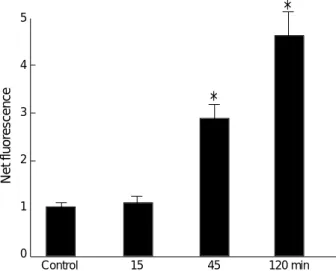
Mean fluorescence of 2′,7′-dichlorofluorescein was detect- ed by flow cytometry. At 15 min and 120 min post-stimu- lation, mean fluorescence was 27.8±8.16 and 213.1±35.18, respectively. Mean fluorescence of fMLP-stimulated PMNs significantly increased with time (Fig. 1). Net fluorescences, comparing fMLP-stimulated PMNs with non- stimulated (buffer only) control PMNs, also increased significantly with time (Fig. 2).
Release of IL-8 and TNF- from fMLP-stimulated human PMNs
Isolated PMNs (4-5×10⁶PMN/mL) were stimulated with 10 nM fMLP for the indicated times up to 20 hr. As shown in Table 1, control (non-stimulated) PMNs generated 3.1
±1.42 pg/mL to 496.7±344.09 pg/mL of IL-8 at the
SSC-H\SSC-Height >
FSC-H\FSC-Height >
FL1-H\FL1-Height
15, 30, 45, 120 min
1 1000
800
600
400
200
0
200
100
0
0 200 400 600 800 1000 100 101 102 103 104
Fig.1 A: Following pre-incubation with 2′,7′-dichlorofluorescein diacetate, PMNs were exposed to fMLP for time indicated. Mean fluores- cence was detected by flow cytometer. Electronic gates were established to include >95% PMNs. B: Fluorescence of PMNs increased with time after stimulation of 10 nM fMLP.
A B
time course, whereas stimulation with fMLP resulted in a significant increase of constitutive PMNs-derived IL-8 by 123.4 times (472.9±220.34 pg/mL), 95.6 times (2741.6
±941.16 pg/mL) at 30 min and 2 hr after stimulation, respectively (Table 1). When PMNs were stimulated with fMLP, substantial up-regulation of IL-8 production was observed (Fig. 3).
The release of TNF- from PMNs was slight compared to the IL-8 release. However, TNF- release from control (non- stimulated) PMNs were on the increase with time. When PMNs were stimulated with fMLP, substantial down-regu- lation of TNF- production was observed, as fMLP signifi- cantly suppressed PMNs-derived TNF- release by 54%
(0.32±0.160 pg/mL), by 68% (0.25±0.069), at 4 hr and 20 hr post-stimulation, respectively (Fig. 3).
Effect of IL-4 and IL-10 on human PMNs-derived IL-8
We postulated that IL-4 and IL-10 may have an inhibito- ry effect on the production of PMNs-derived IL-8. To test this hypothesis, PMNs were pretreated with 10 ng/mL of IL-4 for 1 hr or 100 U/mL of IL-10 for 45 min, and then stimulated with 10 nM fMLP for the indicated times.
PMNs pretreated with IL-4 resulted in a significant sup-
IL-8 140
120
100 80
60 40
20
1 Control 0.5 2 4 20 hr
*
*
0.20
-0.2
-0.4
-0.6
-0.8 Control 0.5 2 4 20 hr
×100 (%) ×100 (%) TNF
Fig. 3.Time courses of IL-8 and TNF- release by human PMNs stimulated with 10 nM fMLP and by control (not stimulated with fMLP).
PMNs (4, 5×10⁶/mL) were stimulated with 10 nM fMLP for the times indicated. And IL-8 and TNF- were determined in the cell-free supernatants by ELISA. *: p<0.05.
*
*
5
4
3
2
1
0
Control 15 45 120 min
*
*
Fig. 2.Time course of the net effects of intracellular hydrogen peroxide production by fMLP. *: p<0.05.
Net fluorescence
0 hr 0.5 hr 2 hr 4 hr 20 hr
IL-8 (pg/mL) Control* 3.1±1.42 4.1±1.16 47.8±34.55 120.4±96.93 496.7±344.09
fMLP� 472.9±220.34 2741.6±941.16 1798.9±1619.77 3334.8±1953.59
TNF- (pg/mL) Control 0.17±0.028 0.17±0.019 0.34±0.047 0.68±0.091 0.95±0.568
p=0.001
fMLP 0.19±0.055 0.33±0.131 0.32±0.160 0.25±0.069
p=0.209 p=0.871 p=0.097 p=0.0003
Table 1.IL-8 and TNF- release by human PMNs after fMLP stimulation
*PMNs (4-5×10⁶/mL) were cultured without stimulation. �PMNs (4-5×10⁶/mL) were stimulated for the times indicated with 10 nM fMLP. IL-8: Inter- leukin-8. TNF- : Tumor nectosis factor-alpha. fMLP: formyl methionyl leucyl phenylalanine.
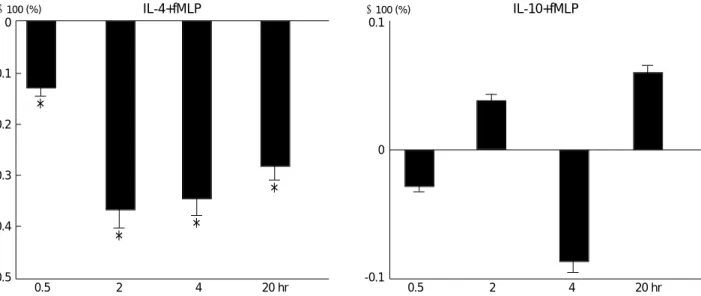
pression of fMLP-stimulated PMNs-derived IL-8 release by 64% at 30 min post-stimulation and by 61% at 2 hr (Fig. 4).
fMLP-stimulated PMNs-derived IL-8 release from PMNs were not significantly suppressed by pretreatment with IL- 10 (Fig. 4).
Effect of IL-4 and IL-10 on human PMNs-derived TNF-
We next postulated that IL-4 and IL-10 may have an anti- inflammatory effect by suppression of the release of PMNs-
derived TNF- . To test this hypothesis, PMNs were pretreated with IL-4 or IL-10, and then stimulated with fMLP in the same manner.
PMNs pretreated with IL-4 resulted in a significant sup- pression of fMLP-stimulated PMNs-derived TNF- release by 12.9%, 36.8%, 34.4%, and 28.1%, in the indicated times (Fig. 5).
There were no differences in TNF- release between IL- 10-pretreated PMNs and only fMLP-stimulated PMNs through the experiment.
IL-4+fMLP*
0
-0.2
-0.4
-0.6
-0.8 0.5 2 4 20 hr
* *
0.6
0.4
0.2
0
-0.2
-0.4 0.5 2 4 20 hr
IL-10+fMLP�
×100 (%) ×100 (%)
Fig. 4.Net effects of IL-4 and IL-10 on IL-8 release by human PMNs through the experiment. Amounts of IL-8 from IL-4- and IL-10- pre- treated PMNs were divided by the amounts of IL-8 from PMNs without pretreatment. *PMNs (4-5×10⁶/mL) were pretreated with 10 ng/mL IL-4 for 1 hr and then stimulated with 10 nM fMLP for the times indicated. �PMNs (4-5×10⁶/mL) were pretreated with 100 U/mL IL- 10 for 45 min and then stimulated with 10 nM fMLP for the times indicated. IL-8 was then determined in the cell-free supernatants by ELISA. *: p<0.05.
IL-4+fMLP 0
-0.1
-0.2
-0.3
-0.4
-0.5 0.5 2 4 20 hr
*
0.1
0
-0.1 0.5 2 4 20 hr
IL-10+fMLP
* *
*
×100 (%) ×100 (%)
Fig. 5.Net effects of IL-4 and IL-10 on TNF- release by human PMNs through the experiment. Amounts of TNF- from IL-4 and IL-10 pretreated PMNs were divided by the amounts of TNF- from PMNs without pretreatment. PMNs (4-5×10⁶/mL) were pretreated with 10 ng/mL IL-4 for 1 hr and with 100 U/mL IL-10 for 45 min and then stimulated with 10 nM fMLP for the times indicated. TNF- was deter- mined in the cell-free supernatants by ELISA. *: p<0.05
DISCUSSION
When the human body’s defense mechanisms are over- whelmed and can no longer control their own inflammatory and immunosuppressive response, the conditions are called systemic inflammatory response syndrome (SIRS), sepsis, and septic shock. The current hypothesis of the pathogene- sis of SIRS, sepsis, and septic shock is an excessive produc- tion/release of inflammatory mediators by excessive activation of inflammatory cells and a parallel induction of immuno- suppressive mediators with a depression of cellular immunity (19-21). The production of many pro-inflammatory media- tors and nitric oxide increase vascular permeability and decrease effective circulating volume, disturbing the micro- circulation to body tissues. The main consequences of these responses to many organs are the onset of shock and the development of a multiple organ dysfunction syndrome (MODS) accompanied by a high mortality rate. The produc- tion of pro-inflammatory mediators from inflammatory cells is inhibited by immunosuppressive mediators, such as IL-4, IL-10, IL-13, and prostagrandin E2, all of which have anti- inflammatory characteristics (22). Therefore, a coordination of pro-inflammatory mediators and anti-inflammatory medi- ators is essential to regulate and to recover from inflamma- tion (20, 21).
This hypothesis has prompted the introduction of new treatment modalities for inflammation, which include anti- endotoxin strategies to neutralize one of the proposed major causal factors, an anti-inflammatory approach to modulate the excessive systemic inflammatory response, and immu- noaugmentation concepts to increase the potency of host defense system (23). Anti-inflammatory approaches include transcriptional and post-translational control of mediator synthesis, control of release from inflammatory cells, bind- ing with neutralizing molecules in the circulation, and thus blocking the interaction between mediators and cell-surface receptors. These approaches appeared to be effective in in vitro studies. In clinical studies, however, they were not shown to be operative. And there has been little attempt to modify hyper-inflammatory reaction of PMNs with biologic anti- inflammatory mediators (24).
fMLP is a traditional chemoattractant. In our study, fMLP activated PMNs by increasing intracellular H2O2production and by releasing IL-8 after stimulation. This inflammatory reaction is mediated by fMLP receptors on the PMNs sur- face. fMLP and fMLP-receptor complex induces oxygen-free radical production and the expression of IL-8 mRNA and the synthesis of IL-8 protein (6, 25). IL-8 is described as a chemoattractant for neutrophils or neutrophil chemotactic factor (26, 27). IL-8 enhances the emigration of neutrophils from the vascular compartment to the site of tissue inflam- mation and activates neutrophils; causes degranulation and increases respiratory burst and lysosomal enzyme release (28).
In addition to the historic role of the neutrophil as a phago-
cyte, PMNs are important in augmenting inflammation by production of IL-8. IL-8 release was increased with time.
Without stimulation with fMLP, only test tube adhesion induced IL-8 and TNF- release from PMNs. When stimu- lated with fMLP, IL-8 release was explosively increased at 30 min and 2 hr post-stimulation. These results are similar to the report of Cassatella et al. (1). They reported that the release of IL-8 in response to fMLP was acute and transient.
In our study, fMLP did not induce TNF- release from human PMNs. On the contrary, fMLP down-regulated test- tube adhesion-induced TNF- release. This result is differ- ent from the previous reports that showed the capability of PMNs to secret newly synthesized TNF (1, 13). Those re- searchers used LPS as a stimulant. LPS is the major compo- nent of the outer membrane of gram-negative bacteria. For- mylated peptides are the bacterial products formed and released at the site of infection, and coexist with LPS. There is not enough evidence for the relationships between LPS and formylated bacterial peptides in the triggering and reg- ulating of the immune inflammatory response. fMLP is best characterized as a member of the formylated peptides family.
Vulcano et al. reported that fMLP reduced the expression of the membrane form of TNF- on the PMNs surface (29) and fMLP, a typical pro-inflammatory agent, could play, at least in the determined conditions, an anti-inflammatory role (30).
In our study, fMLP had two different properties. It up-regu- lated H2O2production and IL-8 release from human PMNs but down-regulated TNF- release.
IL-4 is known to originate from T-cell and causes activa- tion and proliferation of B-cell. It is also a growth factor for T-cell and exerts other effects on granulocytes (28). Other investigators have demonstrated an inhibitory effect of IL-4 on the generation of pro-inflammatory mediators and reac- tive oxygen metabolites from monocytes (31). We demon- strated in this study that IL-4 had a significant suppressive effect on fMLP-induced PMNs-derived IL-8 and TNF- release. In other investigations, IL-4 regulates monocyte- derived IL-8 both by inhibiting gene expression and by accel- erating mRNA degradation (31). These mechanisms may be operative in IL-4 regulation of PMNs-derived IL-8 and TNF- , because PMNs and monocytes share the same stem cell and may have similar conserved regulatory elements. Not only do these results support the notion that the PMNs are an active and dynamic participant of an inflammatory/im- mune response through the activation of oxygen-free radical, along with the production of IL-8 and TNF- , but they also support the notion that these cells are also susceptible to regulation by biologic anti-inflammatory mediators that influence the direction of inflammatory response.
IL-10, a cytokine known to suppress the immune response, originates from human monocytes, macrophages, and T and B lymphocytes (28). IL-10 has been implicated in the regu- lation of the functions of lymphoid and myeloid cells because of its ability to suppress the synthesis of pro-inflammatory
cytokines from T-cells and monocytes/macrophages (7, 32, 33). Cassatella et al. reported that IL-10 diminished the lev- els of TNF- and IL-8 mRNAs long after the onset of stim- ulation of PMNs with LPS, and concluded that IL-10 is a potent inhibitor of pro-inflammatory cytokines released from PMNs stimulated with LPS (10). However, our results dif- fered from theirs. In our study, IL-10 had no significant sup- pressive effects on IL-8 and TNF- release from fMLP-stim- ulated PMN. Cassatella et al. used LPS as a chemoattractant (10). Because the chemotactic effects of LPS are mediated by co-factors, such as LPS binding protein (19), we specu- late that the anti-inflammatory effects of IL-10 might have been mediated by co-factors, such as serum factors. Further studies are necessary to unveil the anti-inflammatory mech- anism of IL-10.
In our study, IL-4 exhibited an anti-inflammatory effects on fMLP-stimulated PMN by inhibition of IL-8 and TNF- release, but IL-10 was not shown to have any biologic anti- inflammatory effects on fMLP-stimulated PMNs. Other investigations have demonstrated anti-inflammatory effects of IL-4 and IL-10, which inhibited the expression of IL-8 and TNF- mRNA and hence the synthesis of IL-8 and TNF- (33). In our study, however, IL-4 and IL-10 showed different anti-inflammatory effects. These results suggest that IL-4 and IL-10 act through different anti-inflammatory pathways.
There are some limitations in our study. We obtained only a part of pro-inflammatory mediators from PMNs. There- fore, we could not completely understand the mechanism of anti-inflammatory reaction. For example, we did not com- pletely understand the relation between fMLP and IL-4 for down regulation effects on TNF release. Further investiga- tion is warranted to determine the anti-inflammatory mech- anisms of biologic anti-inflammatory mediators. We could not directly apply these results to clinical conditions. In clini- cal conditions, outcomes of the patients in the hyper-inflam- matory conditions are subjected to various factors. Further studies on the biologic anti-inflammatory mediators in clin- ical conditions, such as sepsis and SIRS, are needed for clini- cal application. Another limitation is that our attempts to regulate PMNs activities are considered premature and no longer adequate. Inflammatory responses are not simple sequential reactions but chaotic in nature. Inflammatory cells and mediators may have multiple and different proper- ties in certain conditions. Therefore, to understand the com- plex web of causality in hyper-inflammation, computer imple- mentations of causal probabilistic networks combining the learning by experience are needed in future studies.
In conclusion, fMLP induced inflammation on normal human PMNs in vitro. fMLP up-regulated intracellular hydrogen production and IL-8 release from human PMNs.
In contrast, fMLP down-regulated TNF- release. This study has demonstrated that the fMLP-induced PMNs-derived IL-8 and TNF- can be effectively modulated by endogenous
mediators, such as IL-4. Interestingly, in contrast to mono- cytes and LPS stimulated PMNs, fMLP-stimulated PMNs failed to respond to the suppressive effects of IL-10. We sug- gest that the anti-inflammatory effects of IL-10 may be medi- ated by co-factors, such as serum factors. IL-4 and IL-10 have shown different anti-inflammatory effects. One inhibits IL- 8 and TNF- release from fMLP-stimulated PMNs, where- as the other does not. These results suggest that IL-4 and IL-10 have different anti-inflammatory mechanisms.
REFERENCES
1. Cassatella MA, Bazzoni F, Ceska M, Ferro I, Baggiolini M, Berton G. IL-8 production by human polymorphonuclear leukocytes. The chemoattractant formyl-methionyl-leucyl-phenylalanine induces the gene expression and release of IL-8 through a pertussis toxin-sensi- tive pathway. J Immunol 1992; 148: 3216-20.
2. Bazzoni F, Cassatella MA, Rossi F, Ceska M, Dewald B, Baggioni M. Phagocytosing neutrophils produce and release high amounts of the neutrophil-activating peptide1/interleukin 8. J Exp Med 1991;
173: 771-4.
3. Strieter RM, Kasahara K, Allen RM, Showell HJ, Standiford TJ, Kunkel SL. Human neutrophil exhibit disparate chemotactic factor gene expression. Biochem Biophys Res Commun 1990; 173: 725.
4. Strieter RM, Kasahara K, Allen RM, Standiford TJ, Rolfe MW, Becker FS, Chensue SW, Kunkel SL. Cytokine-induced neutrophil- derived interleukin-8. Am J Pathol 1992; 141: 397-407.
5. Djeu YJY, Serbousek D, Blanchard DK. Release of tumor necrosis factor by human polymorphonuclear leukocytes. Blood 1990; 76:
1045-9.
6. Sklar LA, Jesitis AJ, Painter RG. The neutrophil N-formyl peptide receptor: dynamics of ligand-receptor interactions and their rela- tionship to cellular responses. In: Synderman R, ed. Regulation of leukocyte function. Vol 14. New York: Plenum press 1984; 14: 29-82.
7. de Waal Malefyt R, Albrams J, Bennett B, Figdor CG, de Vries JE.
Interleukin 10 (IL-10) inhibits cytokine synthesis by human mono- cytes: an autoregulatory role of IL-10 produced by monocytes. J Exp Med 1991; 174: 1209-20.
8. Bogdan C, Vodovotz Y, Nathan C. Macrophage deactivation by interleukin 10. J Exp Med 1991; 174: 1549-55.
9. Bogdan C, Paik J, Vodovotz Y, Nathan C. Contrasting mechanism for suppression of macrophage cytokine release by transforming growth factor- and interleukin-10. J Biol Chem 1992; 267: 23301-8.
10. Cassatella MA, Meda L, Bonora S, Ceska M, Constantin G. Inter- leukin 10 (IL-10) inhibit the release of proinflammatory cytokines from human polymorphonuclear leukocytes. Evidence for an autocrine role of tumor necrosis factor and IL-1 in mediating the production of IL-8 triggered by lipopolysaccharide. J Exp Med 1993; 178: 2207-11.
11. Vannier E, Miller LC, Dinarello CA. Coordinated antiinflammatory effects of interleukin 4: interleukin 4 suppresses interleukin 1 pro- duction but up-regulates gene expression and synthesis of inter- leukin 1 receptor antagonist. Proc Natl Acad Sci USA 1992; 89:
4076-80.
12. Wang P, Wu P, Anthes JC, Siegel MI, Egan RW, Billah MM. Inter- leukin-10 inhibits interleukin-8 production in human neutrophils.
Blood 1994; 83: 2678-83.
13. Wertheim WA, Kunkel SL, Staniford TJ, Burdick MD, Becker FS, Wilke CA, Gilbert AR, Strieter RM. Regulation of neutrophil- derived IL-8: the role of prostaglandin E2, Dexamethasone, and IL- 4. J Immunol 1993; 151: 2166-75.
14. Hong YS. Selective inhibition of polymorphonuclear leukocytes by immunosuppressive concentration of prostaglandin E2. J Korean Med Sci 1996; 11: 8-16.
15. Derevaanko A, D’Amico R, Simms H. Polymorphonuclear leuko- cyte (PMN)-derived inflammatory cytokines--regulation by oxygen tension and extracellular matrix. Clin Exp Immunol 1996; 106:
560-7.
16. Boyum A. Separation of leukocytes from blood and bone marrow.
Scand J Clin Invest 1968; 97: 7.
17. Ferrante A, Thong YH. Optimal conditions for simultaneous purifi- cation of mononuclear and polymorphonuclear leukocytes from human peripheral blood by the Hypaque-Ficoll method. J Immunol Methods 1980; 36: 109-17.
18. Bass DA, Parce JW, Dechatelet LR, Szejda P, Seeds MC, Thomas M. Flow cytometric studies of oxidative product formation by neu- trophil. A graded response to membrane stimulation. J Immunol 1983; 130: 1910-7.
19. Matuschak GM. Multiple organ system failure: clinical expression, pathogenesis, and therapy. In: Hall JB, Schmidt GA, Wood LDH, et al., ed. Principles of Critical Care. 2nd ed. New York: McGraw-Hill 1998: 221-48.
20. Shapiro L, Gelfrand JA. Cytokines in disease. In: Grenvik A, Ayres SM, Holbrook PR, Shoemaker WC. ed. Textbook of Critical care.
4th ed. Philadelphia: W.B. Saunders Company 2000: 578-86.
21. Neugebauer E, Schafer U, Rixen D, Bouillon B. Modulation of the humoral mediator network in the treatment of shock and sepsis. In:
Okada K, Ogata H. ed. Shock, from molecular and cellular level to whole body, Proceedings of the third international shock congress- Shock ′95, Hamamatsu, Japan, 21-23 October, 1995. Amsterdam:
Elsevier Science B.V. 1996: 275-88.
22. de Waal Malefyt R, Figdor CG, Huijbens R, Mohan-Peterson S, Bennett B, Culpepper J, Dang W, Zurawski G, de Vries JE. Effect of IL-13 on phenotype, cytokine production, and cytotoxic function of human monocytes. J Immunol 1993; 151: 6370-81.
23. Doecke WD, Syrbe U, Meincke A, Platzer C, Makki A, Asadullah K,
Klug C, Zuckermann H, Reinke P, Brunner H, Von Baehr R, Volk HD. Improvement of monocyte function-A new therapeutic approach.
In: Reinhard K, Eyrich K, Sprung C, ed. Sepsis-current perspectives in pathophysiology and therapy. Berlin, Heidelberg, New York:
Springer 1994: 473-500.
24. Re F, Mengozzi M, Muzio M, Dinarello CA, Mantovani A, Colotta F. Expression of interleukin-1 receptor antagonist (IL-1ra) by human circulating polymorphonuclear cells. Eur J Immunol 1993; 23: 570-3.
25. McPhail LC, Synderman R. Mechanisms of regulating the respira- tory burst in leukocytes. In: Synderman R, ed. Regulation of leuko- cyte function. Vol 14. New York: Plenum press 1984: 247-81.
26. Roberts PJ, Pizzey AR, Khwaja A, Carver JE, Mire-Sluis AR, Linch DC. The effects of interleukin-8 on neutrophil fMetLeuPhe receptors, CD11b expression and metabolic activity, in comparison and combination with other cytokines. Br J Haematol 1993; 84:
586-94.
27. Wozniak A, Betts WH, Murphy GA, Rokicinski M. Interleukin-8 primes human neutrophils for enhanced superoxide anion produc- tion. Immunology 1993; 79: 608-15.
28. Hamblin AS. Cytokines and cytokine receptors. 2nd edition, New York: Oxford University Press 1993: 21-43.
29. Vulcano M, Alves Rosa MF, Minnucci FS, Chernavsky AC, Isturiz MA. N-formyl-methionyl-leucyl-phenylalanine (fMLP) inhibits tumour necrosis factor-alpha (TNF- ) production on lipopolysac- charide (LPS)-stimulated human neutrophils. Clin Exp Immunol 1998; 113: 39-47.
30. Balazovich KJ, Suchard SJ, Remick DG, Boxer LA. Tumor necro- sis factor-alpha and FMLP receptors are functionally linked during FMLP-stimulated activation of adherent human neutrophils. Blood 1996; 88: 690-6.
31. Standiford TJ, Strieter RM, Chensue SW, Westwick J, Kasahara K, Kunkel SL. IL-4 inhibits the expression of IL-8 from stimulated human monocytes. J Immunol 1990; 145: 1435-9.
32. Yssel HR, de Waal Malefyt R, Roncarolo MG, Abrams JS, Lahes- man R, Spitz H, de Vries JE. IL-10 is produced by subsets of CD4+
T cell clones and peripheral blood T cell. J Immunol 1992; 149:
2378-84.
33. de Waal Malefyt R, Haanen J, Spit H, Roncarolo MG, te Velde A, Figdor C, Johnson K, Kastelein R, Yssel H, de Vries JE. IL-10 and viral IL-10 strongly reduce antigen-specific human T cells prolifer- ation by diminishing the antigen-presenting capacity of monocytes via downregulation of class II major histocompatibility complex expression. J Exp Med 1991; 174: 915-24.