INTRODUCTION
Intracranial tuberculoma is a rare condition and is one of the causes of intracranial mass lesions. However, its reported incidence varies, from 0.15% to 30% in intracranial mass lesions (1, 2). Intracranial tuberculoma is a serious and fatal disease, but its frequency has increased in recent years due to its occurrence in immune-compromised hosts (3). The diagnosis of intracranial tuberculoma is often difficult because conventional magnetic resonance (MR) imaging of tubercu- loma reveals various findings. In addition, it requires differ- ential diagnosis with metastatic tumors, abscesses, malignant brain tumors, and other brain lesions that can be associated with acute myeloid leukemia (AML) (4). Moreover, neuro- logical symptoms are not typical (5).
We describe an interesting case of intracranial tuberculo- ma that subsided after anti-tuberculous medication in a pa- tient with AML.
CASE REPORT
A 52-yr-old man visited our hospital and was diagnosed
with AML in June 2006. Bone marrow aspiration revealed 75% blasts, and the French American British classification was M2. Cytogenetic study showed normal chromosomes, and the patient was classified into the intermediate-risk group.
Treatment with idarubicine (12 mg/m2 day 1-day 3) and Ara-C (100 mg/m2, day 1-day 7) remission induction chemo- therapy was initiated. During induction chemotherapy, the patient experienced a neutropenic fever, and empirical anti- biotics were administered to treat anal abscesses. After the first course of induction chemotherapy, the patient achieved complete remission, and we treated him with three times high-dose Ara-C (2 g/m2 Bid, days 1, 3, 5) consolidation chemotherapy sequentially. After the third cycle consolida- tion treatment, he visited the outpatient department because of right-side paresthesia in November 2006. The complete blood count showed WBC 4,200/mL (normal, 4,800-10,800), Hgb 11.3 g/dL (normal, 12-18), and platelet 160,000/mL (normal, 130,000-450,000), and a leukemic relapse was not suspected. For further evaluation of neurologic symptoms, MR images of the brain were checked. The gadolinium-en- hanced brain MR imaging showed multiple ring-enhanced lesions with mild perilesional edema in the brain; the lesions were located in the cerebrum, cerebellar hemispheres, and
Jae-Sook Ahn, Duk-Hwan Yang, Yoe-Kyeoung Kim, Sang-Hee Cho, In-Young Kim*, Je-Jung Lee, Ik-Joo Chung, Hyeoung-Joon Kim
Departments of Internal Medicine and Neurosurgery*
Chonnam National University Medical School, Gwangju, Korea
Address for correspondence Hyeoung-Joon Kim, M.D.
Department of Internal Medicine, Chonnam National University Hwasun Hospital, 160 Ilsimri, Hwasun-eup, Hwasun-gun, Jeollanam-do 519-809, Korea Tel : +82.61-379-7637, Fax : +82.61-379-7628 E-mail : hjoonk@chonnam.ac.kr
S171 J Korean Med Sci 2007; 22 (Suppl): S171-3
ISSN 1011-8934
Copyright � The Korean Academy of Medical Sciences
Multiple Intracranial Tuberculomas Mimicking Granulocytic Sarcomas in Acute Myeloid Leukemia
The diagnosis of incracranial tuberculoma in immune-compromised hosts is often difficult because conventional magnetic resonance (MR) imaging of tuberculoma reveals various findings and neurologic symptoms are not typical. Here, we report a case of a 54-yr old man with multiple intracranial tuberculoma who was treated for acute myeloid leukemia. He complained of right-side paresthesia after the third consolidation chemotherapy without leukemic relapse and fever. MR imaging of the brain showed multiple ring-enhanced lesions in the cerebrum, cerebellar hemi- sphere, and pons. The lesions appeared to mimic a metastatic tumor or abscess.
Cerebrospinal fluid analysis showed no abnormal cells, but the level of adenosine deaminase was elevated (28.8 IU/L, normal 0-8). Stereotactic brain biopsy was performed, but only reactive gliosis was observed. To confirm diagnosis, an open brain biopsy was performed. The histopathology demonstrated chronic granuloma- tous inflammation with caseous necrosis. Tuberculous-polymerase chain reaction of the biopsy showed a positive result. He was treated with anti-tuberculosis medi- cation and a high dose of steroid. Paresthesia improved, and follow-up brain MR imaging showed the decreased size and numbers of ring-enhanced lesions and improvement of perilesional edema 1 month after treatment. Here, we report on an interesting case of intracranial tuberculoma in acute myeloid leukemia.
Key Words : Acute Myelogenous Leukemia; Intracranial Tuberculoma; Immunocompromised Patient
Received : 9 April 2007 Accepted : 22 June 2007
S172 J.-S. Ahn, D.-H. Yang, Y.-K. Kim, et al.
pons. They were mostly located at the gray-white matter junction (Fig. 1). For differential diagnosis of granulocytic sarcomas, a bone marrow biopsy was performed. On the bone marrow biopsy, focal abnormal cell clusters were observed on the paratrabecular area, and showed a non-remission state.
Cerebrospinal fluid (CSF) analysis showed: pH 7.799, WBC negative, protein 100 mg/dL (normal, 15-50), glucose 49 mg/
dL (normal, 40-70), and adenosine deaminase 28.8 IU/L (nor- mal, 0-8). Mycobacterium tuberculosis polymerase chain reaction (PCR) and India ink staining showed negative find- ings.
To confirm diagnosis, a stereotactic brain biopsy was per- formed, but only reactive gliosis was observed. An open brain biopsy was then conducted. The gross finding showed a mass with a central necrotic portion surrounded by hard wall. The histopathological findings demonstrated chronic granuloma- tous inflammation with tuberculous-PCR positivity, but acid- fast bacilli staining was negative (Fig. 2). A routine chest radiograph showed non specific findings. High-resolution computed tomography (HRCT) was assessed because of the possibility of pulmonary tuberculosis. However, HRCT show- ed no abnormalities. The patient was treated with isoniazid, rifampin, ethambutol, pyrazinamide, and high-dose steroid.
After treatment for 1 week, paresthesia improved. A follow- up brain MR imaging after one month showed decreased size and number of ring-enhanced lesions and improving perile- sional edema. Therefore, we planned for anti-tuberculosis maintenance treatment to be prolonged for about 9 months as well as additional reinduction chemotherapy.
DISCUSSION
Mortality and morbidity in patients with acute leukemia is mainly related to relapse or infection. The cellular immu- nity is extremely disrupted following high-dose chemother- apy. Opportunistic infections from bacterial, viral, and fun- gal pathogens can occur at this time (6). In areas where tuber- culosis is endemic, the general population is exposed to the tubercle bacillus throughout life. The dysregulation of im- mune T-cells would render these patients immunologically unable to defend themselves against Mycobacterium tuber- culosis. The incidence of microbiologically tuberculous in- fection in acute leukemia was reported about 2.2%, and the most common site of tuberculous infection was the lung (6).
The intracranial tuberculomas in acute leukemia was not a
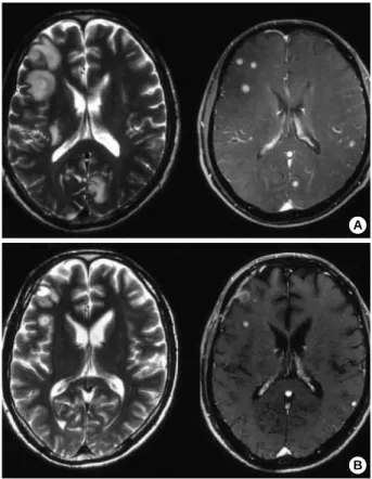
Fig. 1.T2-weighted axial image shows multiple ring-enhanced masses with a high signal in the central portion and the perilesional edema (left upper), and a gadolinium-enhanced T1-weighted image shows multiple ring enhancement (right upper) (A). One month after the initiation of anti-tuberculosis treatment, multiple lesions in the brain showed decreased size and number with im- proving perilesional edema (B).
A
B
Fig. 2.Photomicro- graph of histologic examination dem- onstrated granulo- matous inflamma- tion (A) and myco- bacterium tubercu- losis polymerase chain reaction sho- wed positive find- ings (B).
Sample PC NC Size Marker
132 bp
B A
Intracranial Tuberculomas in Acute Myeloid Leukemia S173
common infection site. Only 1% of immune-competent pa- tients with tuberculosis develop an intracranial tuberculoma, usually as part of miliary tuberculosis (7-9).
The diagnosis of intracranial tuberculomas is difficult to make in immune-compromised patients, as misleading results may arise from non specific symptoms (e.g., headache, fever, weight loss, and weakness) (10). It is especially difficult to distinguish symptoms of a brain tumor from those of an ab- scess (e.g., seizure, headache, visual disturbance, and hemi- paresis) (1, 11), and diagnosis of intracranial tuberculomas based on radiographic and bacteriologic investigations is also quite difficult (3, 12). Cultures of CSF of tuberculoma are usually found to be negative (5), but positive PCR results in the biopsied tissue are found in 60% to 80% of cases. In most cases of intracranial tuberculomas, the initial clinical diagnosis was a neoplasm. For this reason, clinicians must always be aware of neoplasm, and should make the differen- tial diagnosis from various tumorous conditions in the brain (7, 10).
An association with pulmonary tuberculosis was reported from 25% to 83% of cases, and 40% of patients had a his- tory of tuberculosis (1, 2, 5, 11). Therefore, the patient his- tory and findings on chest radiology should be taken together in the diagnosis of intracranial tuberculomas.
Conventional MR imaging is very useful to detect small lesions and to distinguish tuberculomas from other inflam- matory lesions or brain tumors. A slightly hyperintense rim with perilesional edema on T1-weighted images was also described in pyogenic abscesses, and these findings are simi- lar to those of tuberculomas. However, the hypointensity or isointensity that is frequently seen in the central portions of tuberculomas on T2-weighted images can be used to differ- entiate tuberculomas from pyogenic abscesses (5). Diffusion- weighted imaging of the brain showed homogeneous high signals in tuberculomas. In our case, MR imaging of the brain showed a similar finding of tuberculomas, but multi- ple brain lesions and patient disease status (non-remission state) were needed for differential diagnosis between tuber- culomas to other brain tumors (metastatic tumors or granu- locytic sarcomas) (4, 13, 14).
Many brain lesions could be associated with myeloid leu- kemia, and the diagnosis was difficult to establish in this case because of diffuse multiple parenchymal lesions mimicking granulocytic sarcomas or abscesses (15). The case described in this report was an interesting case of intracranial tubercu- losis in AML.
REFERENCES
1. Park SP, Choi JH, Doh JO. Clinical analysis of intracranial tuber- culomas. J Korean Neurosurg Soc 1984; 13: 425-31.
2. Dastur DK, Manghani DK, Udani PM. Pathology and pathogenetic mechanisms in neurotuberculosis. Radiol Clin North Am 1995; 33:
733-52.
3. Tsugawa J, Inoue H, Tsuboi Y, Takano K, Utsunomiya H, Yamada T. Serial MRI findings of intracranial tuberculomas: a case report and review of the literature. No To Shinkei 2006; 58: 225-30.
4. Chi-Shing Z, John LG, Paul K, Hervey DS, Jamshid A. Cerebral infections and inflammation. In: Haaga JL, Lanzieri CF, Gilkeson RC, eds. CT and MR imaging of the whole body. 4th, Vol(1), Cleve- land, USA: Mosby, 2002; 220-4.
5. Kim CY, Kim DG. Central nervous system tuberculoma. J Korean Neurosurg Soc 1998; 27: 21-8.
6. Jagarlamudi R, Kumar L, Kochupillai V, Kapil A, Banerjee U, Thul- kar S. Infections in acute leukemia: an analysis of 240 febrile epi- sodes. Med Oncol 2000; 17: 111-6.
7. Guzel A, Tatli M, Aluclu U, Yalcin K. Intracranial multiple tuber- culomas: 2 unusual cases. Surg Neurol 2005; 64 (Suppl 2): S109-12.
8. Klossek A, Dannenberg C, Feuerhahn MR, Korholz D. Pulmonary tuberculosis in a child receiving intensive chemotherapy for acute myeloblastic leukemia. J Pediatr Hematol Oncol 2004; 26: 64-7.
9. Ku SC, Tang JL, Hsueh PR, Luh KT, Yu CJ, Yang PC. Pulmonary tuberculosis in allogeneic hematopoietic stem cell transplantation.
Bone Marrow Transplant 2001; 27: 1293-7.
10. Bayindir C, Mete O, Bilgic B. Retrospective study of 23 pathologi- cally proven cases of central nervous system tuberculomas. Clin Neurol Neurosurg 2006; 108: 353-7.
11. Farrell VJ. Brain stem tuberculoma in adult patients: diagnosis and treatment. Surg Neurol 1990; 34: 383-9.
12. O'Brien NC, van Eys J, Baram TZ, Starke JR. Intracranial tubercu- loma in children: a new look at an old problem. South Med J 1988;
81: 1239-44.
13. Obara H, Nishimura S, Hayashi N, Numagami Y, Inoue T, Kubo K, Kaimori M, Nishijima M. Intracranial granulocytic sarcoma in a patient with acute myeloid leukemia. No To Shinkei 2006; 58: 797- 801.
14. Tatsui CE, Koerbel A, Prevedello DM, Araujo JC, Ditzel LF, Bleg- gi-Torres LF. Central nervous system granulocytic sarcoma after bone marrow transplant: case report. Arq Neuropsiquiatr 2002; 60:
852-5.
15. Weiser MA, O’Brien S, Escalante C, Manzullo E. Tuberculosis me- ningitis in a patient with acute myelogenous leukemia. Leuk Lym- phoma 1999; 33: 187-92.