Received on April 13, 2009. Revised on April 27, 2009. Accepted on May 4, 2009.
*Corresponding Author. Tel: 82-2-3399-1601; Fax: 82-2-3399-1617; E-mail: [email protected] Keywords: cordycepin, type 2 diabetes, pro-inflammatory cytokines, immunomodulator
Cordycepin Suppresses Expression of Diabetes Regulating Genes by Inhibition of Lipopolysaccharide-induced
Inflammation in Macrophages
Seulmee Shin1, Sungwon Lee1, Jeonghak Kwon1, Sunhee Moon1, Seungjeong Lee2, Chong-Kil Lee2, Kyunghae Cho3, Nam-Joo Ha1 and Kyungjae Kim1*
1College of Pharmacy, Sahmyook University, Seoul, 2College of Pharmacy, Chungbuk University, Cheongju, 3Department of Biology, Seoul Women’s University, Seoul, Korea
Background: It has been recently noticed that type 2 dia- betes (T2D), one of the most common metabolic diseases, causes a chronic low-grade inflammation and activation of the innate immune system that are closely involved in the pathogenesis of T2D. Cordyceps militaris, a traditional me- dicinal mushroom, produces a component compound, cordy- cepin (3’-deoxyadenosine). Cordycepin has been known to have many pharmacological activities including immuno- logical stimulating, anti-cancer, and anti-infection activities.
The molecular mechanisms of cordycepin in T2D are not clear. In the present study, we tested the role of cordycepin on the anti-diabetic effect and anti-inflammatory cascades in LPS-stimulated RAW 264.7 cells. Methods: We confirmed the levels of diabetes regulating genes mRNA and protein of cytokines through RT-PCR and western blot analysis and fol- lowed by FACS analysis for the surface molecules. Results:
Cordycepin inhibited the production of NO and pro-in- flammatory cytokines such as IL-1β, IL-6, and TNF-α in LPS-activated macrophages via suppressing protein ex- pression of pro-inflammatory mediators. T2D regulating genes such as 11β-HSD1 and PPARγ were decreased as well as expression of co-stimulatory molecules such as ICAM-1 and B7-1/-2 were also decreased with the increment of its concentration. In accordance with suppressed pro-in- flammatory cytokine production lead to inhibition of diabetic regulating genes in activated macrophages. Cordycepin sup- pressed NF-κB activation in LPS-activated macrophages.
Conclusion: Based on these observations, cordycepin sup- pressed T2D regulating genes through the inactivation of NF-κB dependent inflammatory responses and suggesting that cordycepin will provide potential use as an immunomod-
ulatory agent for treating immunological diseases.
[Immune Network 2009;9(3):98-105]
INTRODUCTION
Obesity is the most common metabolic disease in the in- dustrial world (1). The prevalence of obesity is increasing at an alarming rate along with its associated morbidities, such as atherosclerosis and type 2 diabetes (2). Diabetes mellitus is a metabolic disorder of the endocrine system that is found in all parts of the world and is rapidly increasing in most parts of the world (3).
Type 2 diabetes mellitus, non-insulin-dependent diabetes mellitus (NIDDM), accounts for more than 90% of diabetes patients. Insulin resistance in peripheral tissues leads to com- pensatory hyperinsulinemia, followed by β-cell failure, leading first to prandial and later to overt fasting hyperglycemia (4).
Recent studies have indicated that obesity is associated with a chronic inflammation state, suggesting that inflammation is a potential mechanism by which obesity leads to insulin re- sistance (5). The authors reported that adipocytes, especially in the obese, secrete a number of pro-inflammatory cyto- kines, some of which have been shown to directly inhibit in- sulin signaling. Adipocytokines probably act through master pro-inflammatory regulators, such as those of the NF-κB and JNK/AP-1 signaling pathways to modulate the expression of genes coding for many inflammatory proteins and to alter in-
Figure 1. Chemical structure of cordycepin.
sulin signaling.
Macrophages, which are a type of differentiated tissue cell that originate as blood monocytes, play an important role in immune and allergic reactions as well as in inflammation (6).
These cells have several functions including the killing and removal of pathogenic microbes and the processing and pre- sentation of antigens ingested by lymphocytes (7).
Cordyceps militaris is known as the rare Chinese caterpillar fungus (8) and has benefits to the human body including the circulatory, immune, respiratory, and glandular systems. Cordy- cepin (Fig. 1), 3’-deoxyadenosine, is a major component of cordyceps millitaris and a derivative of the nucleoside ad- enosine only differing from the latter by the absence of oxy- gen in the 3’ position of its ribose entity (9). Cordycepin has been studied for anti-tumor (10), anti-inflammatory (11), an- ti-diabetic (12), and renoprotective effects (13). Even though cordycepin demonstrates a number of pharmacological prop- erties, further studies are necessary to address these pharma- cological differences.
The manner by which macrophages induce insulin resist- ance in inflammatory responses has not been established, as yet. Macrophages secrete factors induce inflammation in adi- pose tissue and influence insulin sensitivity, but the specific factors involved, and mechanisms by which they exert these effects, remain unknown. In this study, we tested the role of cordycepin on the NF-κB-dependent inflammation cas- cades and inhibition of diabetes regulating genes in lip- opolysaccharide (LPS)-stimulated RAW 264.7 cells.
MATERIALS AND METHODS Reagents
Cordycepin and lipopolysaccharide (LPS) were purchased from Sigma (St. Louis, USA). The cell culture media DMEM, antibiotic-penicillin/streptomycin solution and fetal bovine se-
rum (Hyclone, Logan, USA) were used for the cell culture.
Cell culture
Murine macrophages cell line (RAW 264.7) was obtained from the American Type Culture Collection (ATCC). Cells were cultured in Dulbecco’s Modified Eagle Medium (DMEM) supplemented with high glucose, L-glutamine, 110 mg/L so- dium pyruvate, 10% fetal bovine serum (FBS), and 1% (v/v) penicillin (10,000 U/ml)/ streptomycin (10,000 U/ml) (P/S).
The cells were stimulated with LPS (100 ng/ml) in the pres- ence of cordycepin for 24 hr at a concentration 2×10⁵ cells/well/200μl of media on 96-well plates for the NO assay.
MTT assay for cell viability
A commercially-available cell viability assay was employed to evaluate the cytotoxic effect of cordycepin using thiazolyl blue tetrazolium bromide (Sigma, St. Louis, USA). RAW264.7 cells (2×10⁵ cells/well) were plated with various concen- trations of cordycepin in 96-well microtiter plates (Nunc, Roskilde, Denmark) and were then cultured overnight at 37oC in a 5% CO2 incubator. Afterwards, 50μl of MTT solution was added to each well, and the cells were then cultured for 4 hrs at 37oC in a 5% CO2 incubator. 100μl of solubilized sol- ution were added to each well. The plate was allowed to stand overnight in the incubator after evaluation for complete solubilization of the purple formazan crystals and the meas- urement of the optical density (OD) at 560 nm by a micro- plate reader (Molecular Devices corporation, Sunnyvale, USA).
Measurement of NO content
To assay the total production of NO, 100μl of each culture supernatant were incubated at room temperature for 10 min with 100μl of Griess reagent (stock-I: 0.2% N-(1-naphthyl) ethylenediamine-HCl, stock-II: 2% sulfanilamide in 5% H2PO4).
The O.D values of samples were read at 540 nm. A standard are curve using NaNo2 was then used to calculate the NO2−
concentration.
Isolation of total RNA and RT-PCR
Total RNA was extracted from RAW 264.7 cells using the RNeasy Mini kit (QIAGEN, USA) in an RNase-free environment.
RNA was quantified by reading the absorbance at 260 nm as previously described (14). The reverse transcription of 1μg RNA was carried out using M-MLV reverse transcriptase (Promega, USA), oligo (dT) 16 primer, dNTP (0.5μM) and 1 U RNase inhibitor. After incubation at 65oC for 5 min and
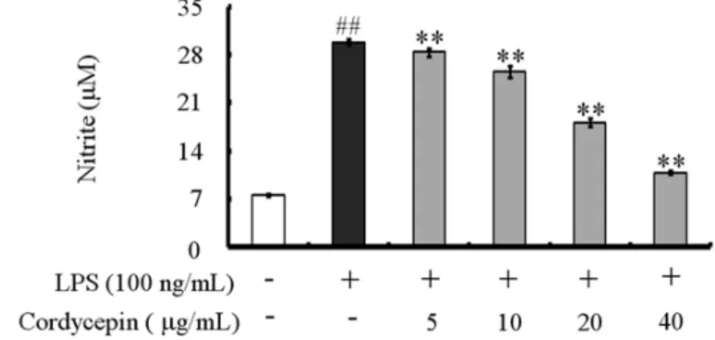
Figure 2. Effect of cordycepin on NO production in RAW 264.7 cells. Cells were treated with different concentrations of cordycepin; nitrite concentrations in the culture media were determined using Griess reagent assay. The results are reported as mean±S.D. of 3 independent experiments. ##p
<0.01 vs. cells only based on Student’s t-test. **p<0.01 vs.
cells only based on Student’s t-test.
37oC for 60 min, M-MLV reverse transcriptase was inactivated by heating at 70oC for 15 min. The polymerase chain reaction (PCR) was performed in 50 mM KCl, 10 mM Tris-HCl (pH 8.3), 1.5 mM MgCl2 and 2.5 mM dNTPs with 5 units of Taq DNA polymerase and 10 pM of each primer set for 11β- htdroxysteroid dehydrogenase type 1 (11β-HSD1), perox- isome proliferators-activated receptor γ (PPARγ), and regu- lated upon activation normal T-cell expressed and secreted (RANTES). The cDNA was amplified by 35 cycles of denatur- ing at 94oC for 45 s, annealing at 62oC for 45 s, and extension at 72oC for 1 min. Final extension was performed at 72oC for 5 min. The PCR products were electrophoresed on a 1.5%
agarose gels and stained with ethidium bromide. The primers used were 5' CAAGGCGGGAAAGCTCATGG 3' (forward) and 5' GGAGGAGATGACGGCAATGC 3' (reverse) for 11β-HSD1, 5' ATCATCCTCACTGCAGCCGC 3' (forward) and 5' CACACT- TGGCGGTTCCTTCG 3' (reverse) for RANTES, 5' GAGCCT- GTGAGACCAACAGC 3' (forward) and 5' GATTCCGAAGT- TGGTGGGCC 3' (reverse) for PPARγ, and 5' GTGGGCC- GCCCTAGGACCAG 3' (forward) and 5' GGAGGAAGAGGA- TGCGGCAGT 3' (reverse) for β-actin. β-actin was used as an internal control.
Preparation of nuclear extracts
After culture the cells were collected and washed twice with cold PBS, resuspended in hypotonic buffer (10 mM HEPES, pH 7.9, 10 mM KCl, 1.5 mM MgCl2, 0.2 mM PMSF, 0.5 mM DTT, 10μg/ml aportinin). After 15 min incubation on ice, the cells were lysed by the addition of 0.1% NP-40 and vigorous vortexing for 1 min. The nuclei were pelleted by cen- trifugation at 12,000×g for 1 min at 4oC and resuspended in high salt buffer (20 mM HEPES, pH 7.9, 25% glycerol, 400 mM KCl, 1.5 mM MgCl2, 0.2 mM EDTA, 0.5 mM DTT, 1 mM NaF, 1 mM sodium orthovanadate). The supernatant fluid was
stored in aliquots at −70oC.
Western blot analysis
RAW 264.7 cells were washed with phosphate-buffered saline (PBS) and lysed by lysis buffer (1% SDS, 1.0 mM sodium va- nadate, 10 mM Tris-Cl buffer, pH 7.4) for 5 min. 20μg of protein from the cell lysates was applied to 8∼12% SDS-poly- acrylamide gels and then transferred to nitrocellulose mem- branes. The membranes were blocked with 5% skim milk in PBST solution for 1 hr. They were then incubated with an- ti-IL-1β, anti-IL-6, anti-TNF-α, anti-i-NOS, anti-COX-2, and anti-NF-κB monoclonal antibody for 2 hrs and washed 3 times with PBST. After incubation with alkaline phospha- tase-labeled secondary antibody for 2 hrs, the bands were vi- sualized using a Western Blot Kit with alkaline phosphatase substrate (Vector, Burlingame, USA).
Flow cytometry
RAW 264.7 cells (1×10⁶ cells/ml) were cultures in Petri-dishes.
The cells were treated with various concentration of cordyce- pin (10, 20, 40μg/ml) in the presence of LPS (100 ng/ml).
The dishes were incubated at 37oC for 24 hrs in humidified 5% CO2 incubator under standard conditions. The cells wash- ed with PBS. The washed cells blocked with staining buffer containing 10% normal mouse serum (NMS) for 20 min on ice. The blocked cells were incubated with co-stimulatory molecules such as ICAM-1, B7-1 and B7-2 antibody for 20 min on ice. The incubated cells were washed with staining buffer at 3 times. The washed cells fixed by 1% paraformalde- hyde in PBS. The fixed cells were measured by flow cy- tometry (Beckman coulter, Brea, USA).
Data analysis
Data are expressed as mean±standard deviation. Statistical
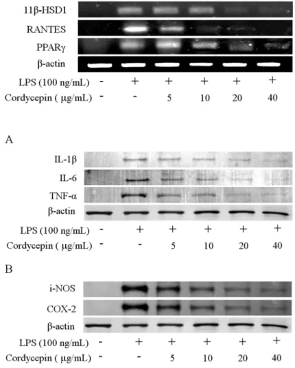
Figure 3. Effect of cordycepin on the expression of T2D regulating genes in RAW 264.7 cells. Levels of 11β- HSD1, RANTES, and PPARγ mRNA in RAW 264.7.
Cells were incubated with various concentrations of cordycepin in the presence of LPS (100 ng/mL) for 24 hrs. The mRNA levels of T2D regulating genes were determined by RT-PCR analysis. β-actin was used as a control.
Figure 4. Effect of cordycepin on the expression of pro-inflammatory cytokines (A) and related proteins (B) in RAW 264.7 cells. Levels of IL-1β, IL-6, and TNF-α (A) and i-NOS and COX-2 (B) in RAW 264.7 cells. Cells were incubated with various concentrations of cordy- cepin in the presence of LPS (100 ng/ml) for 24 hrs.
Protein (20μg) from each sample was resolved in 8∼
12% SDS-PAGE and then analyzed by Western blotting.
β-actin was used as a control.
significance between the groups was determined by paired t-test and one-way ANOVA for repeated measures. Results with p<.05 were considered statistically significant. Data were assessed using an SPSS program (version 15.0, SPSS Inc., Chicago, Illinois).
RESULTS
Effect of cordycepin on cell viability
To rule out the toxic effect of cordycepin, we tested its effect on the viability of RAW 264.7 by MTT assay. The exposure of cells to cordycepin at 5∼40μg/ml for 24 hr showed no significant adverse effect on the cell viability versus the un- treated control (data not shown).
Reduction of NO production in LPS-stimulated RAW 264.7 by cordycepin
In an effort to investigate the effect of cordycepin, we first confirmed whether cordycepin inhibits NO production in acti- vated macrophages. The macrophages did not release NO in response to the medium alone; LPS (100 ng/ml) was used as a positive control for macrophage activation (Fig. 2). When various concentration of cordycepin (5, 10, 20, 40μg/ml) were added to the culture media in the presence of LPS (100 ng/ml) at the time of cell stimulation (18 hrs), NO production was decreased in a cordycepin concentration-dependent manner.
Inhibition the gene expression of T2D regulating protein and chemokine by cordycepin
To further investigate to important role of cordycepin on T2D,
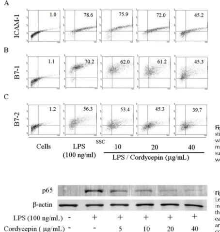
Figure 5. Effects of cordycepin on the expression co- stimulatory molecule. RAW 264.7 cells were cultured with various concentrations of cordycepin (10, 20, 40μg/
ml) in the presence of LPS (100 ng/ml) for 24 hours. The surface ICAM-1 (A), B7-1 (B), and B7-2 (C) molecules were labeled with either anti-ICAM-1, anti-B7-1/-2.
Figure 6. Effect of cordycepin on NF-κB activation.
Levels of NF-κB protein in RAW 264.7 cells. Cells were incubated with various concentrations of cordycepin in the presence of LPS (100 ng/ml) overnight. Protein from each sample was resolved in 12% SDS-PAGE and then analyzed by Western blotting. β-actin was used in as a control.
murine macrophage cells with cordycepin (5∼40μg/ml) in the presence of LPS (100 ng/ml) for 24 hrs decreased T2D regulating genes. As shown in Fig. 3, cordycepin suppressed 11β-HSD1, RANTES, and PPARγ expression dose-depen- dently.
Effect of cordeycepin on pro-inflammatory cytokines and related proteins
We determined the intracellular levels of pro-inflammatory cytokines, and related proteins by western blot analysis, showing that cordycepin decreased IL-1β, IL-6, TNF-α, i-NOS, and COX-2 in a dose-dependent manner (Fig. 4).
Regulated the surface expression levels of co-sti- mulatory molecules
The RAW 264.7 cell surface expression of ICAM-1, B7- 1, and B7-2 was examined by flow cytometry. As shown in Fig. 5, cordycepin inhibited cell surface molecules such as ICAM-1, B7-1, and B7-2 in a dose-dependent manner. LPS-stimulated
RAW 264.7 treated with a high concentration of cordycepin (40μg/ml) had a greater reduction than other concentration.
Suppressed NF-κB activation by cordycepin To investigate whether cordycepin could affect nuclear trans- location of NF-κB, western blot analysis for NF-κB p65 was carried out with cell lysate in macrophages (Fig. 6). Amount of NF-κB p65 was markedly increased upon exposure to LPS alone, but cordycepin decreased NF-κB p65.
DISCUSSION
The data presented in this paper indicated that cordycepin can exert significant anti-diabetic effects on macrophage-me- diated immune responses. The present study demonstrated that cordycepin suppressed NO generation, cytokine (IL-1β, IL-6, and TNF-α) expression, and co-stimulatory molecules in RAW 264.7 cells. M1 macrophages exposed to the classic activation signals IFN-γ and LPS express opsonic receptors,
whereas M2 macrophages are characterized by abundant lev- els of non-opsonic receptors (15). Inflammation is a complex process involving numerous mediators of cellular and plasma origins. M1 macrophages fuse their lysosomes more effi- ciently to phagosomes, exposing intracellular or recently in- gested extracellular microbes to a variety of microbiocidal ly- sosomal enzymes. M1 macrophages also produce oxygen rad- icals and NO, both of which have potent antimicrobial activity.
Arginine metabolism is characterized by high levels of iNOS in M1 macrophages, whereas the arginase pathway predom- inates in M2 polarized macrophages. NO is synthesized via the oxidation of arginine by a family of NOS and plays a vital role in regulating physiological processes, such as blood ves- sel tone and neurotransmission, as well as in host defense and immunity (16,17). However, increasing evidence in- dicates that NOS plays a complex role in modulating in- flammatory response (18). Among these cytokines, IL-1β, IL-6, and TNF-α, have attracted more attention in that they can be localized to the infected tissue, manifested systemically throughout the body and cause vasodilation as well as symp- toms of inflammation, such as redness, swelling, heat, and pain (19).
Cordycepin down-regulated the expression of pro-in- flammatory molecules likewise NO in LPS-stimulated RAW 264.7 cells (Fig. 2) and when we examined the morpho- logical changes that took place in macrophages treated with cordycepin and LPS, cells treated with LPS and low concen- trations of cordycepin (5∼10μg/ml) were similar those ex- posed to LPS alone. However, cells treated with high concen- tration of cordycepin (40μg/ml) in combination with LPS were smoother than those treated with LPS alone (data not shown). Also it inhibited the activation of pro-inflammatory cytokines and related proteins in both of the LPS-activated cell types (Fig. 4).
Macrophages secreted anti-inflammatory cytokines by cor- dycepin, it differentiate M2 macrophages. M2 macrophages are generally characterized by low production of pro-in- flammatory cytokines and high expression of scavenger re- ceptors (15). Cordycepin increased TG accumulation in mac- rophages (Supplementary Fig. 1). It indicated that M2 macro- phages uptake TG from circulation, then are used for fuel (20).
Expression of cytokines requires the activation of NF-κB.
NF-κB, a nuclear transcription factor, regulates the ex- pression of various genes, including IL-1β, i-NOS, and COX-2
that play critical roles in apoptosis and autoimmune diseases.
Its activation requires phosphorylation of IκB, by which lead IκB to ubiquitination and degradation. As shown in Fig. 6, cordycepin decreased pro-inflammatory mediators via sup- pression of NF-κB activation in murine macrophages.
Growing evidence has pointed to a correlative and causa- tive relationship between inflammation and insulin resistance.
The pro-inflammation cytokines such as TNF-α and IL-6 have been demonstrated to mediated insulin resistance as a result of obesity in many rodent obesity models (21,22). TNF- α was overexpressed in white adipose tissue (WAT) in obese an insulin-resistance states; mice lacking the TNF-α li- gand or the p55 TNF receptor were partially protected from obesity-induced insulin resistance (23).
Increasingly, insulin resistance has been recognized as the integral feature of the so-called metabolic syndrome, which includes glucose intolerance, insulin resistance, obesity, hy- pertriglyceridemia, hypertension, and accelerated athero- sclerosis (24).
Glucocorticoids regulate adipocyte differentiation, function and distribution, and in excess, cause visceral fat obesity and convergence of metabolic disease (25). Glucocorticoid re- ceptor (GR) is controlled by isozymes of 11β-HSD. Although two isoforms have been identified, it is 11β-HSD1 that has attracted attention with respect to therapeutic inhibition. 11β- HSD1 is a bidirectional enzyme that resides within the endo- plasmic reticulum and is widely expression in many gluco- corticoid target tissues including liver, adipose tissue where it acts locally to regenerate active cortisol from inactive corti- sone and thereby amplify GR activation (26).
RANTES is increased in WAT in the setting of murine and human obesity. Both mRNA and protein levels of RANTES were increased in a gender-dependent fashion in WAT of obesity. RANTES levels were particularly elevated in male mice in the stromal/vascular fraction of WAT as compared with its adipocyte fraction. In addition, monoclonal anti- bodies directed against RANTES reduced T-cell chemotaxis in- duced by media conditioned by WAT isolated from obese male mice. These findings underscore the role of RANTES-in- duced T-cell chemotaxis by WAT in obesity and suggest an opportunity for pharmacological interventions (27).
The transcription factor PPAR is a member of steroid re- ceptor superfamily and has three subtypes named α, δ/β, γ (28). Specially, PPARγ is characterized originally as a key regulation of adipocyte differentiation and lipid metabolism.
And it has been shown in macrophage foam cells in athero-
sclerotic plaques (29).
As shown in Fig. 3, cordycepin decreased diabetes regulat- ing genes such as 11β-HSD1, RANTES, and PPARγ in acti- vated macrophages.
IL-1 receptor antagonist (IL-1ra) is a antagonist to the pro-inflammatory cytokine IL-1 receptor, anti-inflammatory cytokine, and it is commonly thought to play an important role in the regulation of inflammatory responses, as corrobo- rated by the enhanced sensitivity of IL-1ra knock-out mice to septic shock and their predisposition to the spontaneous de- velopment of inflammatory disorders (30,31). IL-1ra mRNA may contribute to regulation of IL-1ra synthesis that another mechanism may involve inefficient functioning of poly- adenylation determinants present in the distal portion of the IL-1ra 3’- untranslated region (3’-UTR) (32).
Cordycepin is an inhibitor of transcription and poly- adenylation (33), so that gene expression of IL-1ra in our data remained unprocessed (data not shown).
Intracellular adhesion molecules (ICAMs), ICAM-1, ICAM-2 and ICAM-3, are cell surface ligands for leukocyte integrins.
They are crucial in the binding of lymphocytes and other leu- kocytes to certain cells including antigen-presenting cells (APCs). Cordycepin consistently suppressed expression of ICAM-1 surface molecule in macrophages (Fig. 5A). The B7 family plays an important role as a co-stimulatory factor in APCs. Cordycepin treatment had another major effect on co-stimulatory molecules B7-1/-2 by strongly down-regulating the surface levels of B7-1/-2 molecules in macrophage cells (Fig. 5B, C). All these molecules have been described to be of major importance in APC function (34,35).
In conclusion, we have demonstrated that cordycepin pos- sessed anti-inflammatory and anti-diabetic effects on macro- phages. Our findings are supported by inhibitory activity of cordycepin on the NF-κB pathway and other bioactive sub- stances, as well as inhibited expression of diabetes regulating genes.
ACKNOWLEDGEMENTS
This paper was supported by the Sahmyook University Research fund in 2009.
CONFLICTS OF INTEREST
The authors have no financial conflict of interest.
REFERENCES
1. Hotamisligil G, Arner P, Caro J, Atkinson R, Spiegelman B: Increased adipose tissue expression of tumor necrosis factor-alpha in human obesity and insulin resistance. J Clin Invest 95;2409-2415, 1995
2. Ogden C, Carroll M, Curtin L, McDowell M, Tabak C, Flegal K: Prevalence of overweight and obesity in the United States, 1999-2004. Am Med Assoc 295;1549-1555, 2006 3. Li W, Zheng H, Bukuru J, De Kimpe N: Natural medicines
used in the traditional Chinese medical system for therapy of diabetes mellitus. J Ethnopharmacol 92;1-21, 2004 4. DeFronzo R: Pathogenesis of NIDDM. A balanced overview.
Diabetes care 15;318-368, 1992
5. sjöholm A, Nyström T: Endothelial inflammation in insulin resistance. The Lancet 365;610-612, 2005
6. Ross JA, Auger MJ, In Burke B, Lewis CE: The biology of the macrophage. In: Burke B, Lewis CE, editors. The mac- rophage. 2nd ed. Oxford: Oxford Medical Publications;
p1-72, 2002
7. Kinne R, Brauer R, Stuhlmuller B, Palombo-Kinne E, Burmester G: Macrophages in rheumatoid arthritis. Arthritis Research 2;189-202, 2000
8. Gai G, Jin S, Wang B, Li Y, Li C: The efficacy of Cordyceps militaris capsules in treatment of chronic bronchitis in com- parison with Jinshuibao capsules. Chin J New Drugs 13;
169-171, 2004
9. Paterson R: Cordyceps: a traditional Chinese medicine and another fungal therapeutic biofactory? Phytochemistry 69;
1469-1495, 2008
10. Yoo H, Shin J, Cho J, Son C, Lee Y, Park S, Cho C: Effects of Cordyceps militaris extract on angiogenesis and tumor growth. Acta Pharmacologica Sinica 25;657-665, 2004 11. Kim H, Shrestha B, Lim S, Yoon D, Chang W, Shin D, Han
S, Park S, Park J, Park H: Cordycepin inhibits lip- opolysaccharide-induced inflammation by the suppression of NF-kappaB through Akt and p38 inhibition in RAW 264.7 macrophage cells. Eur J Pharmacol 545;192-199, 2006 12. Yun Y, Han S, Lee S, Ko S, Lee C, Ha N, Kim K: Anti-dia- betic effects of CCCA, CMESS, and cordycepin from Cordyceps militaris and the immune responses in streptozo- tocin-induced diabetic mice. Nat Pro Sci 9;291-298, 2003 13. Cho M, Lee D, Kim M, Sung J, Ham S: Antimutagenicity
and cytotoxicity of cordycepin isolated from Cordyceps militaris. Food Sci Biotechnol 12;472-475, 2003
14. Majumder N, Dey R, Mathur R, Datta S, Maitra M, Ghosh S, Saha B, Majumdar S: An unusual pro-inflammatory role of interleukin-10 induced by arabinosylated lipoarabi- nomannan in murine peritoneal macrophages. Glycoconju- gate J 23;675-686, 2006
15. Mantovani A, Sica A, Sozzani S, Allavena P, Vecchi A, Locati M: The chemokine system in diverse forms of macro- phage activation and polarization. Trends in immunology 25;677-686, 2004
16. Furchgott R, Cherry P, Zawadzki J, Jothianandan D:
Endothelial cells as mediators of vasodilation of arteries. J
Cardiovasc Pharmacol 6;S336-343, 1984
17. Rausch-Fan X, Matejka M: From plaque formation to perio- dontal disease, is there a role for nitric oxide? Eur J Clin Invest 31;833-835, 2008
18. Wei X, Charles I, Smith A, Ure J, Feng G, Huang F, Xu D, Muller W, Moncada S, Liew F: Altered immune re- sponses in mice lacking inducible nitric oxide synthase.
Nature 375;408-411, 1995
19. Haddad JJ, Safieh-Garabedian B, Saadé NE, Kanaan SA, Land SC: Chemioxyexcitation (delta pO2/ROS)-dependent release of IL-1 beta, IL-6 and TNF-alpha: evidence of cyto- kines as oxygen-sensitive mediators in the alveolar epithe- lium. Cytokine 13;138-147, 2001
20. Moestrup S, Moller H: CD163: a regulated hemoglobin scavenger receptor with a role in the anti-inflammatory response. Annals of Medicine 36;347-354, 2004
21. Hotamisligil G, Shargill N, Spiegelman B: Adipose ex- pression of tumor necrosis factor-alpha: direct role in obe- sity-linked insulin resistance. Science 259;87-91, 1993 22. Rotter V, Nagaev I, Smith U: Interleukin-6 (IL-6) Induces
Insulin Resistance in 3T3-L1 Adipocytes and Is, Like IL-8 and Tumor Necrosis Factor-α, Overexpressed in Human Fat Cells from Insulin-resistant Subjects. J Bio Chem 278;
45777-45784, 2003
23. Uysal KT, Wiesbrock SM, Hotamisligil GS: Functional analy- sis of tumor necrosis factor (TNF) receptors in TNF-al- pha-mediated insulin resistance in genetic obesity. Endo- crinology 139;4832-4838, 1998
24. Xu H, Barnes G, Yang Q, Tan G, Yang D, Chou C, Sole J, Nichols A, Ross J, Tartaglia L: Chronic inflammation in fat plays a crucial role in the development of obesity-re- lated insulin resistance. J Clin Invest 112;1821-1830, 2003 25. Bujalska I, Kumar S, Stewart P: Does central obesity reflect
Cushing's disease of the omentum? Lancet (British edition) 349;1210-1213, 1997
26. Tomlison J, Stewart P: 11β-Hydroxysteroid dehydro- genase type I as a therapeutic target in the metabolic syndrome. Drug Discov Today 2;93-96, 2005
27. Wu H, Ghosh S, Perrard X, Feng L, Garcia G, Perrard J, Sweeney J, Peterson L, Chan L, Smith C: T-cell accumu- lation and regulated on activation, normal T cell expressed secreted upregulation in adipose tissue in obesity. Circulat- ion 115;1029, 2007
28. Issemann I, Green S: Activation of a member of the steroid hormone receptor superfamily by peroxisome proliferators.
Nature 347;645-650, 1990
29. Marx N, Sukhova G, Murphy C, Libby P, Plutzky J:
Macrophages in human atheroma contain pparγ different- iation-dependent peroxisomal proliferator-activated re- ceptor γ (PPARγ) expression and reduction of MMP-9 ac- tivity through PPARγ activation in mononuclear phagocy- tes in vitro. ASIP 153;17-23, 1998
30. Nicklin M, Hughes D, Barton J, Ure J, Duff G: Arterial in- flammation in mice lacking the interleukin 1 receptor antag- onist gene. J Exp Med 191;303-312, 2000
31. Horai R, Saijo S, Tanioka H, Nakae S, Sudo K, Okahara A, Ikuse T, Asano M, Iwakura Y: Development of chronic inflammatory arthropathy resembling rheumatoid arthritis in interleukin 1 receptor antagonist-deficient mice. J Exp Med 191;313-320, 2000
32. Yamshchikov V, Mishina M, Cominelli F: A possible role of IL-1ra 3’-untranslated region in modulation of protein production. Cytokine 17;98-107, 2002
33. Beach L, Ross J: Cordycepin. An inhibitor of newly synthe- sized globin messenger RNA. J Bio Chem 253;2628-2632, 1978
34. Dazzi F, D'andrea E, Biasi G, De Silvestro G, Gaidano G, Schena M, Tison T, Vianello F, Girolami A, Caligaris-Cappio F: Failure of B cells of chronic lymphocytic leukemia in presenting soluble and alloantigens. Clin Immunol Immu- nopathol 75;26-32, 1995
35. Deeths MJ, Mescher MF: ICAM-1 and B7-1 provide similar but distinct costimulation for CD8+ T cells, while CD4+ T cells are poorly costimulated by ICAM-1. Eur J Immunol 29;45-53, 1999