Introduction
The advent of cone-beam computed tomography(CBCT) changed the patterns of imaging use in the field of dental and maxillofacial imaging. The field of view(FOV) and resolution obtained on most CBCT scanners provide a comprehensive depiction of critical anatomic structures in the maxillofacial region, extending into the calvarium and the neck. Hence, in addition to evaluation of any pa- thology associated with the hard tissues of the jaws and sino-nasal structures, the physiologic and pathologic cal-
cifications associated with the soft tissues of the head and neck can be explicitly evaluated with CBCT. Vascular and intracranial calcifications are among the most studied soft tissue calcifications in the head and neck area.1-3 Pineal gland calcifications have been studied in both the medi- cal and dental literature,2,4-8 but these calcifications have largely been reported as incidental observations, with no specific clinical relevance.
The pineal gland is a neuronal structure located between the 2 cerebral hemispheres that produces melatonin, a hormone that modulates sleep patterns in both circadian and seasonal cycles.9 The pineal gland typically calcifies over time.9,10 Studies have shown that pineal gland calci- fication can be noted in children as young as 5 years.9,11 The degree and frequency of calcification have been noted to increase with age.10 Pineal gland calcification also de-
Prevalence of pineal gland calcification as an incidental finding in patients referred for implant dental therapy
Sunil Mutalik1, Aditya Tadinada1,*
1Section of Oral and Maxillofacial Radiology, School of Dental Medicine, University of Connecticut Health Center, Farmington, CT, USA
ABSTRACT
Purpose: Pineal gland calcification has been proposed to play a role in the pathogenesis of Alzheimer disease. This study evaluated the prevalence and extent of pineal gland calcification in cone-beam computed tomography(CBCT) scans of patients referred for dental implant therapy who could possibly be a vulnerable group for this condition.
Materials and Methods: A retrospective evaluation of 500 CBCT scans was conducted. Scans that showed the area where the pineal gland was located were included. The scans were initially screened by a single observer to record the prevalence and extent of calcification. Six weeks following the completion of the study, another investigator randomly reviewed and selected 50 scans to investigate inter-observer variation, which was evaluated using reli- ability analysis statistics. The prevalence and measurements of the calcifications were reported using descriptive statistics. The chi-square test was used to compare the prevalence between males and females.
Results: The prevalence of pineal gland calcification was 58.8%. There was no statistically significant correlation between age and the extent of the calcification. The prevalence of calcification was 58.6% in females and 59.0% in males. The average anteroposterior measurement was 3.73±1.63mm, while the average mediolateral measurement was 3.47±1.31mm. The average total calcified area was 9.79±7.59mm2.
Conclusion: The prevalence of pineal gland calcification was high in patients undergoing implant therapy. While not all pineal gland calcifications lead to neurodegenerative disorders, they should be strongly considered in the presence of any symptoms as a reason to initiate further investigations.(Imaging Sci Dent 2017; 47: 175-80) KEY WORDS: Pineal Gland; Cone-Beam Computed Tomography; Alzheimer Disease; Dental Implants
Copyright ⓒ 2017 by Korean Academy of Oral and Maxillofacial Radiology
This is an Open Access article distributed under the terms of the Creative Commons Attribution Non-Commercial License(http://creativecommons.org/licenses/by-nc/3.0) which permits unrestricted non-commercial use, distribution, and reproduction in any medium, provided the original work is properly cited.
Imaging Science in Dentistry·pISSN 2233-7822 eISSN 2233-7830 Received April 27, 2017; Revised July 21, 2017; Accepted July 31, 2017
*Correspondence to : Prof. Aditya Tadinada
Department of Oral and Maxillofacial Radiology, School of Dental Medicine, University of Connecticut Health Center, 263 Farmington Avenue, Farmington, CT 06030, USA
Tel) 1-860-679-2579, Fax) 1-860-679-4760, E-mail) [email protected]
pends on environmental factors, such as altitude and sun- light exposure.10 It has been hypothesized that increased pineal gland calcification results in decreased melatonin production.5,12
Reduced melatonin production has been implicated in a variety of neurodegenerative disorders such as migraine, stroke, schizophrenia, and Alzheimer disease.4,5,7,13,14 Pa- tients referred for dental implant therapy usually tend to be individuals in their late 50s to early 60s, and therefore may be a vulnerable group for Alzheimer disease. To fa- cilitate easy identification of the location of the gland and any associated calcification on CBCT scans, the pineal gland is usually noted as a midline calcification in the posterior cranial fossa in axial sections. The calcification is posterior and superior to the level of the sella turcica.
As mentioned earlier, in a large-FOV CBCT scan, pineal calcification is usually an incidental finding. Hence, cli- nicians can contribute to the early diagnosis and preven- tion of challenging neurodegenerative conditions. Since most dental implant patients are middle-aged or older, the possibility of studying this phenomenon in these patients is ideal. The objective of this study was to analyze the CBCT scans of patients referred for dental implant ther- apy to evaluate the prevalence and extent of pineal gland calcification according to age and gender.
Materials and Methods
The present cross-sectional study consisted of the retro- spective evaluation of CBCT scans of patients referred to the University of Connecticut School Of Dental Medicine for advanced imaging. An institutional review board(IRB)
exemption was granted for this study by the University of Connecticut IRB. The inclusion criteria for the study were scans with a large FOV(170mm×120mm), which gen- erally extended from the bregma(forehead) to the menton (lower tip of the bony chin) superoinferiorly and from the anterior nasal spine to the cervical spine anteroposterior- ly, without any metallic or motion artifacts. The exclu- sion criteria were scans that did not adequately show the pineal gland area, had image artifacts that compromised the quality of the scans, or did not have associated details such as gender and age. All scans were acquired with a single CBCT machine(Accuitomo-170; J Morita Corp., Kyoto, Japan), and scans were obtained at 90 kVp and 10 mA. Initially, 1000 scans were screened on the basis of the inclusion and exclusion criteria, and ultimately, 500 scans were selected based on the inclusion criteria. The selected scans were de-identified by a research assistant to remove all patient identifiers. The investigator had ac- cess to the patients’ age and gender from the Digital Im- aging in Communications and Medicine data. The scans were imported and evaluated in a CBCT reconstruction program, Invivo5(Anatomage Inc., San Jose, CA, USA).
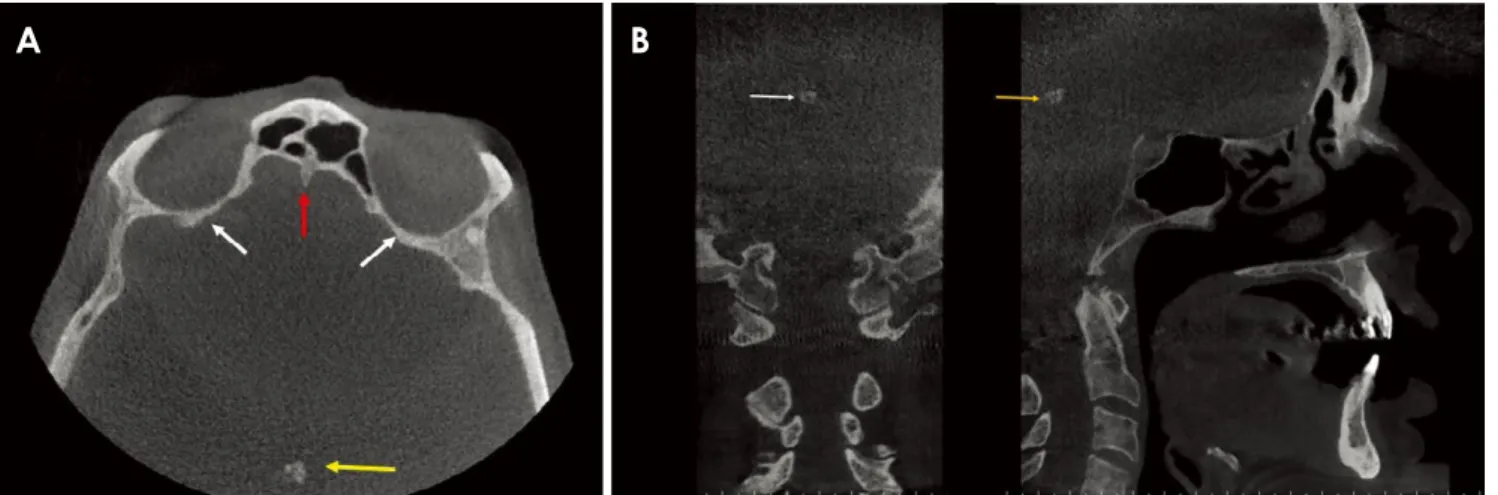
Pineal gland calcification was identified as a midline cal- cification in the posterior cranial fossa within the cranium (Fig. 1). Owing to its small size, pineal gland measure- ments were difficult in the coronal and sagittal planes.
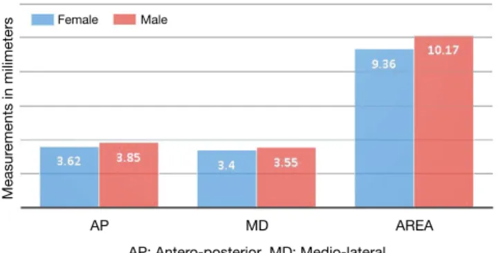
Hence, the size of the calcification was measured in the axial scans in the anteroposterior and lateral directions (linear measurements, Fig. 2). The area was calculated by marking the perimeter of the calcification(areal mea- surements, Fig. 2). The data were entered into Microsoft Excel(Microsoft Corp., Seattle, WA, USA). Statistical
Fig. 1. A. Axial cone-beam computed tomography(CBCT) section. Arrows depict the pineal gland calcification(yellow), the crista galli (red), and the orbital cavity(white). B. Coronal(left) and sagittal(right) CBCT sections depict pineal gland calcification(white arrow in the coronal section; yellow arrow in the sagittal section).
A B
analysis was performed with SPSS version 17.0(SPSS Inc., Chicago, IL, USA). Six weeks following the com- pletion of the study, another investigator randomly re- viewed 50 scans to investigate interobserver variation. A reliability analysis with interclass correlation coefficients was performed to assess interobserver variation. The data were analyzed using descriptive statistics to the calculate mean values and prevalence of pineal gland calcification.
The difference between the prevalence among males and females was analyzed using the chi-square test. The cor- relations between the age of the patient and the linear and areal measurements of pineal calcifications were analyzed using the Pearson correlation coefficient.
Results
Of the 500 scans that were evaluated, 290(58%) were from female patients and 210(52%) were from male pa-
tients. Pineal gland calcification was noted in 294 scans, with a prevalence of 58.8%. Of these scans, 170 were from females(58.6%) and 124 were from males(59.0%).
The chi-square statistic for the difference between male and female subjects was 0.0092(P=.92). The difference was not statistically significant. The mean linear and areal measurements were higher in males than females(Fig. 3).
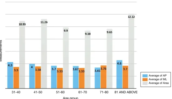
The patients ranged from 32 to 85 years of age, with a mean age of 63.42±10.43 years. The degree of calcifi- cation in different age groups is shown in Figure 4. The mean anteroposterior measurement was 3.73±1.63mm, with a range of 1.14-8.55mm. The mean lateral mea- surement was 3.47±1.31mm, with a range of 0.88-8.01 mm. For each of the measures, the interclass correlation coefficient was measured, as well as the coefficient for linear measurements, which was 0.826(0.604-0.923) for the anteroposterior measurements and 0.853(0.667-0.935) for the mediolateral measurements. The mean area of cal- cification was 9.79±7.59mm2, with a range of 1.20-55.87 mm2. For area, the interclass correlation coefficient was 0.986(0.969-0.994). This basically means that the most consistent agreement was for the areal measurements and that there was good agreement for the anteroposterior and mediolateral measurements. There was no linear rela- tionship between age and the measurements. In addition, there were no correlations between age and the areal or linear measurements(P=.454, r=0.232). However, it is interesting to note that all the measurements were high in patients 81 years of age and above. The difference in the average mediolateral dimension according to age was minimal.
Fig. 2. Pictures depict linear(left) and areal(right) measurements of the pineal gland.
Measurements in milimeters
AP MD AREA
AP: Antero-posterior MD: Medio-lateral Female Male
Fig. 3. Average linear and areal measurements in males and fe- males.
Discussion
Pineal gland calcification, melatonin secretion, and their relationship to numerous neurodegenerative disorders have been a topic of interest to the medical community.15,16 Although the prevalence of pineal calcification has been studied by many authors in the dental literature, few stud- ies have investigated the correlations among age, gender, and the extent of pineal gland calcification.1,2,8 The pres- ent study evaluated the feasibility of CBCT for evaluating pineal gland calcification, and investigated its relation- ship to age and gender. The major limitation of this study is the lack of clinical and laboratory data on the function of the pineal gland, making it difficult to extrapolate these findings to neurodegenerative changes in a clinically rele- vant way.
As stated in the introduction, patterns of pineal gland calcification in younger individuals are different from those in older subjects.9 Pineal gland calcification may also depend on environmental factors, such as altitude and the amount of exposure to sunlight.11 In the current study, we did not observe any specific patterns of calcifi- cation. A possible explanation for this observation could be that none of our patients were younger than 31 years.
Another clear limitation of this study is that a majority of our study population was from the northeast United States, which has sparse sunlight for several months each year.
The prevalence of pineal calcification in the present
study was 58.8%. This prevalence is higher than has been reported in most previous studies using CBCT.1,2 Sedghi- zadeh et al.1 reported that the prevalence of intracranial physiologic calcifications in their study was 35.2%, and that 80% of cases were accounted for by pineal calcifica- tion. Barghan et al.2 reported a prevalence of 11.18% in their study. Studies of multi-slice CT have reported prev- alence rates as high as 60%-80%, depending on the age group studied.11 In the dental literature, pineal calcifica- tion has been most often detected as an incidental finding, and has been disregarded as a physiologic process relat- ed to age.1,2,8 This is likely to be because many patients who receive care at dental clinics are ambulatory and are not often assessed for neurodegenerative disorders. With this study, the authors would like to emphasize that when such calcifications are present, it is important to obtain a thorough medical history from the patient and to assess any other signs and symptoms that might hint at a neuro- degenerative pattern. While we do not suggest that every patient with pineal calcification will have a neurodegen- erative disorder or should be referred for an evaluation of such conditions, it is nevertheless a consideration on the diagnostic list.
Males in general are known to have a higher degree of pineal calcification than females.6,10 In our study, the pre- valence of calcification was slightly higher in female pa- tients. However, this difference was not statistically sig- nificant. Many authors link such differences to the effect of melatonin on gonadotropins.6,17,18 Melatonin antagoniz-
Fig. 4. Distribution of pineal gland measurements in different age groups.
Measurements
31-40 41-50 51-60 61-70 71-80 81 AND ABOVE
Age group
Average of AP Average of ML Average of Area
es the effect of estrogen by stimulating the production of progesterone, while increased levels of testosterone cause a reduction in melatonin levels.6,17,18 This is probably a reason why the role of melatonin has been extensively studied in the pathogenesis of endometrial carcinomas.18
The most important finding of the study was the ab- sence of a correlation between gland calcification and the age of the patients. In addition, the average measurements did not vary significantly according to age. This may have been due to a lack of a sufficient sample size with calci- fications in each age group. More than 200 patients who had calcifications were between 51 and 85 years of age.
Since the size of the pineal gland varies across individu- als, the degree of gland calcification has been suggested to be a more specific indicator of the gland’s function.5,12 It has been shown that the degree of gland calcification is higher in patients with Alzheimer disease than in controls and patients with other types of dementia.5 Calcification has also been proposed to decrease the capability of the gland to produce melatonin.12 The uncalcified gland vol- ume has been directly associated with the production of melatonin. This has been proven using magnetic reso- nance imaging, which is known to have better soft reso- lution than computed tomography.16 Melatonin receptors are found in a variety of tissues in the body, such as the retina, vasculature, immune system, skin, pancreas, and endometrium.17 Due to its effects on these tissues, mel- atonin has been implicated in neurodegenerative, malig- nant, and immunological disorders.17 Since melatonin ex- erts important functions in numerous tissues and is related to functional gland volume, it is important for studies to correlate the degree of gland calcification to melatonin production. As this study was a retrospective analysis, it did not address melatonin levels. Although the area of pineal calcification correlated positively with age in our study, this relationship was not statistically significant. It would be interesting to study the extent of calcification on CBCT and its correlation to the production of melatonin in a larger sample in future research in order to under- stand this correlation better.
The authors conclude that CBCT was helpful in detect- ing the presence of pineal gland calcification. The preva- lence of pineal gland calcification was high as an inciden- tal finding in patients undergoing implant therapy. Pineal gland calcification in males was more common than in females in the current study. However, the study did not show any correlations between age and linear measure- ments or the area of pineal gland calcification. Future re- search should focus on correlating melatonin production
and the extent of calcification on CBCT in patients with clinical signs and symptoms of early neurological degen- eration or deficits.
References
1. Sedghizadeh PP, Nguyen M, Enciso R. Intracranial physiolog- ical calcifications evaluated with cone beam CT. Dentomaxil- lofac Radiol 2012; 41: 675-8.
2. Barghan S, Tahmasbi Arashlow M, Nair MK. Incidental find- ings on cone beam computed tomography studies outside of the maxillofacial skeleton. Int J Dent 2016; 2016: 9196503.
3. Damaskos S, Tsiklakis K, Syriopoulos K, van der Stelt P. Ex- tra- and intra-cranial arterial calcifications in adults depicted as incidental findings on cone beam CT images. Acta Odontol Scand 2015; 73: 202-9.
4. Kitkhuandee A, Sawanyawisuth K, Johns NP, Kanpittaya J, Johns J. Pineal calcification is associated with symptomatic cerebral infarction. J Stroke Cerebrovasc Dis 2014; 23: 249- 5. Mahlberg R, Walther S, Kalus P, Bohner G, Haedel S, Reis-53.
chies FM, et al. Pineal calcification in Alzheimer’s disease: an in vivo study using computed tomography. Neurobiol Aging 2008; 29: 203-9.
6. Mohammed KA, Adjei Boakye E, Ismail HA, Geneus CJ, Tobo BB, Buchanan PM, et al. Pineal gland calcification in Kurdistan: a cross-sectional study of 480 roentgenograms.
PLoS One 2016; 11: e0159239.
7. Bersani G, Garavini A, Taddei I, Tanfani G, Nordio M, Pancheri P. Computed tomography study of pineal calcification in schizophrenia. Eur Psychiatry 1999; 14: 163-6.
8. Edwards R, Altalibi M, Flores-Mir C. The frequency and nature of incidental findings in cone-beam computed tomo- graphic scans of the head and neck region: a systematic re- view. J Am Dent Assoc 2013; 144: 161-70.
9. Whitehead MT, Oh C, Raju A, Choudhri AF. Physiologic pi- neal region, choroid plexus, and dural calcifications in the first decade of life. AJNR Am J Neuroradiol 2015; 36: 575-80.
10. Turgut AT, Karakaş HM, Ozsunar Y, Altın L, Ceken K, Alıcıoğ- lu B, et al. Age-related changes in the incidence of pineal gland calcification in Turkey: a prospective multicenter CT study. Pathophysiology 2008; 15: 41-8.
11. Doyle AJ, Anderson GD. Physiologic calcification of the pi- neal gland in children on computed tomography: prevalence, observer reliability and association with choroid plexus calci- fication. Acad Radiol 2006; 13: 822-6.
12. Kunz D, Schmitz S, Mahlberg R, Mohr A, Stöter C, Wolf KJ, et al. A new concept for melatonin deficit: on pineal calcifi- cation and melatonin excretion. Neuropsychopharmacology 1999; 21: 765-72.
13. Ozlece HK, Akyuz O, Ilik F, Huseyinoglu N, Aydin S, Can S, et al. Is there a correlation between the pineal gland calcifi- cation and migraine? Eur Rev Med Pharmacol Sci 2015; 19:
3861-4.
14. Sandyk R. The pineal gland and the mode of onset of schizo- phrenia. Int J Neurosci 1992; 67: 9-17.
15. Wu YH, Swaab DF. The human pineal gland and melatonin in
aging and Alzheimer’s disease. J Pineal Res 2005; 38: 145-52.
16. Sigurdardottir LG, Markt SC, Sigurdsson S, Aspelund T, Fall K, Schernhammer E, et al. Pineal gland volume assessed by MRI and its correlation with 6-sulfatoxymelatonin levels among older men. J Biol Rhythms 2016; 31: 461-9.
17. Slominski RM, Reiter RJ, Schlabritz-Loutsevitch N, Ostrom
RS, Slominski AT. Melatonin membrane receptors in periph- eral tissues: distribution and functions. Mol Cell Endocrinol 2012; 351: 152-66.
18. Sandyk R, Anastasiadis PG, Anninos PA, Tsagas N. Is the pineal gland involved in the pathogenesis of endometrial car- cinoma. Int J Neurosci 1992; 62: 89-96.